


Abstract
Background: Walking at various speeds and durations may result in different peak plantar pressure (PPP). However, there is no study comparing the effect of walking speeds and durations on PPP. The purpose of this study was to explore whether different walking speeds and durations significantly change PPP and establish a normal response in healthy people. Methods: An in-shoe plantar pressure system was used to measure PPP under the first toe, first metatarsal, second metatarsal, and heel regions in 12 healthy, young people. All participants performed six walking trials at three speeds (3, 6, and 9 km/h) and for two durations (10 and 20 min). The 3 × 2 two-way analysis of variance was used to examine the main effects of speeds and durations and their interaction. Results: The results showed that walking speeds significantly affected PPP and that walking duration did not. No interaction between the walking speed and duration was observed. Peak plantar pressure values under the first toe and the first metatarsal head were significantly higher (P < .05) at 9 km/h (509.1 ± 314.2 kPa and 591.4 ± 302.4 kPa, respectively) than at 3 km/h (275.4 ± 168.7 kPa and 369.4 ± 205.4 kPa, respectively) after 10-min walking. Conclusions: People at risk for foot ulcers may use slow and brisk walking for exercise to reduce PPP, thus reducing risk for foot ulcers. Our study demonstrated that slow running at 9 km/h significantly increases PPP.
Diabetes mellitus (DM) is a common metabolic disease caused by abnormal insulin secretion or insulin action.1] The International Diabetes Federation reported that the number of people diagnosed with type 2 DM was more than 380 million worldwide in 2013, and may grow to 592 million worldwide by 2035.[2] Diabetic foot ulcers (DFUs) are among the most severe diabetes-related complications,[3] resulting in hospitalization and lower limb amputation.[4] It is estimated that 19% to 34% of the diabetic population will develop DFUs in their lifetime.[5] Diabetic foot management costs $9 to $13 billion per year, which places a considerable economic burden on the medical systems in the United States.[6] Therefore, prevention of DFUs plays an essential role in the care of people with DM.[7,8]
Various preventive interventions of DFUs have been suggested, including exercise, healthy diet, and medications to control blood glucose level.[8] Exercise, either aerobic or resistance exercise, has been considered as the most important intervention to regulate blood glucose.[9] A recent systematic review pointed out that exercise could reduce glycated hemoglobin and improve the ankle-brachial index.[10] Glycated hemoglobin has been recommended by the World Health Organization as a significant index for diagnosing and monitoring DM, particularly in type 2 DM.[11] The ankle-brachial index is often used in diabetic care to assess peripheral arterial health status because a low ankle-brachial index is significantly associated with various diabetic complications, including peripheral arterial disease and DFUs.[12] The American Diabetes Association (ADA) recommends that people with type 2 DM should perform moderate to vigorous intensity physical activity at least three sessions per week, and avoiding more than 2 successive days without exercise. The total amount of exercise time in a week must be more than 150 min.[13] Walking is the most common physical activity in people with DM.[14] However, the evidence regarding whether people with diabetic peripheral neuropathy (DPN) engaging in weightbearing physical activity, such as brisk walking, would the increase risk for DFUs remains insufficient.[7,10,15] People with DPN have abnormally high plantar pressure during weightbearing physical activity (eg, walking), and lack protective responses during walking.[13] Weightbearing physical activity might increase plantar pressure, thus increasing risk for DFUs in this population.
Peak plantar pressure (PPP) during weightbearing activity has been broadly used to assess the risk of skin breakdown in people with DM.[16] Individuals with DM exhibit significantly higher PPP compared to nondiabetic people during walking or other weightbearing activities because of loss of sensory feedback and altered soft-tissue biomechanical property of the foot.[17,18,19,20] Peak plantar pressure has been shown to predict DFUs.[21,22,23] A regression model derived from a sample of 380 participants showed that PPP is strongly associated with development of DFUs, concluding that plantar pressure assessment should be adopted in clinical practice.[24] Therefore, PPP may be used to evaluate the foot ulcer risk associated with various intensities of weightbearing exercise.
Until now, there have been no data comparing PPP patterns at various walking speeds and durations in either healthy people or people with DM. From the aspect of PPP and foot ulcer risk, the evidence is weak concerning whether people with DPN should perform moderate intensity physical activity for a longer duration or vigorous intensity physical activity for a shorter duration; both physical activities are recommended by the ADA.[13] Before the relationship between weightbearing physical activity and risk of DFUs has been established in people with DM, there is a need to explore these relationships in healthy people without DM. These findings may help shed light on how weightbearing physical activity behavior could change risk for foot ulcers. In the literature, walking speed has been shown to be related to PPP, with faster walking speed causing higher PPP.[25,26,27] Rosenbaum et al[27] conducted the first study investigating the effect of walking speed on plantar pressure and found that plantar pressure under the medial forefoot significantly increased with increasing speed. However, these previous studies were conducted in a short period of walking ranging from 2 to 7 min. Prolonged walking may gradually increase skin temperature and mechanical stress, which might increase the risk of ulceration.[28,29,30] According to the latest Physical Activity Guidelines for Americans, the minimum duration of physical activity must be more than 10 min.[31] However, it is unclear whether the duration of physical activity would cause different PPP. For example, it is unclear whether longer walking (eg, 20-min walking) would increase PPP compared to shorter walking (eg, 10-min walking). Considering the effect of time on plantar pressure, some previous studies used the pressure-time integral as a variable when examining the risk of foot ulceration.[32] However, studies investigating the pressure-time integral only consider a short walking period rather than a walking duration more than 10 min.[33,34] Given the fact of insufficient information regarding the walking duration effect on PPP, it becomes essential to measure PPP after walking at different speeds and durations.
To the best of our knowledge, there is no study investigating the relationship between PPP and walking durations and speeds. Different combinations of walking speeds and durations may have different impacts on PPP. For developing a proper weightbearing exercise program for people with DPN, it is important to understand the effect of walking speeds and durations on PPP in healthy people first. Before conducting a study in people with DM, the relationship between PPP and walking speeds and durations in healthy people should be established first. We hypothesized that: 1) a faster walking speed would increase PPP; 2) a longer duration would increase PPP; and 3) an interaction occurs between the walking speed and duration on PPP.
Methods
A 3 × 2 factorial design, including three speeds (slow at 3 km/h, moderate at 6 km/h, and fast at 9 km/h) and two durations (10 and 20 min), was used in this study.
Participants
Healthy participants who were aged 18 to 35 years and able to walk independently without any device were recruited from the university and adjacent community. Plantar pressure pattern is not significantly affected by age between 18 and 35 years.[35] The participant was not eligible for the study if he or she had active ulcers on the foot, DM, pain over any lower extremity joints, obvious foot deformities (flat foot or high arch[36]), or a history of foot amputation or other lower extremity operations. The study was approved by the University of Illinois at Urbana-Champaign Institutional Review Board (No. 19225). Before screening and examination procedures, all participants signed the informed consent. Twelve healthy participants (five men and seven women; mean ± standard deviation age, 25.7 ± 5.5 years; height, 1.71 ± 0.09 m; body weight, 64.2 ± 13.5 kg) met the inclusion criteria and volunteered for this study.
Procedure
Three speeds (ie, 3 km/h [slow walking], 6 km/h [fast walking)], and 9 km/h [slow running]) and two durations (10 and 20 min) were tested in this study. These three speeds were common in people with DM in their daily life.[37] All participants were asked to walk at a speed of 3 km/h on a treadmill in the first week, 6 km/h in the second week, and 9 km/h in the third week in a room with the temperature of 24 ± 2°C. For each week, participants were randomly assigned into 10 or 20 min of walking and then the other. For avoiding carryover effects and muscle fatigue, a participant was allowed to rest for at least 20 min between two walking trials. The plantar pressure data were continuously recorded during walking using the F-scan system (Tekscan, South Boston, Massachusetts).[19,38] A suitable pair of standard shoes and socks (Altrex, Teaneck, New Jersey) with F-scan in-shoe sensors between the socks and the insoles were prepared for the participants. An F-scan in-shoe sensor contains 960 sensing elements. The size of each sensing element is 5.08 × 5.08 mm. Before the walking trial, participants would walk for 3 to 5 min to get familiarized with the standard shoes. The sampling rate was 300 Hz.
Statistical Analysis
The plantar pressure data from the last minute of each trial (either 10 or 20 min of walking) were analyzed using Matlab (Natick, Masschusetts) codes to calculate PPP under the first toe (T1), first metatarsal head (M1), second metatarsal head (M2), and heel regions (HL).[19,38] Peak plantar pressure was defined as PPP = max(p), where p is the pressure distribution in the four plantar regions of interest.[19,38] All statistical analyses were performed using IBM SPSS Version 26.0 (IBM Corp, Armonk, New York). The baseline demographic data were presented as mean and standard deviations. Two-way (three speeds and two durations) analysis of variance with repeated measures was used to analyze PPP during six walking trials. The Fisher least significant difference test was used as the post hoc test. The significant level was set as .05.
Results
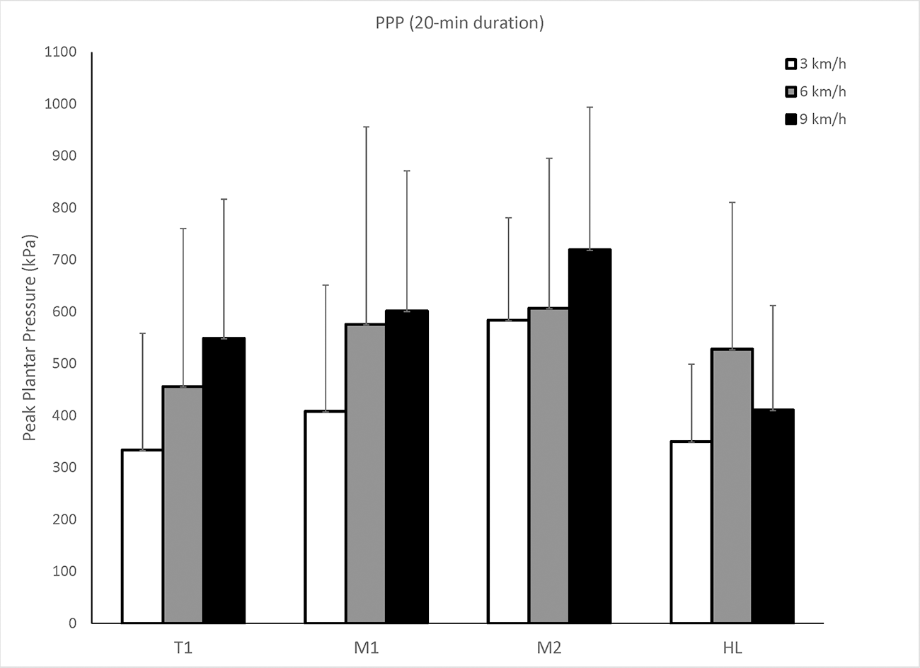
The means and standard deviations of PPP under four plantar areas (ie, T1, M1, M2, and HL) during three speeds (ie, 3, 6, and 9 km/h) and two durations (ie, 10 and 20 min) are provided in Table 1. The result showed that the walking speed affected PPP under the area of T1, M1, and HL (P < .05) and the walking duration did not. The results showed that there was no interaction between the speed and duration factors on PPP. When comparing PPP under a plantar region at a speed between 10-min and 20-min durations, there were no statistical differences (Table 1).
Table 1.
PPP Under Four Plantar Areas During Walking at Three Speeds and Two Durations
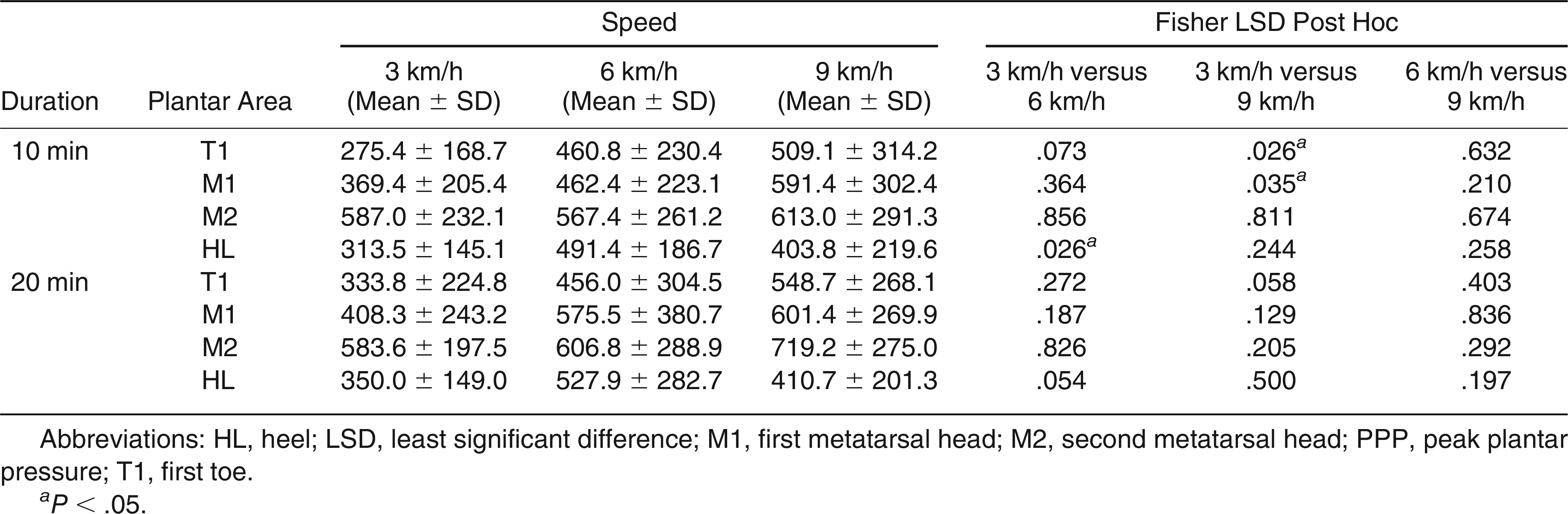
Figure 1 shows PPP under four plantar areas (ie, T1, M1, M2, and HL) after walking at 3 speeds (ie, 3, 6, and 9 km/h) for 10 min. The results showed that walking at 9 km/h significantly increased PPP compared to walking at 3 km/h on T1 (P < .05) and M1 (P < .05). Walking at 6 km/h significantly increased PPP compared to 3 km/h on HL (P < .05).
Figure 1.
Peak plantar pressure (PPP) values under the four plantar areas during walking at three speeds for 10 min. The results show that walking at 9 km/h significantly increases PPP compared to walking at 3 km/h under the first toe (T1) (P < .05) and the first metatarsal head (P < .05). LSD, least significant difference; T1, first toe; M1, first metatarsal head; M2, second metatarsal head; HL, heel.
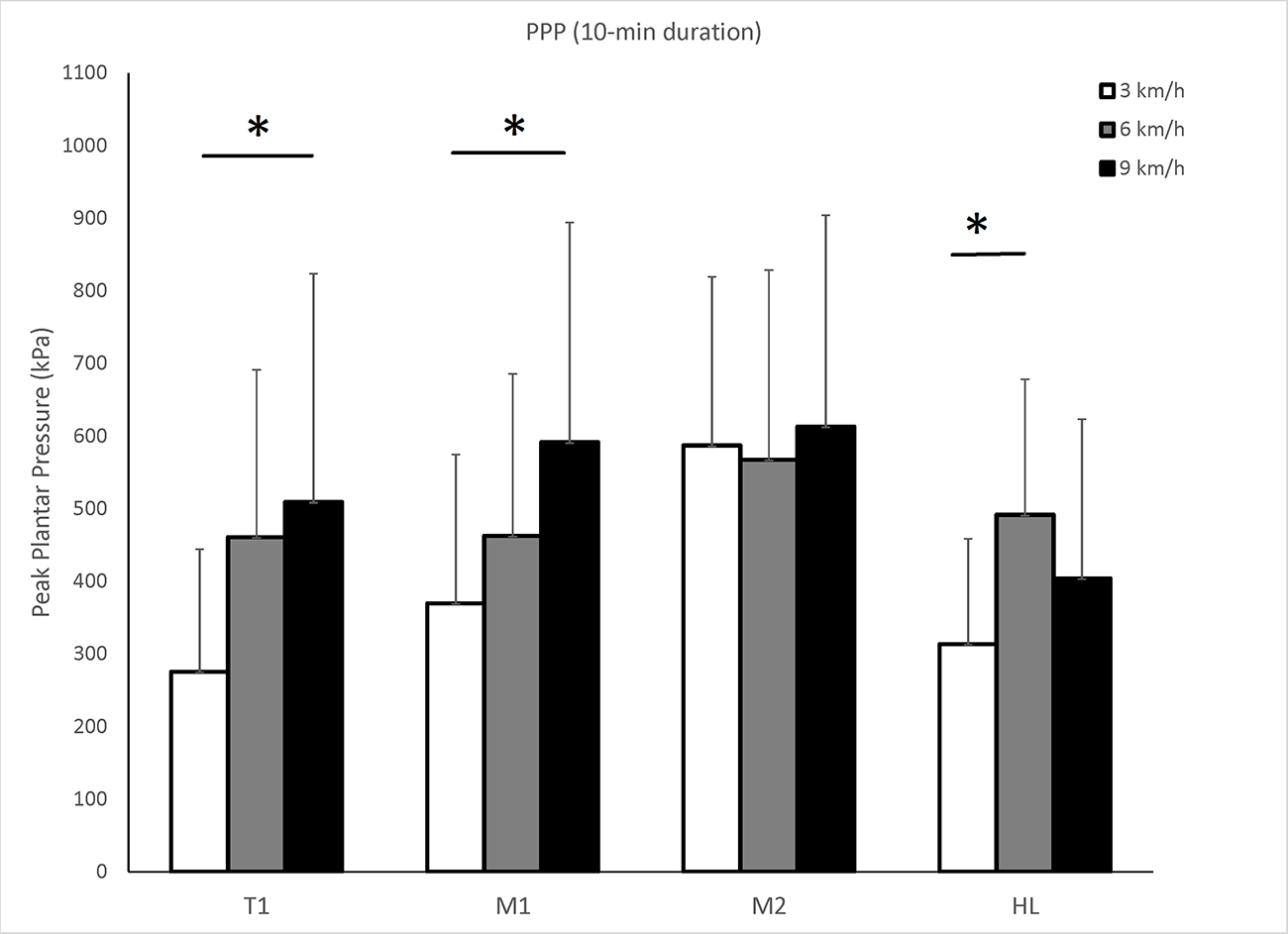
Figure 2 shows PPP under four plantar areas (ie, T1, M1, M2, and HL) during walking at 3 speeds (ie, 3, 6, and 9 km/h) for 20 min. Although there were no statistical differences, there was a trend that PPP increased at a faster walking speed on the big toe, first metatarsal head, and second metatarsal head except the heel.
Figure 2.
Peak plantar pressure (PPP) values under the four plantar areas during walking at three speeds for 20 min. Although there are no statistical differences, there is a trend that PPP increases at a faster walking speed under the first toe, first metatarsal head, and second metatarsal head except the heel. LSD, least significant difference; T1, first toe; M1, first metatarsal head; M2, second metatarsal head; HL, heel.
Discussion
This study aimed to explore whether different walking speeds and walking durations affect individuals’ PPP patterns in people without DM. Traditionally, clinicians assessed an individual’s PPP at the preferred walking speed for a duration of less than 1 min. The lack of consideration of the influence of exercise intensity (in our case, various walking speeds and durations) on PPP partly reflects on the ADA’s exercise guidelines without considering the effect of weightbearing physical activity on the risk level of foot ulcers in people with DPN. In the literature, there are only a few studies investigating the effect of walking speeds on PPP in healthy people. To the best of our knowledge, our study is the first study investigating the effect of various walking speeds and walking durations on PPP patterns. We intended to demonstrate the influence of walking speeds and walking durations on PPP values in healthy people first. Our results may provide a foundation to explore the influence of various walking speeds and durations on the risk of DFUs in people with DM. As discussed in a recent systematic review and meta-analysis,[10] the field focuses on the effect of physical activity on reducing blood glucose level and improving vascular health, and ignores whether weightbearing physical activity (eg, brisk walking) may increase risk for DFUs. In this study, we demonstrated the effect of the walking speeds and durations on PPP values.
The current result demonstrates that walking at 9 km/h for 10 min significantly increased PPP under the first toe and the first metatarsal region compared with 3 km/h. Our finding indicates that faster walking speed (from 3 to 6 to 9 km/h) causes higher PPP. Rosenbaum et al[27] investigated the effect of three walking speeds (1.8, 2.7, and 3.8 mph) on plantar pressure patterns in healthy subjects and found that faster walking speeds significantly increased plantar pressures over most plantar regions except for the midfoot. Segal et al[26] studied walking at five different speeds (1.7, 2.2, 2.8, 3.4, 3.9, and 4.5 mph) and found that PPP under the first toe and the heel increased at higher speeds. A study conducted by Koo et al[25] showed that faster walking speed led to significantly higher PPP of the medial forefoot and heel regions in 20 healthy participants. However, these findings were derived from walking less than 10 min, which is below the minimum exercise time recommended by the ADA. It is unclear whether a longer walking duration (eg, 10 min) would change PPP responses. Our results suggest that after 10-min walking, PPP under the first toe and the first metatarsal head were significantly higher at 9 km/h than at 3 km/h. After 20-min walking, there was a trend that PPP under the first toe was higher at 9 km/h than at 3 km/h. This implies that if a person performs fast walking (brisk walking) or slow running, PPP would significantly increase under the first toe and first metatarsal head, whereas PPP over the heel may decrease. The reason is that participants ran at 9 km/h but walked at 6 km/h in this study. Running patterns vary with different people, including rearfoot, midfoot, and forefoot striking patterns.[39] Although rearfoot strikers still contact the ground with heels during running, all types of striking patterns would increase pressure under the forefoot not under the rearfoot compared to walking.[40] The forefoot area is one of the most common sites of developing DFUs; thus, unloading the pressure from the forefoot has been an important intervention for preventing and treating DFUs.[41] Because of the immediate increase of plantar pressure under the forefoot region during running, people at risk for foot ulcers may consider slow walking (3 km/h) or brisk walking (6 km/h) rather than slow running (9 km/h). For the first metatarsal head, PPP was 591.4 kPa while slow running at 9 km/h and 369.4 kPa while walking at 3 km/h. However, if a person has a recently healed ulcer over the heel, walking at 6 km/h for 10 minutes results in a higher PPP under the heel (491.4 kPa) compared to slow running at 9 km/h (403.8 kPa).
Walking duration is another factor this study examined. Peak plantar pressure values at each walking speed were not significantly different between 10-min and 20-min walking. In other words, PPP under each plantar region would not significantly change after walking from 10 min to 20 min. This may imply that people would walk at a similar gait pattern during this period (from 10 min to 20 min). This indirectly supports that people at risk for foot ulcers may consider that walking up to 20 min rather than 10 min is more beneficial for health than walking faster. There is a trend of promoting light-intensity, longer duration exercise, including walking, for frail people. However, whether light-intensity for longer duration or high-intensity for shorter duration (in our case, brisk walking for a shorter duration or slow walking for a longer duration) is better requires more research. Our results demonstrated that longer walking duration (from minute 10 to 20) would not significantly increase PPP. In the literature, there is no study examining the accumulative plantar pressure (ie, the duration effect of PPP) during walking because of technical challenges in a real-life environment,[42,43] which highlights the contribution of the current study. The results derived from the comparison of two walking durations (10-min versus 20-min walking) provide PPP patterns during slow walking, brisk walking, and slow running.
Our results have significant clinical implications. As discussed previously, walking faster (3 to 6 km/h and 6 to 9 km/h) may largely increase PPP, and walking longer (from 10 min to 20 min) may not significantly increase PPP under the first toe and the first metatarsal head. For two of our protocols (3 km/h for 20 min versus 6 km/h for 10 min), the “total exercise volume” may be the same but PPP at 6 km/h for 10 min appears larger compared to 3 km/h for 20 min (T1, 460.8 kPa for 10 min at 6 km/h and 333.8 kPa for 20 min at 3 km/h; M1, 462.4 kPa for 10 min at 6 km/h and 408.3 kPa for 20 min at 3 km/h). For reaching the effective exercise volume on regulating the blood glucose level, a combination of lower physical activity intensity and a longer physical activity period may be better for people at risk for foot ulcers.[44] Our previous work also pointed out that the exercise volume might play an essential role in plantar skin blood flow response.[15] Therefore, people at a higher risk of foot ulcers might choose longer duration at brisk walking speed rather than slow running. From another aspect, if a high-intensity weightbearing physical activity (eg, slow running at 9 km/h in this study) is preferred, a gradual increase in weightbearing and duration for patients at risk for foot ulcers should be proposed.[45,46] The current study further suggests that walking speed rather than walking duration is a more significant factor for increasing PPP and risk for foot ulcers.
There are several limitations in this study. First, this study was conducted in people without DM. Thus, our findings cannot be directly applied to people with DM. Our study demonstrated that fast walking and slow running may significantly increase PPP, and walking from 10 to 20 min may not significantly increase PPP. Our findings imply that people at risk for foot ulcers may choose slow (3 km/h) or fast walking (6 km/h) for a longer duration and may not choose slow running (9 km/h) for a short period from the aspect of PPP. Second, we assessed the duration only between 10 and 20 min. However, people at risk for foot ulcers may pursue a longer physical activity. Future studies may need to study the effect of longer duration of various walking speeds on PPP. Third, the current results should be interpreted cautiously because of a small sample size in healthy volunteers. Our results cannot be applied directly to people at risk for foot ulcers. Also, this study assesses the risk of foot ulcers using the value of PPP. However, there are various factors that contribute to the development of foot ulcers.
Conclusions
Our results showed that walking speed is a significant factor for increasing PPP values and that walking duration is not. Our study demonstrated the effect of walking speeds and durations on PPP values. Our results showed that slow walking (3 km/h) and brisk walking (6 km/h) result in lower PPP compared to slow running (9 km/h). The walking duration at 20 min does not increase PPP compared to walking at 10 min under all three speeds (3, 6, and 9 km/h).
Financial Disclosure
None reported.
Conflicts of Interest
None reported.
Reference
- Riu, FR; Vert, IS; Martin, AL; et al. A prospective study of cardiovascular disease in patients with type 2 diabetes. 6.3 years of follow-up. J Diabetes Complications 2003, 17, 235. [Google Scholar]
- Guariguata, L; Whiting, DR; Hambleton, I; et al. Global estimates of diabetes prevalence for 2013 and projections for 2035. Diabetes Res Clin Pract 2014, 103, 137. [Google Scholar]
- Lim, JZ; Ng, NS; Thomas, C. Prevention and treatment of diabetic foot ulcers. J R Soc Med 2017, 110, 104. [Google Scholar]
- Mills, JL. Lower limb ischaemia in patients with diabetic foot ulcers and gangrene: recognition, anatomic patterns and revascularization strategies. Diabetes Metab Res Rev 2016, 32 suppl 1, 239. [Google Scholar]
- Armstrong, DG; Boulton, AJM; Bus, SA. Diabetic foot ulcers and their recurrence. N Engl J Med 2017, 376, 2367. [Google Scholar]
- Rice, JB; Desai, U; Cummings, AK; et al. Burden of diabetic foot ulcers for Medicare and private insurers. Diabetes Care 2014, 37, 651. [Google Scholar]
- Jan, YK; Liao, FY; Cheing, GLY; et al. Differences in skin blood flow oscillations between the plantar and dorsal foot in people with diabetes mellitus and peripheral neuropathy. Microvasc Res 2019, 122, 45. [Google Scholar]
- van Netten, JJ; Price, PE; Lavery, LA; et al. Prevention of foot ulcers in the at-risk patient with diabetes: a systematic review. Diabetes Metab Res Rev 2016, 32 suppl 1, 84. [Google Scholar]
- Laughlin, MH. Physical activity-induced remodeling of vasculature in skeletal muscle: role in treatment of type 2 diabetes. J Appl Physiol (1985) 2016, 120, 1. [Google Scholar]
- Liao, F; An, R; Pu, F; et al. Effect of exercise on risk factors of diabetic foot ulcers: a systematic review and meta-analysis. Am J Phys Med Rehabil 2019, 98, 103. [Google Scholar]
- Use of Glycated Haemoglobin (HbA1c) in the Diagnosis of Diabetes Mellitus: Abbreviated Report of a WHO Consultation. World Health Organization; Geneva; 2011.
- Alves-Cabratosa, L; Comas-Cufi, M; Ponjoan, A; et al. Levels of ankle-brachial index and the risk of diabetes mellitus complications. BMJ Open Diabetes Res Care 2020, 8, e000977. [Google Scholar]
- Colberg, SR; Sigal, RJ; Yardley, JE; et al. Physical activity/exercise and diabetes: a position statement of the American Diabetes Association. Diabetes Care 2016, 39, 2065. [Google Scholar]
- Fritz, T; Rosenqvist, U. Walking for exercise—immediate effect on blood glucose levels in type 2 diabetes. Scand J Prim Health Care 2001, 19, 31. [Google Scholar]
- Wu, FL; Wang, WTJ; Liao, F; et al. Effects of walking speeds and durations on plantar skin blood flow responses. Microvasc Res 2020, 128, 103936. [Google Scholar]
- Chatwin, KE; Abbott, CA; Boulton, AJM; et al. The role of foot pressure measurement in the prediction and prevention of diabetic foot ulceration—a comprehensive review. Diabetes Metab Res Rev 2019, 36, e3258. [Google Scholar]
- Lavery, LA; Armstrong, DG; Wunderlich, RP; et al. Predictive value of foot pressure assessment as part of a population-based diabetes disease management program. Diabetes Care 2003, 26, 1069. [Google Scholar]
- Mueller, MJ; Zou, D; Lott, DJ. “Pressure gradient” as an indicator of plantar skin injury. Diabetes Care 2005, 28, 2908. [Google Scholar]
- Jan, YK; Lung, CW; Cuaderes, E; et al. Effect of viscoelastic properties of plantar soft tissues on plantar pressures at the first metatarsal head in diabetics with peripheral neuropathy. Physiol Meas 2013, 34, 53. [Google Scholar]
- Pu, F; Ren, WY; Fu, HY; et al. Plantar blood flow response to accumulated pressure stimulus in diabetic people with different peak plantar pressure: a non-randomized clinical trial. Med Biol Eng Comput 2018, 56, 1127. [Google Scholar]
- Caselli, A; Pham, H; Giurini, JM; et al. The forefoot-to-rearfoot plantar pressure ratio is increased in severe diabetic neuropathy and can predict foot ulceration. Diabetes Care 2002, 25, 1066. [Google Scholar]
- Bus, SA; Maas, M; de Lange, A; et al. Elevated plantar pressures in neuropathic diabetic patients with claw/hammer toe deformity. J Biomech 2005, 38, 1918. [Google Scholar]
- Fawzy, OA; Arafa, AI; El Wakeel, MA; et al. Plantar pressure as a risk assessment tool for diabetic foot ulceration in Egyptian patients with diabetes. Clin Med Insights Endocrinol Diabetes 2014, 7, 31. [Google Scholar]
- Hazari, A; Maiya, A; Agouris, I; et al. Prediction of peak plantar pressure for diabetic foot: the regressional model. Foot (Edinb) 2019, 40, 87. [Google Scholar]
- Koo, S; Park, MS; Chung, CY; et al. Effects of walking speed and slope on pedobarographic findings in young healthy adults. PLoS One 2019, 14, e0220073. [Google Scholar]
- Segal, A; Rohr, E; Orendurff, M; et al. The effect of walking speed on peak plantar pressure. Foot Ankle Int 2004, 25, 926. [Google Scholar]
- Rosenbaum, D; Hautmann, S; Gold, M; et al. Effects of walking speed on plantar pressure patterns and hindfoot angular motion. Gait Posture 1994, 2, 191. [Google Scholar]
- Kottner, J; Dobos, G; Andruck, A; et al. Skin response to sustained loading: a clinical explorative study. J Tissue Viability 2015, 24, 114. [Google Scholar]
- Nixon, J; Cranny, G; Bond, S. Pathology, diagnosis, and classification of pressure ulcers: comparing clinical and imaging techniques. Wound Repair Regen 2005, 13, 365. [Google Scholar]
- Bhattacharya, S; Mishra, RK. Pressure ulcers: current understanding and newer modalities of treatment. Indian J Plast Surg 2015, 48, 4. [Google Scholar]
- Piercy, KL; Troiano, RP; Ballard, RM; et al. The Physical Activity Guidelines for Americans. JAMA 2018, 320, 2020. [Google Scholar]
- Hsi, WL; Chai, HM; Lai, JS. Comparison of pressure and time parameters in evaluating diabetic footwear. Am J Phys Med Rehabil 2002, 81, 822. [Google Scholar]
- Waaijman, R; Bus, SA. The interdependency of peak pressure and pressure-time integral in pressure studies on diabetic footwear: no need to report both parameters. Gait Posture 2012, 35, 1. [Google Scholar]
- Bus, SA; Waaijman, R. The value of reporting pressure-time integral data in addition to peak pressure data in studies on the diabetic foot: a systematic review. Clin Biomech (Bristol, Avon) 2013, 28, 117. [Google Scholar]
- Syed, N; Karvannan, H; Maiya, AG; et al. Plantar pressure distribution among asymptomatic individuals: a cross-sectional study. Foot Ankle Spec 2012, 5, 102. [Google Scholar]
- Buldt, AK; Forghany, S; Landorf, KB; et al. Foot posture is associated with plantar pressure during gait: a comparison of normal, planus and cavus feet. Gait Posture 2018, 62, 235. [Google Scholar]
- Bohannon, RW; Andrews, AW. Normal walking speed: a descriptive meta-analysis. Physiotherapy 2011, 97, 182. [Google Scholar]
- Lung, CW; Hsiao-Wecksler, ET; Burns, S; et al. Quantifying dynamic changes in plantar pressure gradient in diabetics with peripheral neuropathy. Front Bioeng Biotechnol 2016, 4, 54. [Google Scholar]
- Hasegawa, H; Yamauchi, T; Kraemer, WJ. Foot strike patterns of runners at the 15-km point during an elite-level half marathon. J Strength Cond Res 2007, 21, 888. [Google Scholar]
- Nuesch, C; Overberg, JA; Schwameder, H; et al. Repeatability of spatiotemporal, plantar pressure and force parameters during treadmill walking and running. Gait Posture 2018, 62, 117. [Google Scholar]
- van Netten, JJ; Raspovic, A; Lavery, LA; et al. Prevention of foot ulcers in the at-risk patient with diabetes: a systematic review. Diabetes Metab Res Rev 2020, 36 suppl 1, e3270. [Google Scholar]
- Maluf, KS; Mueller, MJ. Novel Award 2002. Comparison of physical activity and cumulative plantar tissue stress among subjects with and without diabetes mellitus and a history of recurrent plantar ulcers. Clin Biomech (Bristol, Avon) 2003, 18, 567. [Google Scholar]
- Duan, Y; Ren, W; Xu, L; et al. The effects of different accumulated pressure-time integral stimuli on plantar blood flow in people with diabetes mellitus. BMC Musculosket Disord 2021, 22, 554. [Google Scholar]
- Piercy, KL; Troiano, RP; Ballard, RM; et al. The Physical Activity Guidelines for Americans. 2018, 320, 2020. [Google Scholar]
- Mueller, MJ; Maluf, KS. Tissue adaptation to physical stress: a proposed “physical stress theory” to guide physical therapist practice, education, and research. Phys Ther 2002, 82, 383. [Google Scholar]
- Kluding, PM; Bareiss, SK; Hastings, M; et al. Physical training and activity in people with diabetic peripheral neuropathy: paradigm shift. Phys Ther 2017, 97, 31. [Google Scholar]
© 2022 American Podiatric Medical Association