


Abstract
Background: The amount of intra-Articular displacement of the fracture is the main issue when deciding the treatment method between conservative or surgical means in intra-articular fractures. In this study, we aimed to determine the intraobserver and interobserver reliability of measuring intra-articular displacement and to compare the digital radiographic and computed tomographic (CT) evaluations in distal tibia intra-articular epiphyseal fractures. Methods: Thirty-seven patients with digital radiography and CT scans were included in the study. Four sets were prepared with these images. Two of four sets were prepared as ankle radiographs, and the other two sets were prepared with CT scan views. Five observers were asked to measure the intra-articular displacement of the fractures in millimeters and also to make a decision between displacement amounts over or under 2 mm. Intraclass correlation coefficient scores were calculated for evaluation of intraobserver reliability, and Fleiss kappa values were calculated for interobserver reliability evaluations. A value greater than 0.75 was accepted as excellent agreement; 0.75 to 0.40 as intermediate to good agreement; and below 0.40 as poor agreement. Results: There were 15 Salter-Harris type 3, 13 Salter-Harris type 4, seven triplanar, and two Tillaux-Chaput fractures. Both intraobserver and interobserver reliabilities were slightly higher for CT scan evaluations. Amounts of displacement were found to be measured higher after CT scan evaluations other than radiographs. After the measurement of joint displacements on CT scans, it was observed that the measurements found below 2 mm on the digital radiograph measurement changed to greater than or equal to 2 mm in 16.4% of the patients. Conclusions: This study confirmed that digital radiographs cannot replace CT scans for the measurement of intra-articular displacement in ankle epiphyseal fractures.
The ankle region is one of the most common locations for epiphyseal fractures, probably because of attached strong ligaments [1]. Epiphyseal fractures can be complicated with angular deformities and growth disturbances. These complications can be a result of the effect of initial injury to the physis or because of inappropriate treatment modalities.
The Salter-Harris (SH) classification is the most commonly used system for epiphyseal fractures [2]. Salter-Harris types 3 and 4 in addition to triplanar and Tillaux-Chaput fractures are all intra-articular epiphyseal fractures. As with all intra-articular fractures, displacement over 2 mm is accepted as an indication for reduction and surgical fixation, both for the health of the physis and to prevent probable osteoarthritis in the future [3,4,5,6]. Thus, it is important to decide whether the displacement of the joint surface is less than or greater than 2 mm.
Previously, low reliability of conventional radiographs was reported compared with computed tomographic (CT) scans when deciding the amount of intra-articular displacement [7,8]. Recently, digital radiographs have been available that can be used with software to measure the distances in millimeters with a very low magnification at the ankle [9,10]. To our knowledge, there is only one study that evaluates the reliability of digital radiographs and CT scans for determining the amount of articular displacement in intra-articular distal tibia epiphyseal fractures in the literature [11]. We hypothesized that measuring the amount of articular displacement with software on a digital radiograph can be consistent with measurements on CT scans. The purpose of this study was to evaluate the interobserver and intraobserver reliabilities of digital radiographs and CT imaging in the evaluation of intra-articular displacement in distal tibia epiphyseal fractures.
Methods
This retrospectively designed study was performed after approval of the local ethical committee. Patients with an open physis who had an International Classification of Diseases, 10th Revision code of S82.3, S82.30, or S82.31 (distal tibia fracture) were searched from the hospital’s digital database between January of 2014 and December of 2018. Patients with SH type 3 or 4 fractures, triplane fractures, and Tillaux-Chaput fractures were included in the study. Other types of the epiphyseal fractures and patients who were not evaluated with a CT scan immediately after preoperative radiography were excluded from the study. Apparently displaced intra-articular fractures were also excluded. Overall, digital radiographs and CT scans of 37 patients were included in the study. The flow chart of the study population is shown in Figure 1.
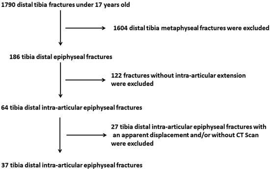
Figure 1.
Flow chart of the study.
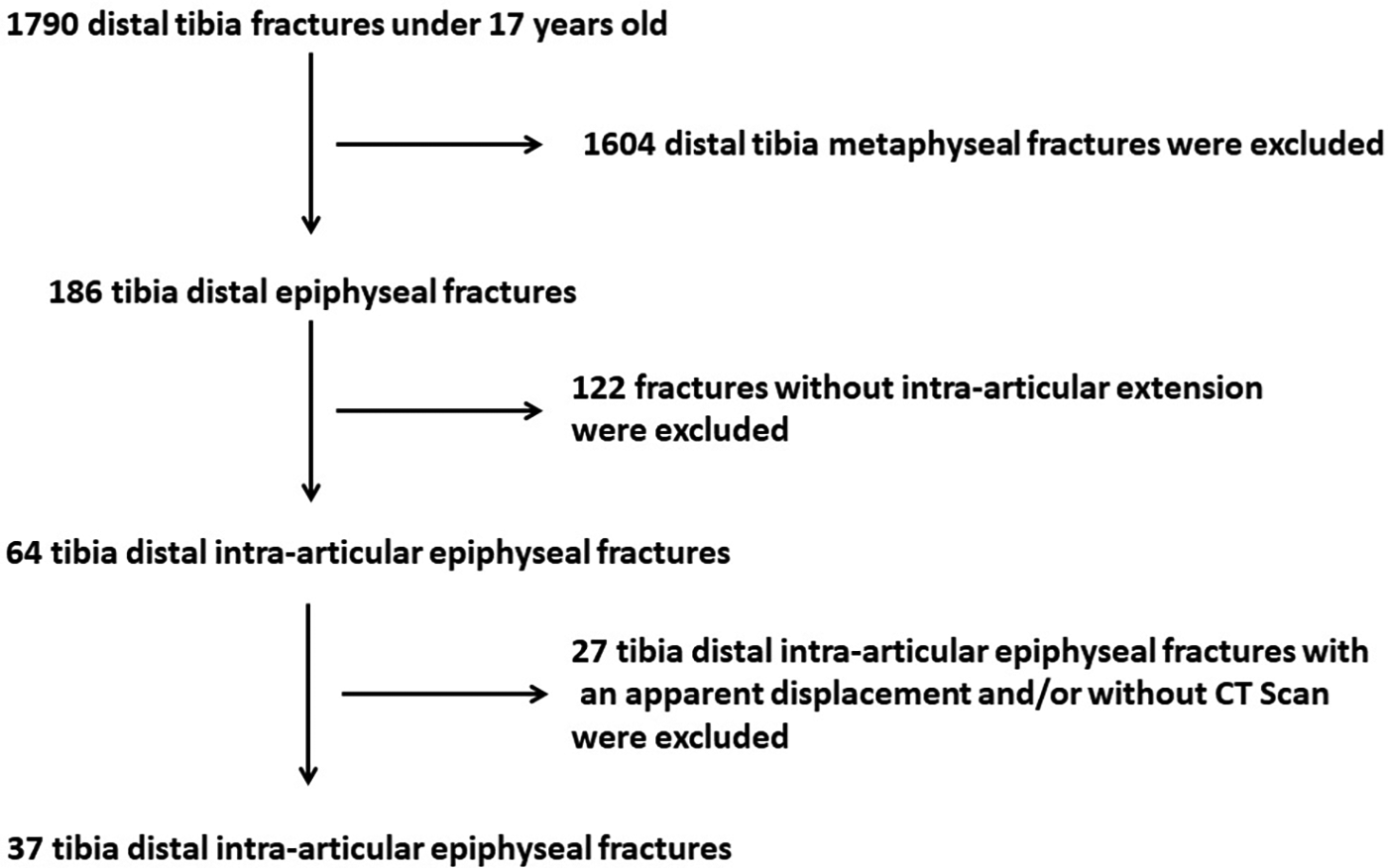
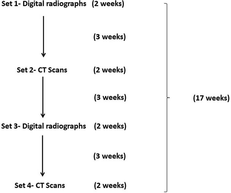
Patients’ digital radiographs and CT scan views were downloaded from the archive and recorded without any identification information but were assigned a number. Ankle radiograph images of the patients were saved as Digital Imaging and Communications in Medicine format. The measurements were performed by the use of the Picture Archiving and Communication System’s tool. Measurements on the CT scans were also performed by the use of the Picture Archiving and Communication System. Two sets were prepared with these radiographs and CT scans (set 1, digital radiographs; set 2, CT scans). The order of the images was changed by an orthopedic surgeon who was not involved in the study, and sets 3 and 4 were developed (set 3, digital radiographs; set 4, CT scans). Five observers consisting of orthopedic specialists who deal mostly with pediatric orthopedic trauma, and with between 2 and 18 years of experience, were asked to measure the maximal intra-articular displacement of the fractures in millimeters and also to make a decision between displacement amount less than or equal to 2 mm (Figure 2, Figure 3 and Figure 4). All observers were blinded to the measurements of the other observers. Timing of the assessments and definition of the sets are summarized in Figure 5. Statistical analyses were carried out using IBM SPSS Version 24.0 (IBM Corp, Armonk, New York). Intraclass correlation coefficient (ICC) values were calculated for the evaluation of intraobserver reliability. Fleiss kappa values other than ICC were calculated for the evaluation of interobserver reliability by using Microsoft Office Excel (Microsoft Corp, Redmond, Washington), because there were more than two observers. A value greater than 0.75 was accepted as excellent agreement; 0.75 to 0.40, intermediate to good agreement; and below 0.40, poor agreement [12,13]. The t test was used for the comparison of measured displacement amounts on radiographs and CT scans after the Shapiro-Wilk test for normality. A value of P < .05 was accepted as statistically significant.
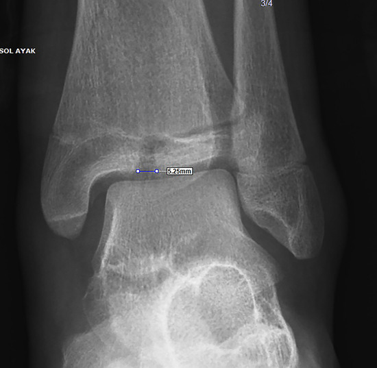
Figure 2.
Measurement on digital radiograph.
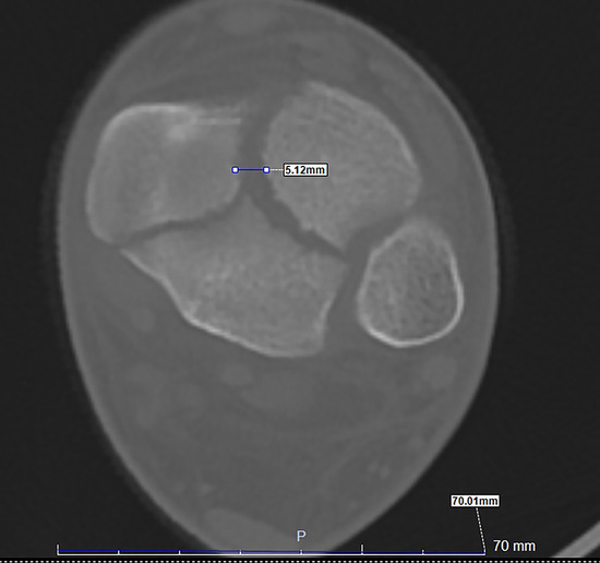
Figure 3.
Measurement on axial cut of computed tomographic scan.
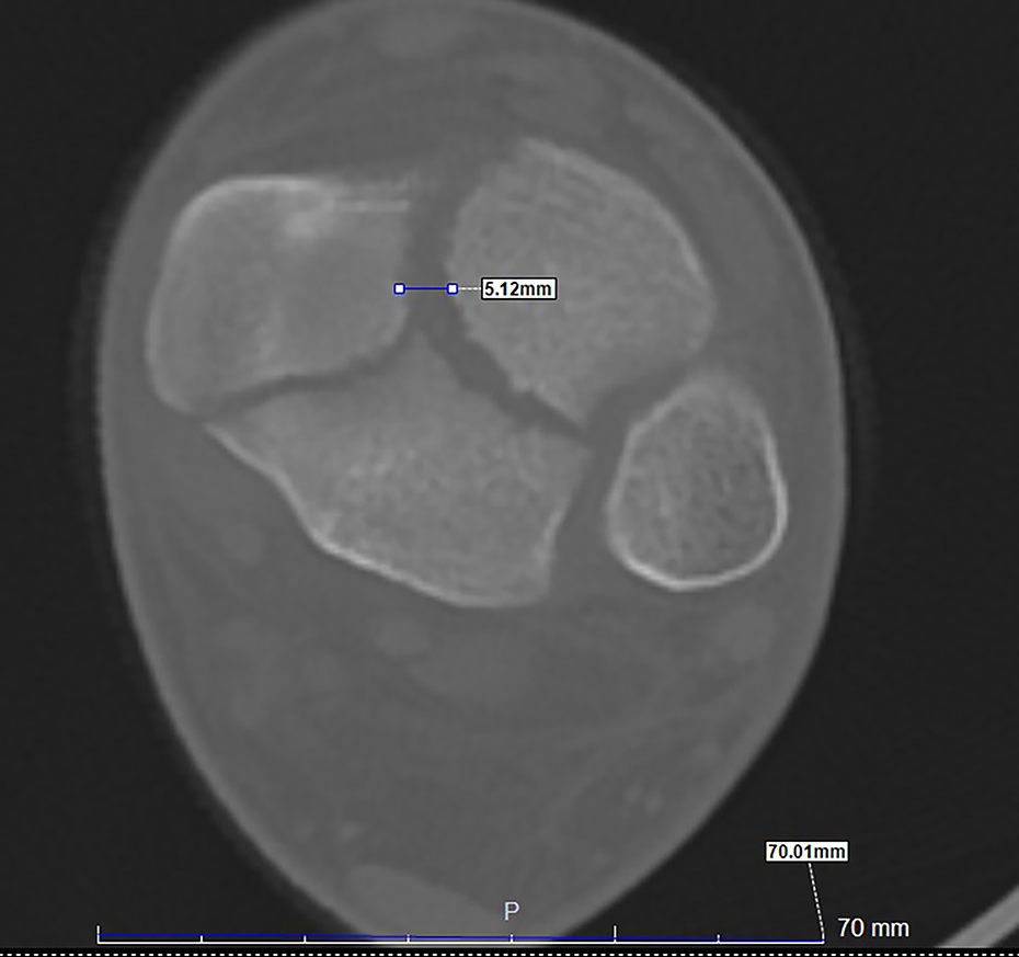
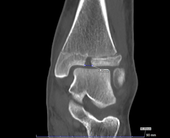
Figure 4.
Measurement on coronal cut of computed tomographic scan.
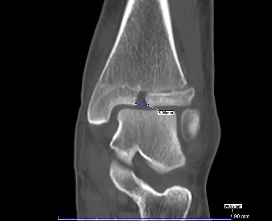
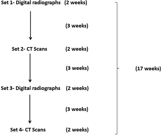
Figure 5.
Timetable for the evaluations.
Results
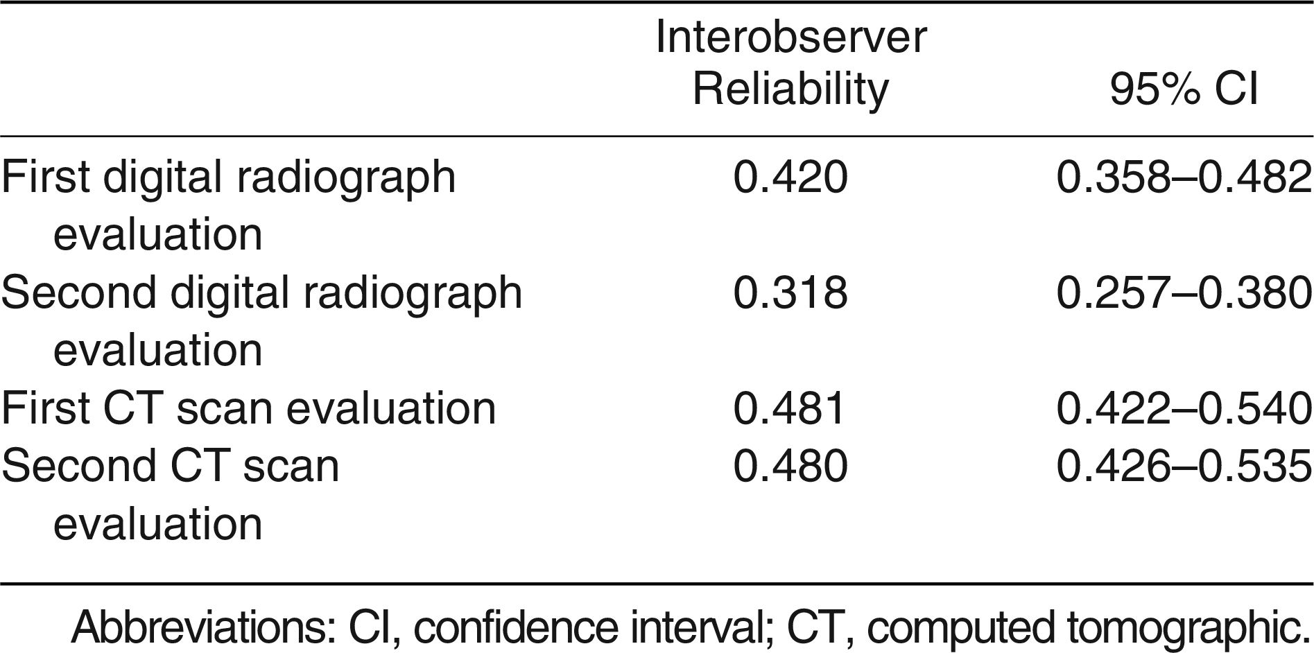
Twenty-seven of the patients were male patients (73%). The mean age of the patients was 11.2 ± 3 years [3,4,5,6,7,8,9,10,11,12,13,14,15,16]. Fifteen of the fractures were SH type 3 (40%), 13 were SH type 4 (35%), seven were triplanar (19%), and two were Tillaux (6%) fractures. Nineteen of the patients (51.3%) were found to be treated surgically by either closed or open means. Interobserver reliability was intermediate to good for digital radiograph and CT scan evaluations for differentiation between under or over 2 mm of displacement (Table 1). Considering the evaluations of the measurements, it was found that interobserver reliability was intermediate to good and poor in the first and second evaluations of digital measurements, respectively (Table 2). Interobserver reliability was found to be intermediate to good in both first and second CT scan evaluations (Table 2).

Table 1.
Interobserver Reliability of Decision Making Between <2-mm or ≥2-mm Intra-articular Displacement on Digital Radiographs and CT Scans
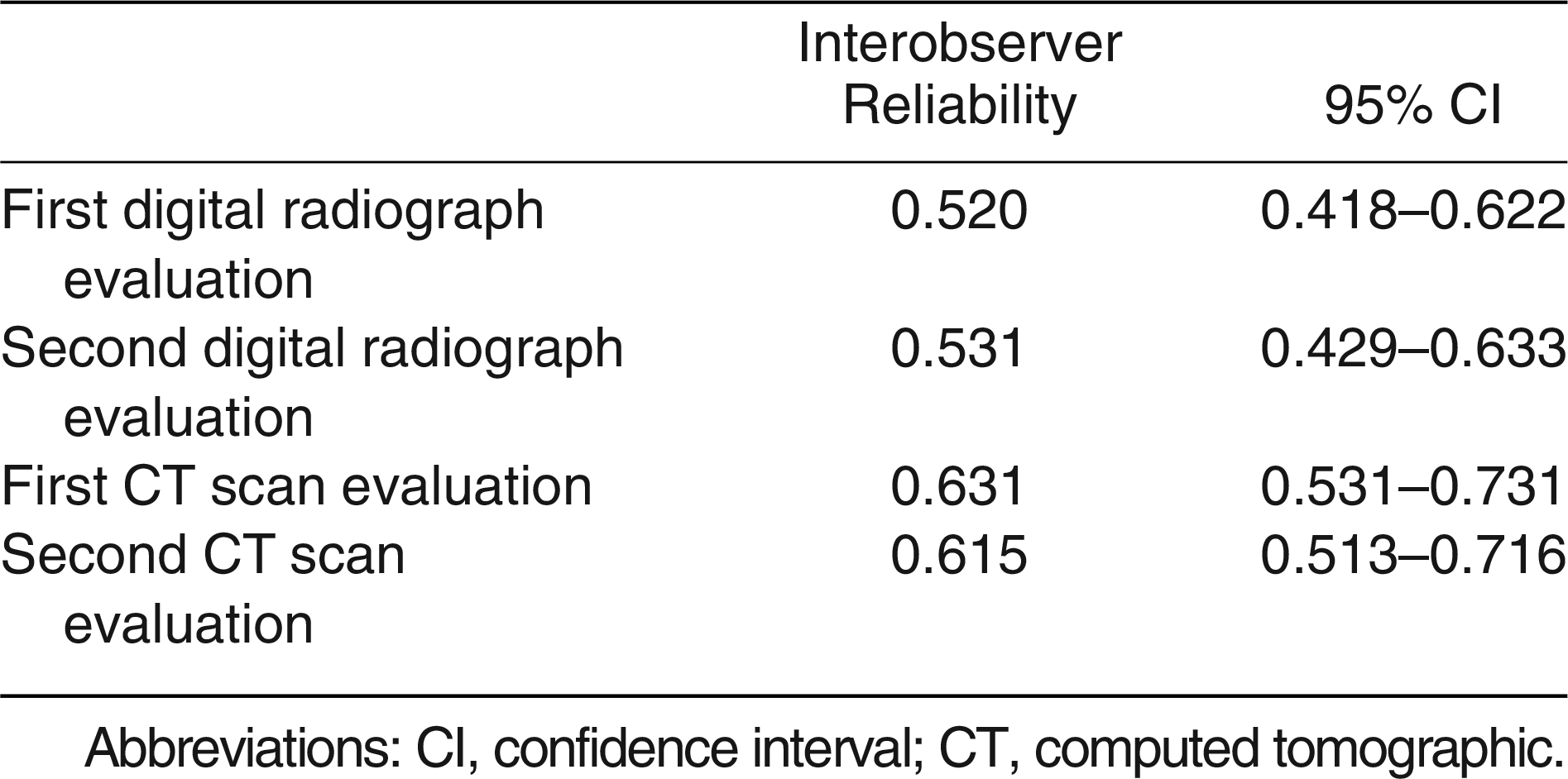
Table 2.
Interobserver Reliability of Intra-articular Displacement Measurements on Digital Radiographs and CT Scans
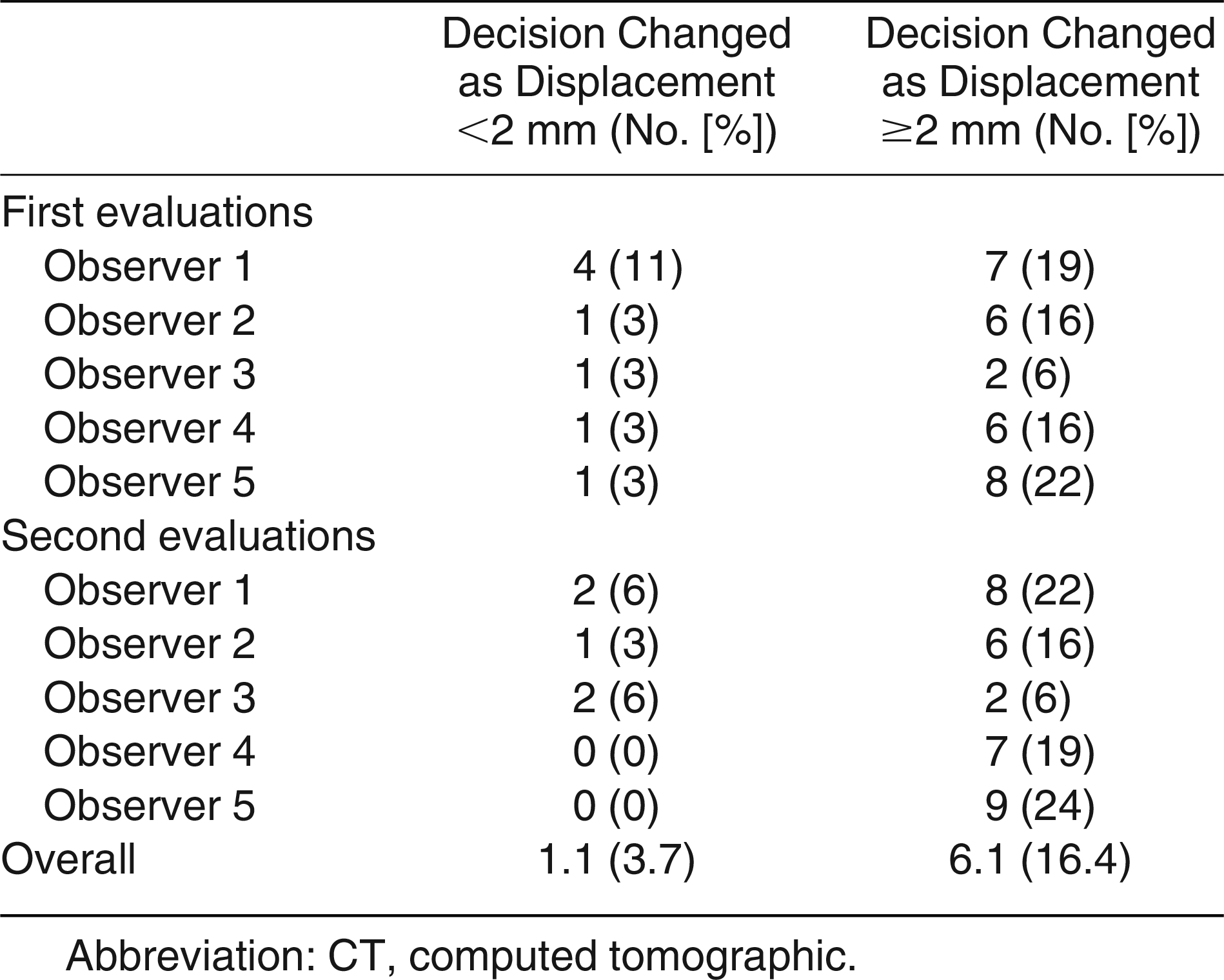
Intraobserver reliabilities were found to be excellent for both measurements and differentiation between under or over 2 mm of displacement for digital radiograph and CT scan evaluations (Table 3 and Table 4). Both intraobserver and interobserver reliabilities were slightly higher for CT scan evaluations. Displacement amounts were found to be measured higher after CT scan evaluations other than after radiographs (1.8 ± 1.2 mm versus 2.2 ± 1.6 mm, and 1.9 ± 1.3 mm versus 2.4 ± 1.8 mm for the first and second evaluations, respectively) (Table 5). There was a statistically significant difference between the measured displacement amounts on radiographs and CT scans (P = .027 and P = .001 for the first and second evaluations, respectively) (Table 5). There was not a statistically significant difference between first and second measurements on radiographs and CT scans (P = .642 and P = .158, respectively) (Table 5). After the evaluation with CT scan, it was found that the observers’ decisions changed to below 2-mm displacement in 3.7% of the evaluations and to greater than or equal to 2-mm displacement in 16.4% of the evaluations (Table 6).
Table 3.
Intraobserver Reliability of Decision Making Between <2-mm or ≥2-mm Intra-articular Displacement on Digital Radiographs and CT Scans
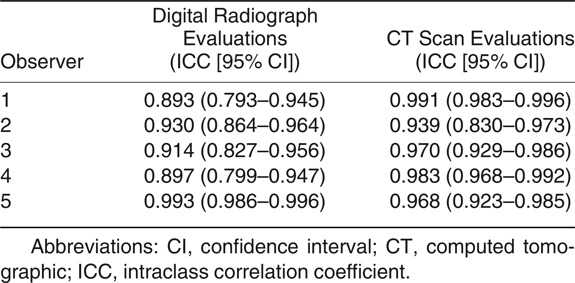
Table 4.
Intraobserver Reliability of Intra-articular Displacement Measurement on Digital Radiographs and CT Scans
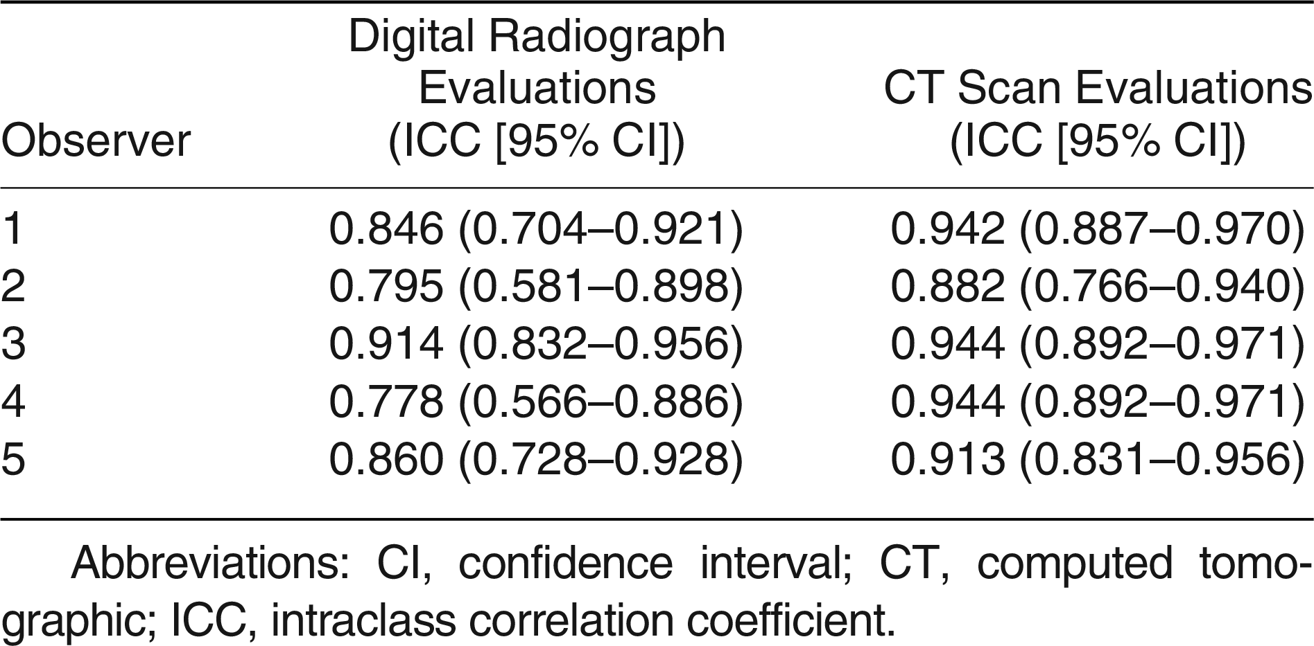
Table 5.
Amount of Measured Intra-articular Displacement
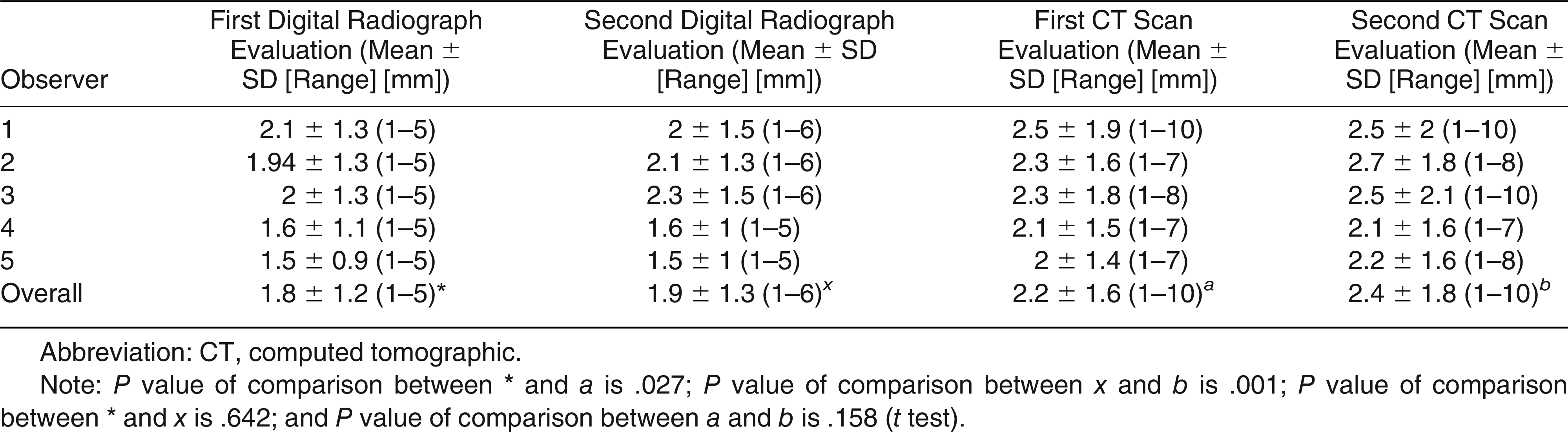
Table 6.
Changes in Decisions of Observers After Evaluating the Intra-articular Displacement on CT Scans
Discussion
As in all intra-articular fractures, the amount of displacement of the joint surface is very important in selecting the treatment modality of intra-articular epiphyseal fractures. Exceeding a displacement limit of 2 mm is very important in deciding surgical treatment [3,4,5,6]. However, there are doubts about the reliability of measuring this displacement. Digital radiography technology enables both image clarity and measurements in millimeters. In this study, we aimed to investigate the reliability of measurement of joint displacement by digital radiography and CT imaging and make a comparison. It was concluded that the reliabilities of digital radiography and CT scan measurement were similar in the determination of joint displacement amount. The displacement amount was measured slightly higher when using computed tomography compared to digital radiography. Overall, 16.4% of the measurements were found to be changed to greater than or equal to 2 mm after CT scan evaluations, which were determined to be under 2 mm on digital radiographs.
Radiography, computed tomography, and magnetic resonance imaging can be used to determine the amount of displacement in intra-articular fractures. Previously, the low reliability of conventional radiographs for measuring joint displacement had been demonstrated in several studies [14,15]. Borrelli et al [8]. evaluated the reliability of articular displacement measurement on acetabular fractures and reported the poor reliability of plain radiographs. The authors also stated that use of CT scans should be continued for preoperative evaluation. Similarly, Cole et al [7]. also reported the better reliability of CT scan evaluation over plain radiographs while evaluating intra-articular displacement of distal radius fractures. Arora et al [16]. evaluated the reliability of distal radial intra-articular fracture displacement after the fracture reduction and concluded that treatment modality could be changed after the measurements on CT scans rather than plain radiographs. The aforementioned studies were performed on localizations such as the acetabulum and distal radius, which can be difficult to evaluate on radiographs. In our study, unlike conventional radiographs, digital radiographs, which can be measured millimetrically with computer programs and can be complemented and reduced on request, were compared with CT imaging. Overall, the decision of the observers was changed, as the displacement was greater than or equal to 2 mm instead of less than 2 mm in 16.4% of the patients after CT evaluations. The proportion of patients where the decision changed confirmed the important role of CT scanning in evaluation of intra-articular displacement in distal tibia epiphyseal fractures.
Eismann et al [11]. performed a study which they evaluated the impact of radiographs and CT scans on fracture classification and treatment planning in pediatric triplane fractures. The authors stated that interobserver reliability was improved to 0.75 from 0.69 (ICC) after the addition of CT imaging. Another finding was that intraobserver reliability improved to 0.77 from 0.72 (ICC) after the addition of CT imaging to the evaluation. In our study, interobserver reliability was found to be increased to 0.631 from 0.520 (Fleiss kappa) for the first evaluation and to 0.615 from 0.531 (Fleiss kappa) for the second evaluation after CT scan use when the observers made a decision between below or greater than or equal to 2-mm displacement. Similarly, after using CT scans, intraobserver reliability was found to be increased to a mean of 0.970 from 0.925 (ICC) when a decision was made between below or greater than or equal to 2-mm displacement. Interobserver reliability was found to be increased to 0.481 from 0.420 (Fleiss kappa) for the first evaluation and to 0.480 from 0.318 (Fleiss kappa) for the second evaluation after CT scan use when the observers measured the displacement in millimeters. After using CT scans, intraobserver reliability was found to be increased to a mean of 0.925 from 0.839 (ICC) after the measurements of displacements. It is apparent that CT scan increases the reliability of displacement measurements. One of the other common findings of the study by Eismann et al [11]. and our study was that the displacements were measured wider on CT scans in comparison with radiographs (2.3 ± 1.6 mm versus 3.2 ± 2 mm for the study by Eismann et al, [11]. and 1.8 ± 1.2 mm versus 2.2 ± 1.6 mm and 1.9 ± 1.3 mm versus 2.4 ± 1.8 mm for the first and second evaluations in our study, respectively). In the study by Eismann et al [11]. and in our study, after CT evaluations, the displacement amounts were changed to greater than or equal to 2 mm in 39% and 16.4% of the patients, respectively. The decisions were changed to less than 2 mm 7% and 3.7% of the patients in the study by Eismann et al and by our study, respectively. El Emam [17]. stated that the minimally targeted level should be 0.62 while using kappa for evaluating interobserver reliability for substantial agreement. The kappa values were 0.520 and 0.523 for the first and second evaluations on digital radiographs, respectively. These kappa values were 0.631 and 0.615 for the first and second evaluations on CT scans, respectively. It is obvious that CT scanning is a very useful tool for preoperative evaluation of displacement in distal tibia intra-articular epiphyseal fractures according to our findings.
This study has several limitations. The number of evaluated patients and observers could be increased. The musculoskeletal radiologists could be added as observers. In addition, conventional and digital radiographs could be compared in terms of measurement of joint displacement amounts, taking CT measurements as reference.
Conclusions
In conclusion, in our study, after the measurement of joint displacements on the CT scans, it was observed that the measurements found below 2 mm on digital radiography measurements changed to less than or equal to 2 mm in 16.4% of the patients. This ratio is considerably high, so we believe that digital radiographs cannot replace CT imaging in intra-articular displacement measurements of distal tibial epiphyseal fractures. One should consider that the amount of intra-articular displacement should be predicted to be slightly higher than the value that is measured on digital radiographs.
Financial Disclosure
None reported.
Conflicts of Interest
None reported.
References
- Mann, DC; Rajmaira, S. Distribution of physeal and nonphyseal fractures in 2650 long-bone fractures in children aged 0–16 years. J Pediatr Orthop 1990, 10, 713. [Google Scholar]
- Salter, RB; Harris, WR. Injuries involving the epiphyseal plate. J Bone Joint Surg Am 1963, 45, 587. [Google Scholar]
- Cai, H; Wang, Z; Cai, H. Surgical indications for distal tibial epiphyseal fractures in children. Orthopedics 2015, 38, e189. [Google Scholar]
- Crawford, AH. Triplane and Tillaux fractures: is a 2 mm residual gap acceptable? J Pediatr Orthop 2012, 32 suppl 1, 69. [Google Scholar]
- Leary, JT; Handling, M; Talerico, M; et al. Physeal fractures of the distal tibia: predictive factors of premature physeal closure and growth arrest. J Pediatr Orthop 2009, 29, 356. [Google Scholar]
- Poyanli, O; Unay, K; Akan, K; et al. Distal tibial epiphyseal fracture (Tillaux) and capsular interposition. JAPMA 2009, 99, 435. [Google Scholar]
- Cole, RJ; Bindra, RR; Evanoff, BA; et al. Radiographic evaluation of osseous displacement following intra-articular fractures of the distal radius: reliability of plain radiography versus computed tomography. J Hand Surg 1997, 22, 792. [Google Scholar]
- Borrelli, J, Jr; Goldfarb, C; Catalano, L; et al. Assessment of articular fragment displacement in acetabular fractures: a comparison of computerized tomography and plain radiographs. J Orthop Trauma 2002, 16, 449. [Google Scholar]
- Drynan, D; McArthur, M; Vohora, A; et al. Accuracy of digital radiography: regional scaling factors for trauma. ANZ J Surg 2018, 88, 341. [Google Scholar]
- King, RJ; Craig, PR; Boreham, BG; et al. The magnification of digital radiographs in the trauma patient: implications for templating. Injury 2009, 40, 173. [Google Scholar]
- Eismann, EA; Stephan, ZA; Mehlman, CT; et al. Pediatric triplane ankle fractures: impact of radiographs and computed tomography on fracture classification and treatment planning. J Bone Joint Surg Am 2015, 97, 995. [Google Scholar]
- Landis, JR; Koch, GG. The measurement of observer agreement for categorical data. Biometrics 1977, 33, 159. [Google Scholar]
- Fleiss, JL. Statistical Methods for Rates and Proportions, 2nd ed; John Wiley & Sons; New York; 1981. [Google Scholar]
- Catalano, LW, III; Barron, OA; Glickel, SZ. Assessment of articular displacement of distal radius fractures. Clin Orthop Relat Res 2004, 423, 79. [Google Scholar]
- Bozentka, DJ; Beredjiklian, PK; Westawski, D; et al. Digital radiographs in the assessment of distal radius fracture parameters. Clin Orthop Relat Res 2002, 397, 409. [Google Scholar]
- Arora, S; Grover, SB; Batra, S; et al. Comparative evaluation of postreduction intra-articular distal radial fractures by radiographs and multidetector computed tomography. J Bone Joint Surg Am 2010, 92, 2523. [Google Scholar]
- El Emam, K. Benchmarking kappa: interrater agreement in software process assessments. Empir Softw Eng 1999, 4, 113. [Google Scholar]
© 2022 American Podiatric Medical Association