


Abstract
Osteoid osteoma is a primary benign bone tumor that consists of a central area (nidus), surrounded by sclerotic bone. The most relevant symptom is pain that increases during the night and improves after salicylates or nonsteroidal anti-inflammatory drug administration. Osteoid osteoma is frequently misdiagnosed because it mimics juvenile idiopathic arthritis, bone infection, or malignancy. A 14-year-old girl presented to our department with a history of chronic pain in her left ankle. Juvenile idiopathic arthritis was diagnosed and anti-inflammatory treatment was prescribed. Because of persistence of ankle pain, the patient underwent further examinations, in particular, bone scintigraphy and computed tomography. As a result, osteoid osteoma of the talar neck was diagnosed. The patient underwent surgical treatment and her condition improved. Osteoid osteoma should also be considered in patients with chronic ankle pain to avoid misdiagnosis and start adequate treatment. This condition should be suspected in a patient with chronic bone pain and normal complete blood count and inflammatory parameters.
Osteoid osteoma (OO) is a primary benign bone tumor [1], representing approximately 10% to 12% of benign skeletal neoplasms [2]. The tumor consists of a central area (nidus) that is surrounded by sclerotic bone [3,4]. This condition was defined by Heine in 1927 and further described by Jaffe in 1935 [5]. Adolescents and young adults are mostly affected, with a female-to-male ratio of 1:2 [6]. The symptomatology is characterized by pain at the tumor site that increases during the night and decreases after administration of salicylates or nonsteroidal anti-inflammatory drugs (NSAIDs) [7].
The pain is attributable to the local inflammation, secondary to the high levels of prostaglandins expressed by the tumor [8]. Radiologically, OO is characterized by a radiolucent or calcified nidus, surrounded by a sclerotic margin [9]. The most frequent locations of OO are the diaphysis or metaphysis of long bones, in particular, the femur (37.5%) and the tibia (20%) [10]. Osteoid osteoma rarely involves the foot and ankle, accounting for approximately 2% to 10% of all cases, and is a rare cause of joint pain [11]. Differential diagnosis most commonly includes ankle sprain, monoarticular arthritis, algodystrophy, osteomyelitis, stress fracture, and eosinophilic granuloma [12]. The delay in diagnosis and treatment of OO in the ankle may be a cause of chronic ankle pain. We describe a case of a girl with OO of the talar neck that was misdiagnosed with juvenile idiopathic arthritis (JIA) with the aim of providing clinicians with the key elements to suspect this condition and to avoid misdiagnoses or late diagnoses.
Case Report
A 14-year-old girl came to our attention with a 1-year history of pain in her left ankle. There was no history of trauma or febrile illness. There was familial history of rheumatoid arthritis. Before coming to our observation, the patient underwent ultrasound, radiography, and magnetic resonance imaging (MRI) of left foot and ankle. Ultrasound revealed joint effusion in the subtalar joint, radiography was unremarkable, and MRI revealed joint effusion and bone marrow edema of the talar neck. Complete blood count, liver and renal function, and inflammatory indices were normal. According to these findings and familial history, JIA was diagnosed. Anti-inflammatory treatment (naproxen, 15 mg/kg/day) was prescribed, but the patient’s symptomatology improved only slightly. Written informed consent was obtained from the patient’s parents for publication of this case report and accompanying images.
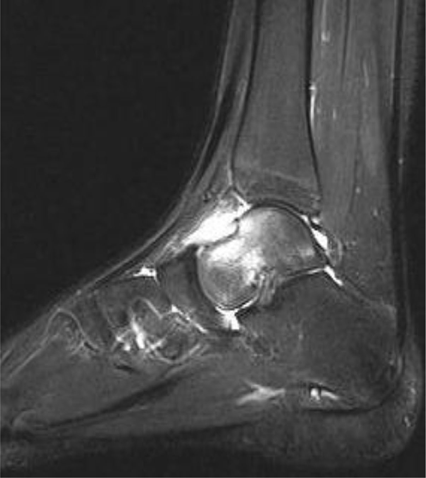
When the patient came to our observation, she reported pain in her left ankle and foot. She was afebrile and in good general condition. The physical examination of the left foot revealed mild soft-tissue swelling and limitation in the range of movement. Blood examinations were normal, with normal values for C-reactive protein and erythrocyte sedimentation rate. Antinuclear antibodies were negative. Ophthalmologic evaluation was negative. An ankle radiograph was normal. The newly acquired T2-weighted MRI scan of the left foot and ankle showed bone marrow edema of the talar neck, joint effusion, and soft-tissue swelling around the joint (Fig. 1).
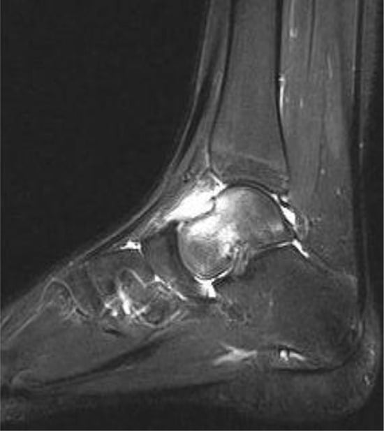
Figure 1.
Left foot and ankle magnetic resonance imaging scan that shows bone marrow edema of the talar neck.
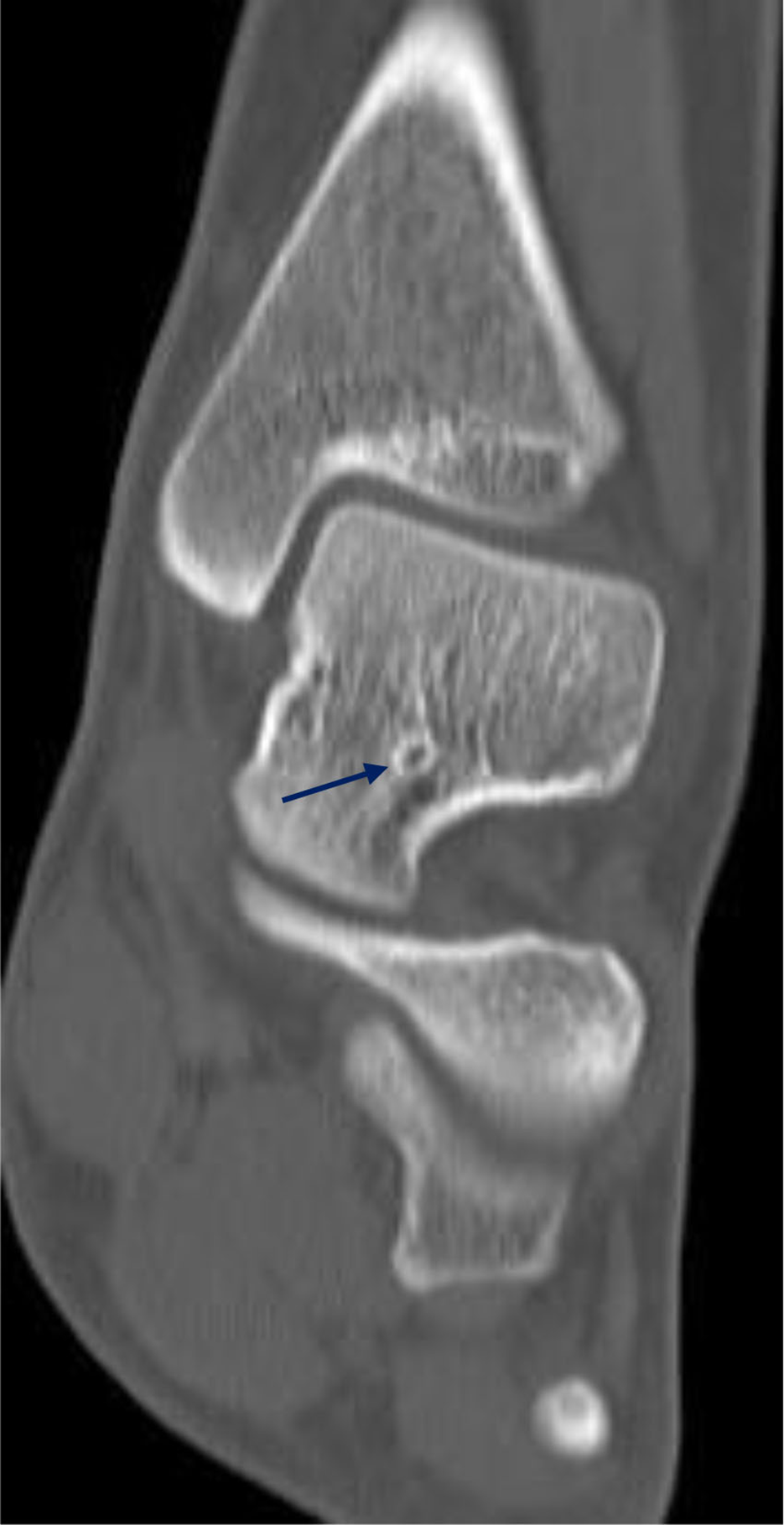
Radionuclide bone scan revealed a diffuse increase in radionuclide uptake at the talus (Fig. 2). Computed tomography (CT) showed a bony lesion surrounded by sclerotic bone at the talar neck (Fig. 3).
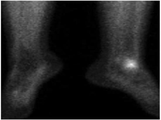
Figure 2.
Radionuclide bone scan revealing a diffuse increase in radionuclide uptake at the left talus.
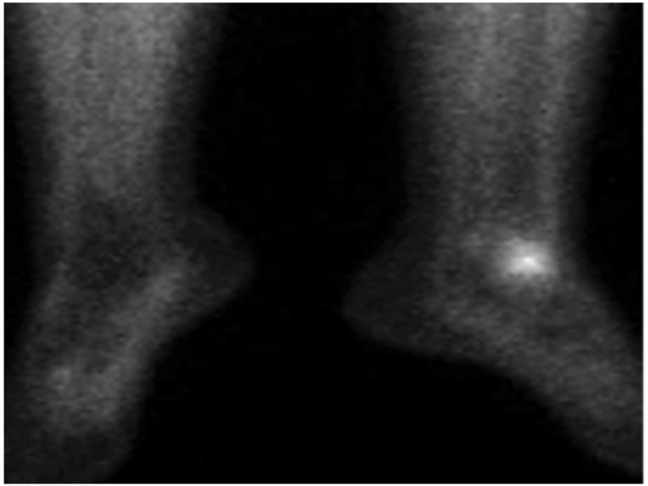
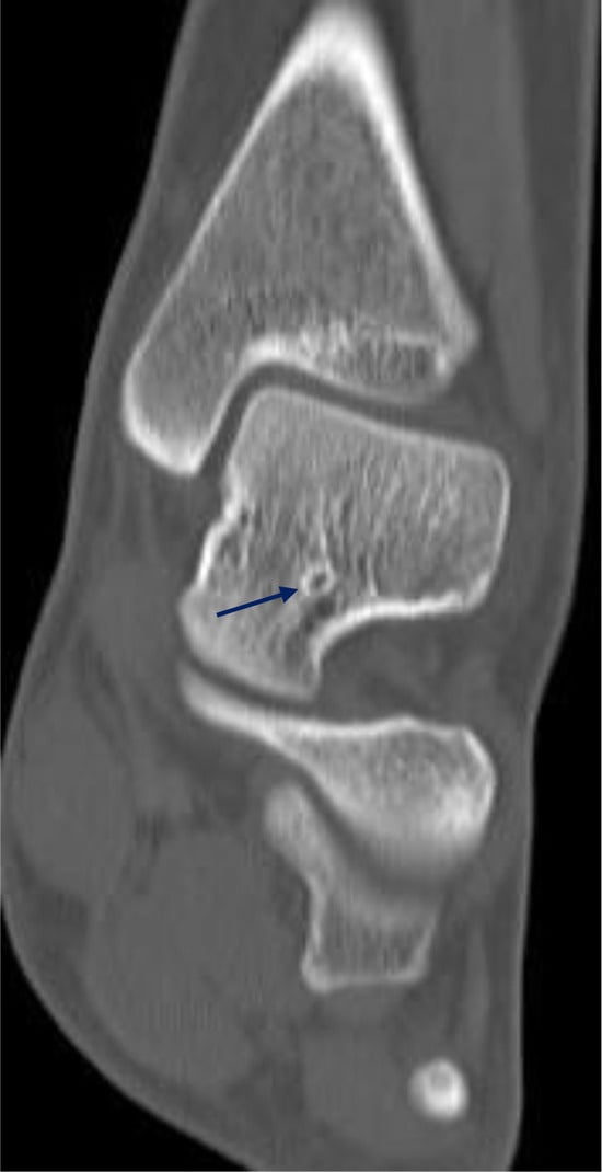
Figure 3.
Left foot and ankle computed tomographic scan showing a bone lesion surrounded by sclerotic bone (blue arrow).
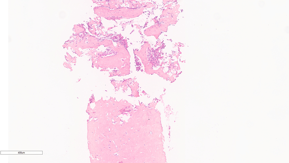
According to the patient’s history, blood tests, and instrumental investigations, OO was diagnosed. The patient subsequently underwent nidus excision with CT-guided radiofrequency. Postoperative pathologic examination showed benign osteoblastic proliferation composed of anastomosing bone trabeculae with variable mineralization, lined by plump osteoblasts, within vascularized connective tissue. Benign giant cells were present, consistent with a nidus of osteoid osteoma (Fig. 4). The patient’s condition improved after treatment. She was able to walk and run when she presented for follow-up 6 months later.
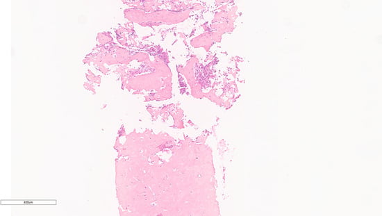
Figure 4.
Postoperative examination of the lesion.
Discussion
Osteoid osteoma is the most common benign bone tumor after osteochondroma and bone cysts. The most common symptom is bone pain that is worse during the night and improves after NSAID administration [13]. The bone pain is secondary to the high levels of prostaglandins expressed by the tumor [14]. The fact that the pain improves after NSAID administration often delays the diagnosis [15].
The femur and tibia are the most frequent locations of OO. Osteoid osteoma of the talus is an infrequent event, and it is frequently misdiagnosed with other conditions such as ankle distortions, monoarticular arthritis, algodystrophy, osteomyelitis, stress fracture, and eosinophilic granuloma [16]. In our case, OO was misdiagnosed as JIA, so the patient started NSAIDs, with a slight improvement. Delayed diagnosis and failed initial management are not uncommon features associated with OO, especially when the lesion localizes to an unusual anatomical site. Considerable delays have also been reported for cases of OO localized to the talus [17]. In the case reported, 1 year elapsed before the diagnosis was made.
Investigations to diagnose OO include conventional radiography, radionuclide bone scan, MRI, and CT [18]. On radiography, OO appears as a radiolucent lesion surrounded by bone scleroses [19]. The tarsal osteoid osteomas, such as those of the talus, may display less reactive sclerosis that can be misleading [20]. In fact, the ankle radiograph of our patient was negative. Radionuclide bone scan shows increased radionuclide uptake at the nidus; the patient’s bone scan revealed increased radionuclide uptake at the left talus [21]. On MRI, the nidus may show a low T1 signal and a variable T2 signal [22]. When talar neck OO is associated with subtalar arthritis, joint effusion, and bone marrow edema, the nidus may be masked, retarding diagnosis, as in our case [23]. We decided to obtain a CT scan to perform a better evaluation of the bone, suspecting OO according to the patient’s symptoms and blood examinations. Computed tomography is considered the gold standard investigation for identifying the anatomical position of the nidus [24]. Treatment of OO can be conservative, with long-term administration of NSAIDs, or can be operative [25]. The goal of the surgery is excision of the lesion, including the nidus. For our patient, operative treatment was chosen because of the duration of symptoms and the poor response to anti-inflammatory treatment. The patient underwent nidus excision. There are three types of surgical approach to remove OO: resection of the nidus with surrounding bones; intralesional resection of the nidus; and percutaneous CT-guided excision with radiofrequency energy, laser, or absolute alcohol (99.8% concentration) [26]. In our patient, percutaneous CT-guided excision with radiofrequency energy was performed. Different studies have demonstrated its superiority compared with other invasive methods [27]. The patient’s condition improved after excision of the nidus.
Conclusions
We present the case of a girl with a 1-year history of ankle pain and a misdiagnosis of JIA. Although OO can be misdiagnosed with JIA, there are some features that allow us to differentiate OO from JIA. First, OO is more common in male patients and JIA is more common in female patients. In OO, the pain occurs at night and improves slightly with anti-inflammatory treatment, whereas in JIA it occurs in the morning and improves with anti-inflammatory treatment. In OO, arthritis or bone pain involves just one joint, whereas more than one joint is involved in the inflammatory process in JIA. Moreover, in OO, blood examinations (eg, antinuclear antibodies, complete blood count, and inflammatory parameters) are in the normal range. Ultrasound and MRI are very useful in JIA to detect joint effusion, tenosynovitis, and cartilage damage. Radiography represents the first-line investigation to detect OO (it appear as a radiolucent lesion surrounded by bone scleroses), but CT is considered the gold standard investigation to identify the anatomical position of the nidus. In a patient with a history of chronic pain with a normal complete blood count and inflammatory parameters, OO should be considered so that a correct diagnosis can be obtained and adequate treatment started.
Acknowledgment:
Kelly Tesone for the written revision of the English of the article.
Financial Disclosure:
None reported.
Conflict of Interest:
None reported.
References
- KITSOULIS, P; MANTELLOS, G; VLYCHOU, M. Osteoid osteoma. Acta Orthop Belg 2006, 72, 119. [Google Scholar] [PubMed]
- PAPATHANASSIOU, ZG; MEGAS, P; PETSAS, T; et al. Osteoid osteoma: diagnosis and treatment. Orthopedics 2008, 31, 1118. [Google Scholar] [CrossRef] [Scilit] [PubMed]
- SONG, MH; YOO, WJ; CHO, TJ; et al. Clinical and radiological features and skeletal sequelae in childhood intra-/juxtaarticular versus extra-articular osteoid osteoma. BMC Musculoskelet Disord 2015, 16, 3. [Google Scholar] [CrossRef] [Scilit] [PubMed]
- JAFFE HL:Osteid osteoma. Proc R SocMed 1953, 46, 1007.
- GIEDON, A; HOLTHUSENW; MASEL, LF; et al. Subacute and chronic “symmetrical” osteomyelitis [in multiple languages]. Ann Radiol (Paris) 1972, 15, 329. [Google Scholar]
- CERASE, A; PRIOLO, F. Skeletal benign bone-forming lesions. Eur J Radiol 1998, 27 (suppl 1), S91. [Google Scholar] [CrossRef] [Scilit]
- LEE, EH; SHAFI, M; HUI, JH. Osteoid osteoma: a current review. J Pediatr Orthop 2006, 26, 695. [Google Scholar] [CrossRef] [Scilit]
- ATESOK, KI; ALMAN, BA; SCHEMITSCH, EH; et al. Osteoid osteoma and osteoblastoma. J Am Acad Orthop Surg 2011, 19, 678. [Google Scholar] [CrossRef] [Scilit]
- SHUKLA, S; CLARKE, AW. SAIFUDDIN A: Imaging of foot osteoid osteoma. Skeletal Radiol 2010, 39, 683. [Google Scholar] [CrossRef] [Scilit]
- UNNI, KK; INWARDS, CY; BRIDGE, JA; et al. Tumors of the Bone and Joints. In AFIP Atlas of Tumor Pathology Series 4; American Registry of Pathology: Washington, DC, 2005; p. 324. [Google Scholar]
- YERCAN, HS; OKCU, G; OZALP, T; et al. Arthroscopic removal of the osteoid osteoma on the neck of the talus. Knee Surg Sports Traumatol Arthrosc 2004, 12, 246. [Google Scholar] [CrossRef] [Scilit]
- SYMEONIDIS, PD; MAROUGIANNIS, D; CHRYSOGLOU, G; et al. Osteoid osteoma of the posterior talar dome. J Foot Ankle Surg 2011, 50, 350. [Google Scholar] [CrossRef] [Scilit] [PubMed]
- GURKAN, V; ERDOGAN, O. Foot and ankle osteoid osteoma. J Foot Ankle Surg 2017, 57, 825. [Google Scholar] [CrossRef] [Scilit] [PubMed]
- MUNK, PL; HUK, ME. Medical management of osteoid osteoma. Can J Surg 2003, 46, 60. [Google Scholar]
- SNOW, SW; SOBEL, M; DI CARLO, EF; et al. Chronic ankle pain caused by osteoid osteoma of the neck of the talus. Foot Ankle Int 1997, 18, 98. [Google Scholar] [CrossRef] [Scilit] [PubMed]
- DIMNJAKOVIC, D; BOJANIC, I; SMOLJANOVIC, T; et al. Osteoma of the ankle: a report of nine arthroscopically treated patients. J Foot Ankle Surg 2014, 54, 89. [Google Scholar] [CrossRef] [Scilit]
- SYMEONIDIS, PD; MAROUGIANNIS, D; CHRYSOGLOU, G; et al. Osteoid osteoma of the posterior talar dome. J Foot Ankle Surg 2011, 50, 350. [Google Scholar] [CrossRef] [Scilit]
- SHUKLA, S; CLARKE, AW. SAIFUDDIN A: Imaging features of foot osteoid osteoma. Skeletal Radiol 2010, 39, 683. [Google Scholar] [CrossRef] [Scilit]
- CHAI, JW; HONG, SH; CHOI, JY; et al. Radiologic diagnosis of osteoid osteoma: from simple to challenging findings. Radiographics 2010, 30, 737. [Google Scholar] [CrossRef] [Scilit]
- SPENCER, EE; BEIRMAN, JS. FEMINO JE: Osteoid osteoma of the fifth metatarsal: a case report and literature review. Foot Ankle Surg 2002, 8, 71. [Google Scholar] [CrossRef] [Scilit]
- FARID, K; EL-DEEB, G. CAILLAT VN: SPECT-CT improves scintigraphic accuracy of osteoid osteoma diagnosis. Clin Nucl Med 2010, 35, 170. [Google Scholar] [CrossRef] [Scilit]
- UYGUR, E; POYANLý, O; KýLýçOÐLU, O. Which emerges first: bone marrow edema or nidus in osteoid osteoma? JAPMA 2018, 108, 523. [Google Scholar] [CrossRef] [Scilit]
- SINGH, JP; SRIVASTAVA, S; SINGH, D. Osteoid osteoma masquerading tubercular arthritis or osteomyelitis on MRI: case series and review of literature. Indian J Radiol Imaging 2015, 25, 261. [Google Scholar] [CrossRef] [Scilit]
- HE, H; XU, H; LU, H; et al. A misdiagnosed case of osteoid osteoma of the talus: a case report and literature review. BMC Musculoskelet Disord 2017, 18, 35. [Google Scholar] [CrossRef] [Scilit] [PubMed]
- GHANEM I: The management of osteoid osteoma: updates and controversies. Curr Opin Pediatr 2006, 18, 36. [CrossRef] [Scilit]
- ASSAFIRI I, SRAJ S: Osteoid osteoma of the talar neck with subacute presentation. Am J Orthop (Belle Mead NJ) 2015, 44, E404.
- HAGE, AN; CHICK, JFB; GEMMETE, JJ; et al. Percutaneous radiofrequency ablation for the treatment of osteoid osteoma in children and adults: a comparative analysis in 92 patients. Cardiovasc Intervent Radiol 2018, 41, 1384. [Google Scholar] [CrossRef] [Scilit] [PubMed]
© 2022 American Podiatric Medical Association