


Abstract
Fracture of an ossified Achilles tendon is a rare clinical entity. Reossification after removal of the bony fragment was reported in only one case previously. In this study, we present a 49-year-old man with a reossified Achilles tendon after the removal of a fractured and ossified Achilles tendon. Treatment of an ossified or fractured Achilles tendon should be selected on a patient-by-patient basis. Surgical treatment can be used when conservative treatment has failed. The possibility of reossification after surgical treatment, especially in patients with risk factors, should be kept in mind, and the patient should be informed about this possibility.
Ossification of the Achilles tendon is a rare clinical entity [1]. Furthermore, fracture of an ossified Achilles tendon (FOAT) is less often reported in the literature [2,3,4,5,6,7,8,9,10,11,12]. Ossification of the Achilles tendon is usually detected incidentally while evaluating heel pain [1,13]. The most common reported causes of ossification are previous trauma and surgery; however, seronegative arthropathies, metabolic diseases, and a few other conditions have also been reported as predisposing factors [1]. The true mechanism of ossification is also unknown [14]. Intratendinous degeneration of the Achilles tendon may lead to ossification and ectopic calcification [15]. In a previously conducted study, the mechanism was explained as follows: decreased oxygen tension caused by tendon degeneration may cause generation of osteoblasts from fibroblasts [14]. However, this hypothesis has not been proved.
The cause of FOAT could be traumatic or nontraumatic [2,3,4,5,6,7,8,9,10,11,12]. Conservative treatment, osteosynthesis, or surgical excision and reconstruction with a different type of flap or grafts have been used for treatment of FOAT [2,3,4,5,6,7,8,9,10,11,12]. The ideal method of treatment is unknown because of the rarity of the cases. The literature contains only one case with a recurring ossification after the removal of the ossified part of the Achilles tendon because of pain [13]. In this study, we report a case with a reossified Achilles tendon after removal of a fractured and ossified Achilles tendon.
Case Report
In 2010, a 39-year-old man presented to the emergency department because of pain in the back of his right heel and difficulty in walking after being kicked on the right calf while playing soccer. On physical examination, there was pain with palpation on the distal calf, but no gap on the course of the Achilles tendon. The Thompson test proved negative; however, the patient felt pain. Plain radiographs revealed calcification approximately 3 cm proximal to the insertion of the Achilles tendon. The patient reported a history of trauma in the right calf 13 years previously (in 1997) but no disease history. Further radiologic examination with computed tomography and magnetic resonance imaging revealed a fractured bony mass in the Achilles tendon, with partial rupture of the tendon (Fig. 1).
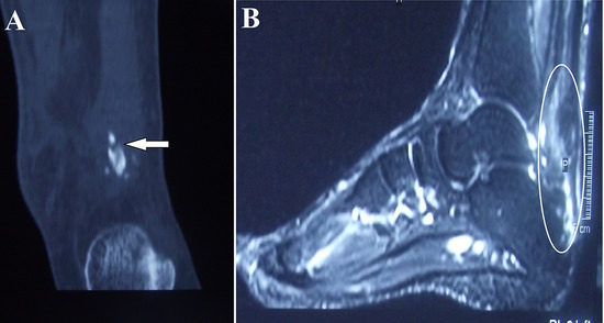
Figure 1.
A, Computed tomographic image of the fractured bone (arrow). B, Sagittal magnetic resonance imaging scan showing partial tear of the Achilles tendon.
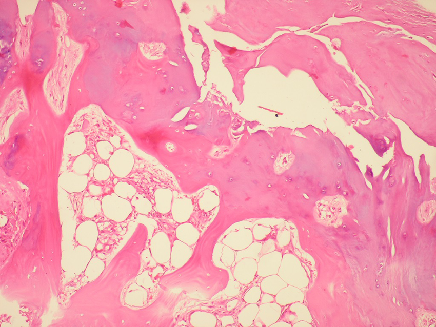
The patient was treated surgically with removal of the fractured bone, and primary repair of Achilles tendon with number 1 absorbable polydioxanone sutures and suturing the fanned out plantaris tendon over the Achilles tendon as a membrane (Fig. 2). The histologic examination of the removed bone showed the formation of lamellar bone with bone marrow (Fig. 3).
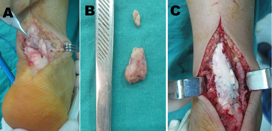
Figure 2.
A, Intraoperative view of fractured bone; B, the removed fractured bone; C, primary repair of the Achilles tendon and suturing the fanned out plantaris tendon.
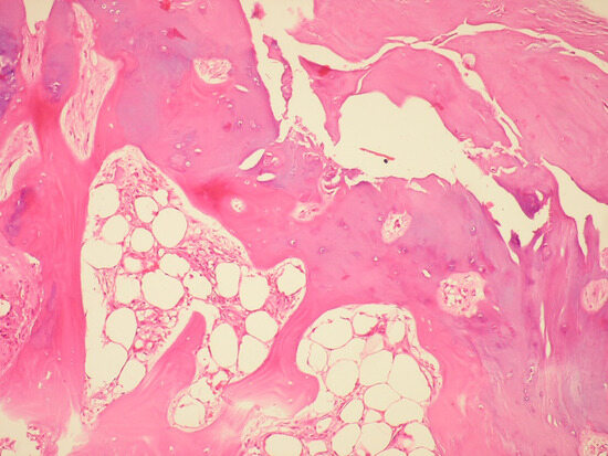
Figure 3.
Microscopic view of the removed bone fragment with lamellar bone formation.
Postoperatively, the patient was placed in a long leg cast for 6 weeks. After removal of the cast, physiotherapy for ankle range of motion was started. After 12 weeks, the patient was able to walk without any restriction. However, the patient was lost to follow-up after 6 months.
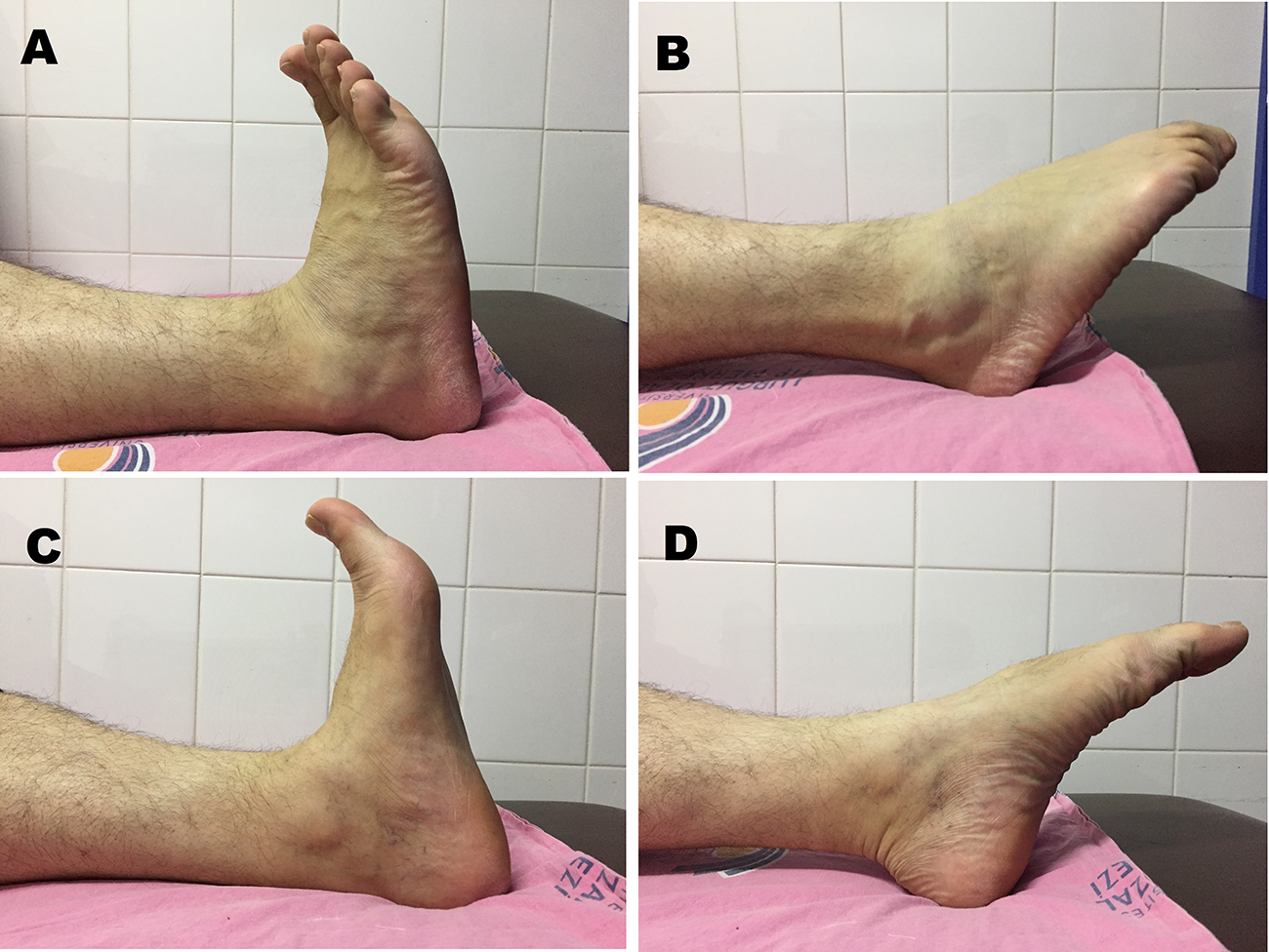
Seven years after surgery, in 2017, the patient was readmitted to the outpatient clinic because of pain in the posterior left heel. On physical examination, there was tenderness at the level of the Achilles insertion to the calcaneus on the left side. A thickening with no discomfort was observed on the right ankle. On inspection of the patient’s medical history, it was realized that the patient was diagnosed with ankylosing spondylitis in 2012 and treated with sulfasalazine. The radiographs and magnetic resonance imaging scans revealed an ossification at the distal aspect of the Achilles tendon in the left foot and a recurring ossification at the proximal aspect at the level of the previously removed bone segment in the right foot (Fig. 4). The patient was treated with nonsteroidal anti-inflammatory drugs (NSAIDs) and physiotherapy for left ankle pain. Three years after the detection of reossification (10 years after the initial surgery), the patient was pain-free, with full range of motion in both ankles, and his American Orthopaedic Foot and Ankle Society score was 95 (Fig. 5).
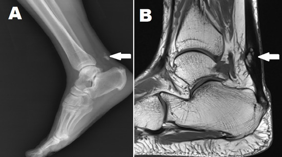
Figure 4.
Radiograph (A) and magnetic resonance imaging scan (B) showing reossification of the Achilles tendon (arrows).
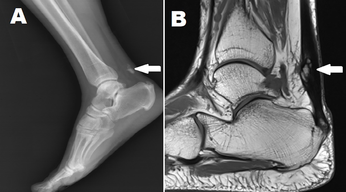
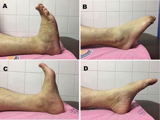
Figure 5.
Photographs showing range of motion in the right (A and B) and left (C and D) ankles.
Discussion
There are few case reports in the literature about FOAT [2,3,4,5,6,7,8,9,10,11,12]. In total, 14 patients have been reported in case reports or case series. To the best of our knowledge, there is only one case report on the reossification of the Achilles tendon after removal of the ossified part, which was treated with immobilization for 6 weeks; however, the authors report no further data about the complaint of the patient and the follow-up period [13]. In our case, we followed our patient for 3 years after detecting the reossification, and the patient had no complaints 10 years after the initial surgery.
Our patient had a history of trauma before initial surgery and was also diagnosed with ankylosing spondylitis 2 years after surgery. Because of this complicated situation, we were unable to determine the cause of the ossification. The cause of ossification and reossification may be trauma resulting from initial ossification and surgery for reossification, or seronegative arthropathy for both. We were unable to make a decision because all these conditions were reported as risk factors [1,14].
The treatment of the ossification of the Achilles tendon differs according to the symptoms of the patients and the presence of a fracture. Conservative management with NSAIDs and physiotherapy may be used as first-line treatment. Surgical removal of the ossified Achilles tendon may be performed if there is persistent pain despite conservative treatment [1,13]. In FOAT cases, conservative treatment, osteosynthesis of the fractured bone, or removal of the bone with repair or reconstruction of the Achilles tendon are the treatment options [1,2,3,4,5,6,7,8,9,10,11,12,13]. The decision regarding repair or reconstruction could be determined according to the size of the bony fragment and the defect that would develop after removal of the bony fragment. In our case, we removed the fractured bony fragment and performed primary repair on the Achilles tendon on the right side. During follow-up, pain on the left side was treated with NSAIDs and physiotherapy; the patient healed uneventfully and showed no symptoms during the 3-year follow-up period.
Conclusions
Ossification of the Achilles tendon and FOAT are rare clinical conditions. We presented the second case of reossification of the Achilles tendon. We followed our case for 3 years after reossification (ie, 10 years after initial surgery). The treatment strategy for the ossified Achilles tendon should be determined according to the symptoms of the patients. If surgical treatment is chosen, the patients should be informed preoperatively about the risk of reossification.
Financial Disclosure: None reported.
Conflict of Interest: None reported.
References
- RICHARDS, PJ; BRAID, JC; CARMONT, MR; et al. Achilles tendon ossification: pathology, imaging and aetiology. Disabil Rehabil 2008, 30, 1651. [Google Scholar] [CrossRef] [Scilit] [PubMed]
- AKSOY, MC; SURAT, A. Fracture of the ossified Achilles tendon. Acta Orthop Belg 1998, 64, 418. [Google Scholar] [PubMed]
- PARTON, MJ; WALTER, DF; RITCHIE, DA; et al. Case report: fracture of an ossified Achilles tendon—MR appearances. Clin Radiol 1998, 53, 538. [Google Scholar] [CrossRef] [Scilit] [PubMed]
- GOYAL, S; VADHVA, M. Fracture of ossified Achilles tendon. Arch Orthop Trauma Surg 1997, 116, 312. [Google Scholar] [CrossRef] [PubMed]
- FINK, RJ; CORN, RC. Fracture of an ossified Achilles tendon. Clin Orthop Relat Res 1982, 169, 148. [Google Scholar] [CrossRef] [Scilit]
- RESNIK, CS; FOSTER, WC. Achilles tendon ossification and fracture. Can Assoc Radiol J 1990, 41, 153. [Google Scholar] [PubMed]
- BROTHERTON, BJ; BALL, J. Fracture of an ossified Achilles tendon. Injury 1979, 10, 245. [Google Scholar] [CrossRef] [Scilit] [PubMed]
- WESELEY, MS; BARENFELD, PA; EISENSTEIN, AL. Fracture of an ossific mass in the Achilles tendon. Bull Hosp Joint Dis 1976, 37, 159. [Google Scholar] [PubMed]
- BATTAGLIA, TC; CHANDLER, JT. Ossific tendonitis of the Achilles with tendon fracture. Orthopedics 2006, 29, 453. [Google Scholar] [CrossRef] [Scilit] [PubMed]
- ISHIKURA, H; FUKUI, N; TAKAMURE, H; et al. Successful treatment of a fracture of a huge Achilles tendon ossification with autologous hamstring tendon graft and gastrocnemius fascia flap: a case report. BMC Musculoskelet Disord 2015, 16, 365. [Google Scholar] [CrossRef] [Scilit] [PubMed]
- YAMAGUCHI, S; ENDO, J; YAMAMOTO, Y; et al. Double-row bridging suture fixation augmented with double Krackow suture for the repair of avulsion of the ossified Achilles tendon: a technical tip. Foot Ankle Int 2015, 36, 849. [Google Scholar] [CrossRef] [Scilit] [PubMed]
- ROMANO, N; BRUNETTI, N; MELANI, EF. Imaging presentation of ossified Achilles tendon fracture. Diagn Interv Imaging 2020, 101, 627. [Google Scholar] [CrossRef] [Scilit] [PubMed]
- YU, JS; WITTE, D; RESNICK, D; et al. Ossification of the Achilles tendon: imaging abnormalities in 12 patients. Skeletal Radiol 1994, 23, 127. [Google Scholar] [CrossRef] [Scilit] [PubMed]
- ARORA, AJ; ARORA, R. Ossification of the bilateral Achilles tendon: a rare entity. Acta Radiol Open 2015, 4, 2058460115599184. [Google Scholar] [CrossRef] [Scilit] [PubMed]
- FREED, L; ELLIS, MB; JOHNSON, K; et al. Fasciotomy and surgical tenotomy for chronic Achilles insertional tendinopathy: a retrospective study using ultrasound-guided percutaneous microresection. JAPMA 2019, 109, 1. [Google Scholar] [CrossRef] [Scilit] [PubMed]
© 2022, Journal of the American Podiatric Medical Association