


Abstract
Background: Diabetic foot ulceration is a severe complication of diabetes characterized by chronic inflammation and impaired wound healing. This study aimed to evaluate the effect of a medical device gel based on adelmidrol + trans-traumatic acid in the healing process of diabetic foot ulcers. Methods: Thirty-seven diabetic patients with foot ulcers of mild/moderate grade were treated with the gel daily for 4 weeks on the affected area. The following parameters were evaluated at baseline and weekly: 1) wound area, measured by drawing a map of the ulcer and then calculated with photo editing software tools, and 2) clinical appearance of the ulcer, assessed by recording the presence/absence of dry/wet necrosis, infection, fibrin, neoepithelium, exudate, redness, and granulation tissue. Results: Topical treatment led to progressive healing of diabetic foot ulcers with a significant reduction of the wound area and an improvement in the clinical appearance of the ulcers. No treatment-related adverse events were observed. Conclusions: The results of this open-label study show the potential benefits of adelmidrol + trans-traumatic acid topical administration to promote reepithelialization of diabetic foot ulcers. Further studies are needed to confirm the observed results.
The diabetic foot ulcer is a debilitating complication of diabetes mellitus, with a 25% lifetime incidence in patients with type 1 or 2 diabetes [1]. Foot ulceration associated with impaired wound healing produces chronic wounds that impair the life quality of diabetic patients, leading to hospitalization and often resulting in lower-extremity amputations [2].
Evidence shows that diabetes is characterized by a systemic and cutaneous chronic inflammatory state, which contributes to impaired wound healing [3–5]. A substantial role in this phenomenon is played by mast cells (MCs), which are immune cells that mature in tissues. In normal conditions, they can contribute to skin injury repair, increasing acute inflammation and enhancing new epidermis regeneration, and new blood vessel formation, through the release of several mediators [6–9]. However, MCs have also been associated with aberrant healing, as shown by the altered number of MCs found during the remodeling phase in the skin injury repair in streptozotocin-induced diabetes mellitus in mice [10]. An increase in the number of degranulated MCs has been reported in both human and mouse diabetic unwounded skin [11]. Noteworthy, an improvement of wound healing in diabetic mice has been reported after systemic MC stabilization with disodium cromoglycate [11] and after the topical treatment of diabetic mice with an indole-type MC stabilizer, either before or after wound induction [12]. In line with these results, photobiomodulation, a treatment with known MC stabilizer properties [13], after surgery significantly improved the tensiometric properties of wound healing alongside a decrease of total and degranulated MC numbers during the remodeling step in a diabetic rat model [14].
Adelmidrol is a diethanolamide derivative of azelaic acid with amphipathic properties, which makes it particularly suitable for topical use. Adelmidrol has been shown to increase the endogenous level of palmitoylethanolamide [15], a physiologic MC modulator endowed with anti-inflammatory and pain-relieving effects [16–18]. Thus, adelmidrol application can enhance the intrinsic cellular protection mechanism of the skin [15]. This assumption is supported by the finding that its use in canine skin wounds reduces the degranulation of cutaneous MCs [19], increases the percentage of reepithelialization, and leads to an increase in the density of elastic fibers [20]. Finally, the ultrasonographic evaluation of the experimental wounds showed that the topical application of adelmidrol significantly reduces the wound volume, thus improving wound healing over time [21].
Trans-traumatic acid, also named “plant wound hormone,” is a monounsaturated dicarboxylic acid produced by enzymatic degradation of fatty acids (linoleic and linolenic), and many plants physiologically secrete it in response to various types of damage [22–24]. Experimental evidence has shown that trans-traumatic acid can induce proliferation and differentiation of plant tissues and stimulate the post-traumatic cicatricial response. Moreover, it can favor the repair of animal tissues, increasing their cellular proliferation [25], with an increase in the reepithelialization and growth of fibroblasts in the injured areas [26]. This pro-reepithelizing effect was attributed to the ability of the trans-traumatic acid to favor the convergence of keratinocytes starting from the margins of the lesions, thus accelerating the reconstruction of epithelial integrity [25].
The purpose of the present investigation is to verify whether the topical application of a medical device gel based on the combination of adelmidrol + trans-traumatic acid allows an improvement in reepithelialization and cicatrization of diabetic foot ulcers.
Materials and Methods
This open-label study was conducted at the outpatient diabetic foot clinic of the Division of Endocrinology, Diabetes, and Metabolism, Azienda Ospedaliera Universitaria Integrata, Verona, Italy.
Patients with a diagnosis of type 1 or type 2 diabetes who have the complication of foot ulcerative lesions were enrolled according to the following inclusion criteria: age 18 years or older; presence of foot ulcers of grade 1 A/B/C or 2 A/B according to the University of Texas diabetic wound classification system [27]; and reliability to compliance. The exclusion criteria were the presence of moderate/severe vascular pathology, ankle-brachial index less than 0.70, tension cutaneous oxygen less than 30 mm Hg [28], presence of moderate/severe infection according to the PEDIS (Perfusion, Extent/Size, Depth/Tissue Loss, Infection, Sensation) scale, and presence of gangrene/dry eschar.
This study was performed following the Good Clinical Practice guidelines and the principles of the Declaration of Helsinki of 1975 and was approved by the departmental review board.
Patients were enrolled between May 2017 and August 2018. After expressing informed consent, patients were treated with one topical application per day of a gel based on adelmidrol + trans-traumatic acid (Nevamast; Epitech Group SpA, Saccolongo, Italy), on the affected area, after careful cleansing of the wound, for 4 weeks.
The following parameters were evaluated for each patient: 1) wound area, measured by drawing a map on a polyurethane film grid, which was scanned and calculated with photo editing software (Photoshop CS6; Adobe Inc, San Jose, California), and 2) clinical appearance of the ulcer, assessed by recording the presence/absence of dry/wet necrosis, infection, fibrin, neoepithelium, exudate, redness, and granulation tissue. Both the wound area and the clinical appearance of ulcers were evaluated weekly. Complete healing was defined as 100% epithelization, and in this case, a value of 0 was attributed to the wound area.
The off-loading device adequacy used by the patient was also assessed weekly. The off-loading device was evaluated as adequate if it was a knee-high device, with an appropriate foot-device interface, worn correctly and continuously all day long [29].
Statistical Analysis
Statistical analysis to evaluate the variation of ulcer area over time was performed using the generalized linear mixed model. Variables such as sex, age, body mass index, diabetes duration, diabetes type, wound duration, presence of neuropathy, vascular complications, and off-loading were included in the model as covariates. The wound area is given as mean ± SE. A responder analysis was also performed. For this purpose, as minimum efficacy criteria (MEC), it established the corresponding minimum percentage reductions in the wound area. In particular, a patient was considered a responder if the wound area decreased by at least 30%, 50%, or 70% after 4 weeks of treatment [30,31]. Percentages of patients reaching the established MEC values were calculated.
The typical ulcer signs were quantified by calculating percentages of patients with dry/wet necrosis, infection, fibrin, neoepithelium, exudate, redness, and granulation tissue at each follow-up.
Results
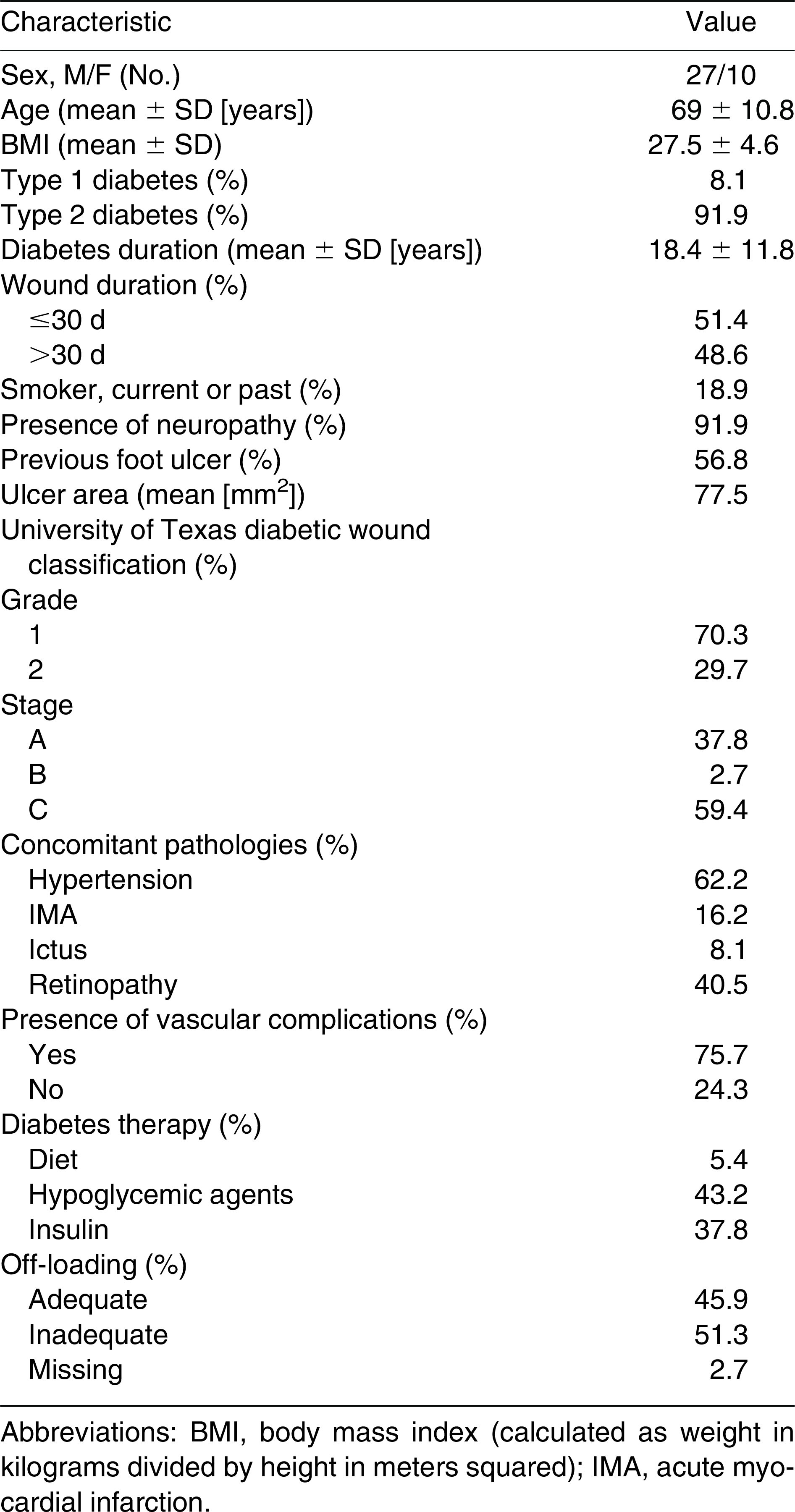
Thirty-seven diabetic patients with foot ulcers were considered for the study. Demographic data and ulcer details of the patients are described in Table 1. Eleven patients dropped out for different reasons: two for stationary symptoms and one for family reasons, one patient died of infarction, and seven patients left because they recovered before the end of the study. Data collected for dropout patients were considered for the analysis. All of the uncollected data were deemed to be missing.

Table 1.
Demographic and Treated Ulcer Details of the 37 Patients Treated with Adelmidrol + Trans Traumatic Acid
Statistical analysis with a generalized linear mixed model showed significant variation of the mean ± SE wound area over time (P < .05), moving from a value of 77.5 ± 14.9 mm2 at baseline to 36.7 ± 10.2 mm2 at week 4 (Figs. 1 and 2). Post hoc analysis performed between each follow-up and its subsequent one showed a significant reduction (P < .05) between baseline and week 1. None of the covariates considered affected the wound area reduction.
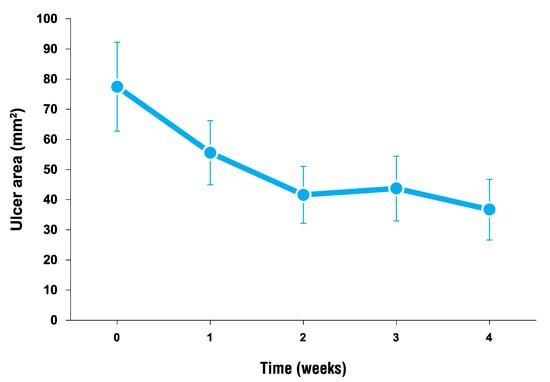
Figure 1.
Foot wound area over time in diabetic patients receiving topical treatment based on adelmidrol + trans-traumatic acid. Data are given as mean ± SE. The mean wound area decreased significantly over time (P < .05); post hoc analysis showed a significant reduction between baseline and week 1 (P < .05). Statistical analysis was performed with a generalized linear mixed model.
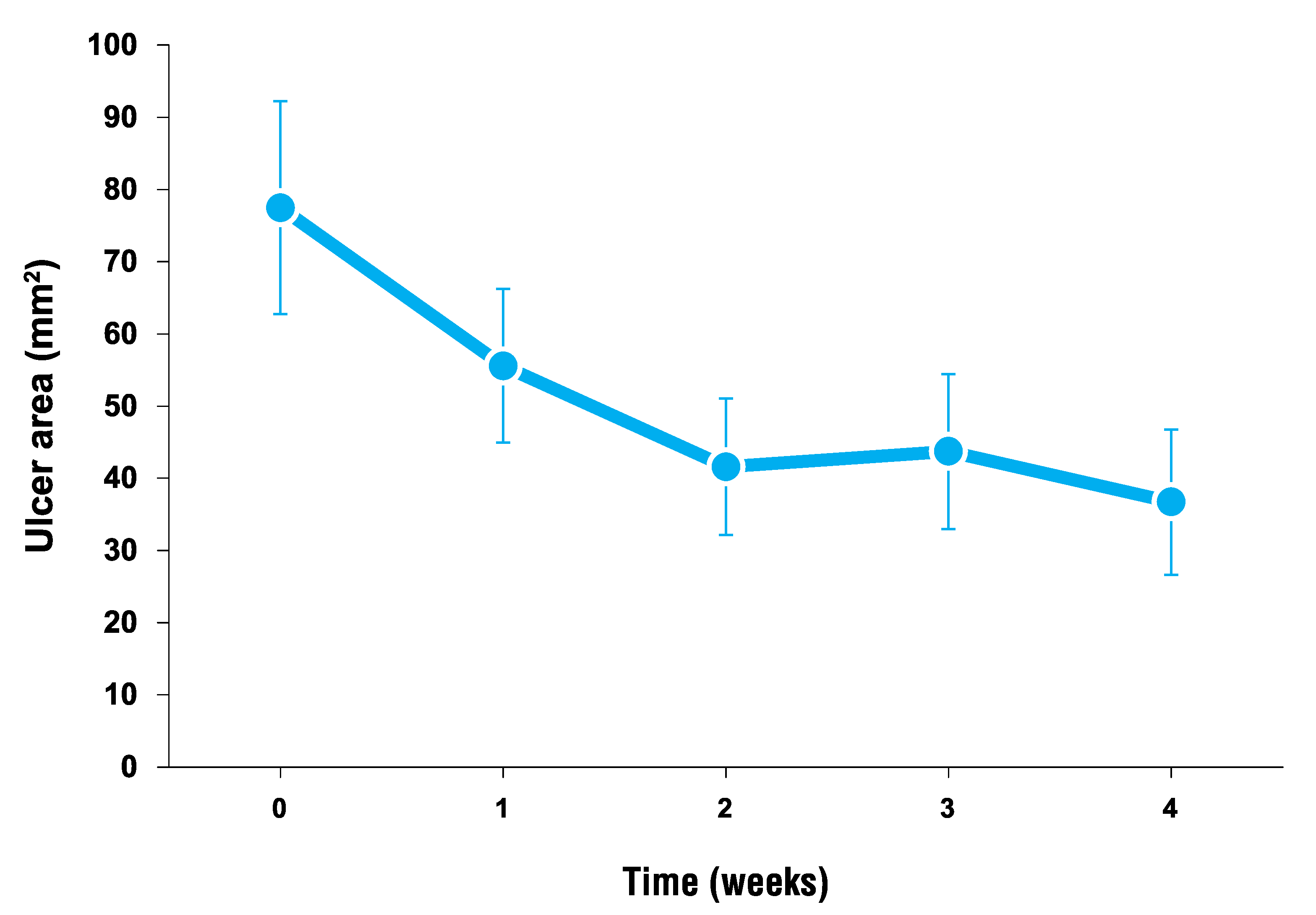
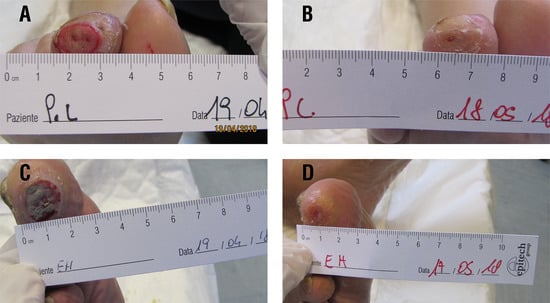
Figure 2.
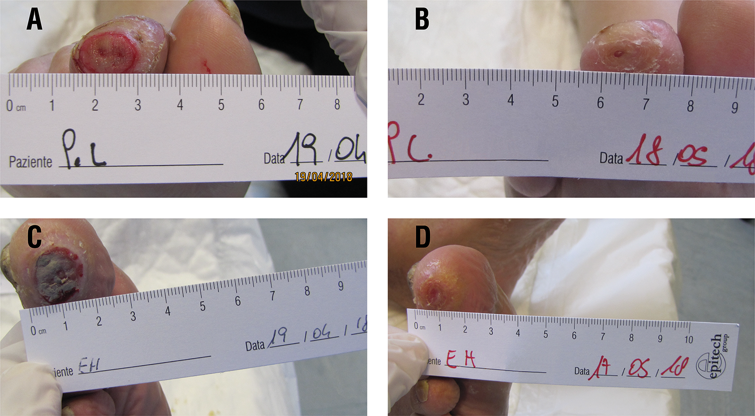
Diabetic foot ulcer examples of two patients at baseline (A and C) and after 4 weeks of topical treatment based on adelmidrol + trans-traumatic acid (B and D).
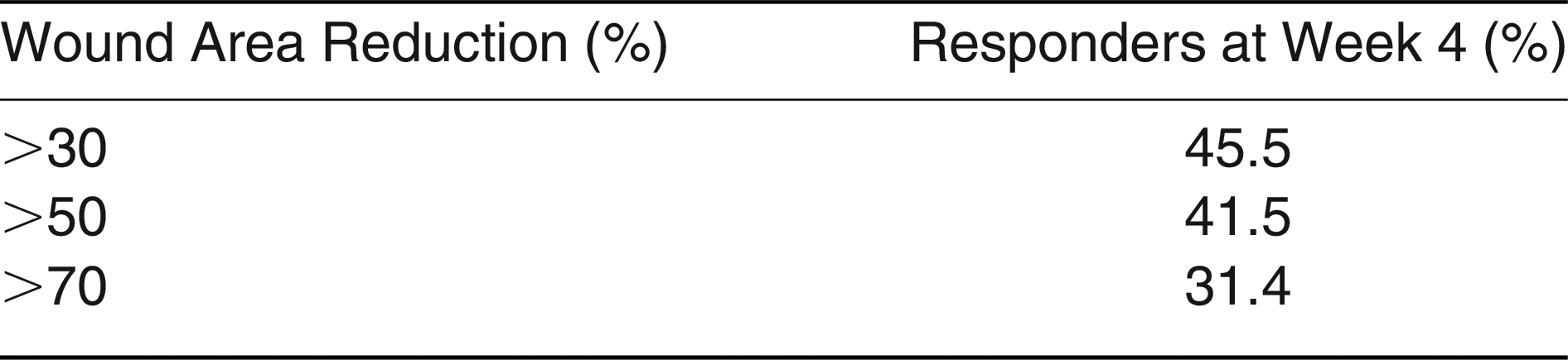
Percentages of responder patients for each established MEC at the end of treatment are given in Table 2.
Table 2.
Responders with a Wound Area Reduction of at Least 30%, 50%, or 70% Calculated at Week 4 After Adelmidrol + Trans-Traumatic Acid Treatment
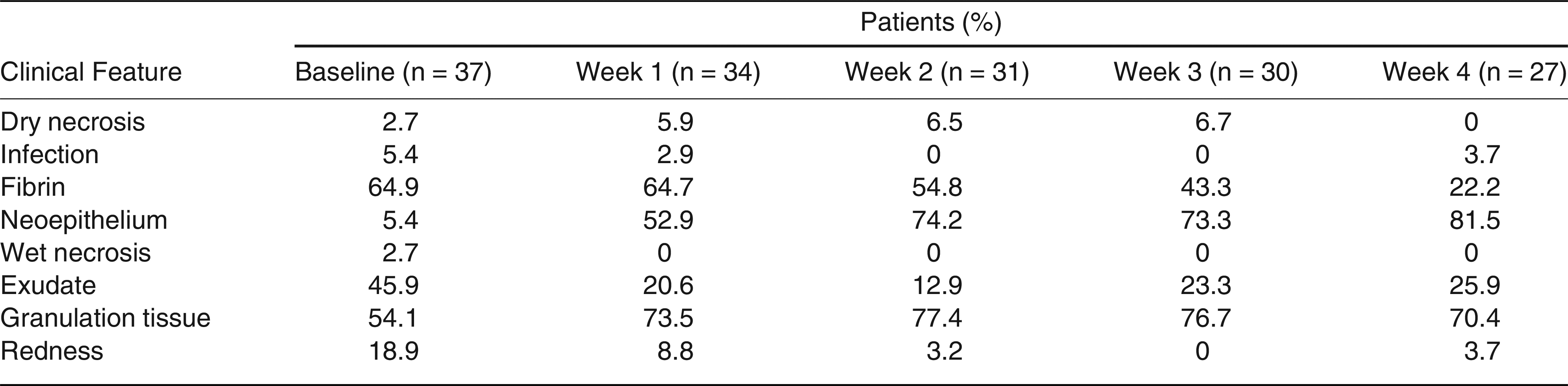
Concerning changes in ulcer appearance, presence of dry/wet necrosis, infection, fibrin, exudate, and redness decreased in patients between baseline and week 4, whereas the presence of neoepithelium and granulation tissue increased between baseline and week 4 (Fig. 3). Percentages of patients presenting the typical clinical features of diabetic ulcers at each follow-up are reported in Table 3.
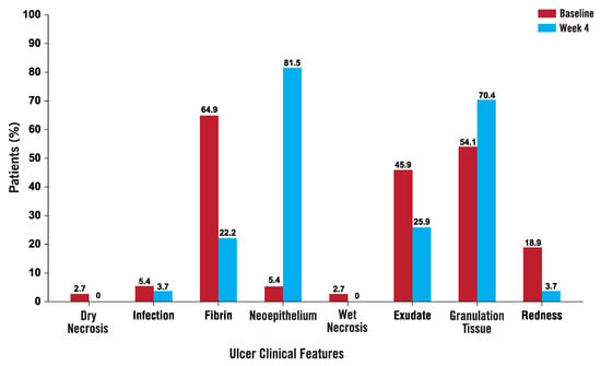
Figure 3.
Typical clinical features of diabetic ulcers at baseline and after 4 weeks of topical treatment based on adelmidrol + trans-traumatic acid. The percentage of patients with dry/wet necrosis, infection, fibrin, exudate, or redness decreased between baseline and week 4; the percentage of patients with neoepithelium or granulation tissue increased between baseline and treatment week 4.
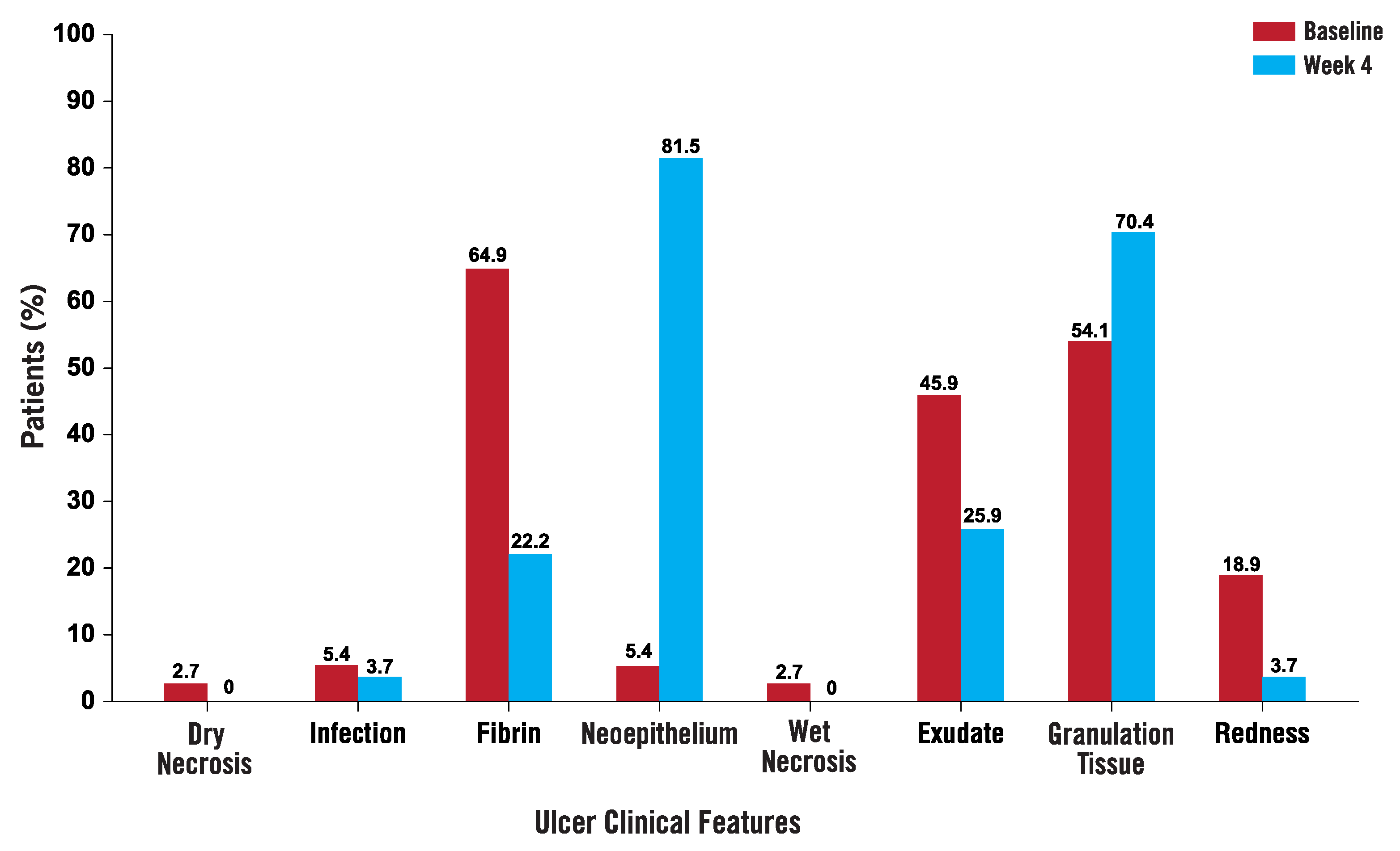
Table 3.
Patients Presenting the Typical Clinical Features of Diabetic Ulcers at Each Follow-up
Discussion
This study shows an improvement in wound healing of diabetic foot ulcers in response to topical treatment with a gel based on adelmidrol + trans-traumatic acid. The wound area significantly decreased during 1 month of treatment, and in particular, the effect was already significant after the first week of treatment (Figs. 1 and 2). At the same time, the percentage of patients with signs of poor wound bed status (presence of dry/wet necrosis, infection, fibrin, exudate, redness) at the end of treatment decreased compared with baseline, whereas the percentage of patients with signs of healing wound bed status (presence of neoepithelium and granulation tissue) increased by the study end (Fig. 3). The present results are in line with a preclinical study showing that adelmidrol + trans-traumatic acid can accelerate the healing process in diabetic mice after wound induction compared with animal vehicle, which shows impaired healing [32].
The diabetic foot usually affects patients with a long duration of illness, with different comorbidities and complex to manage clinically. Moreover, the complexity of this pathologic condition also depends on several variables, such as the presence of neuropathy and vascular complications, but also inadequate off-loading and factors such as age, sex, body mass index, type of diabetes, diabetes duration, and wound duration, that can influence ulcer manifestation. From the statistical analysis, none of the variables mentioned previously herein affects wound area reduction.
The main limitations of the study are the open-label design with the lack of blinding and the small patient size, which increase the difficulty in estimating the effect of the treatment. Furthermore, the small ulcer size considered at baseline led in some cases to rapid healing, which causes an early exit of some patients from the study, therefore increasing missing data.
Notably, treatment with adelmidrol + trans-traumatic acid does not show adverse events, is considered easy to use, and shows potential efficacy in diabetic foot ulcer healing.
According to a recent study, the achievement of a 50% reduction in wound area observed across 4 weeks of treatment is a robust predictor of a long-term healing prognosis [30,31]. In the present study, 41.5% of patients obtained a more than 50% reduction in wound area at the end of treatment, thus encouraging further prospective studies with a double-blind design, larger patient numbers, and larger ulcer sizes at baseline to confirm the findings. Moreover, long-term treatment would allow for better appreciation of the safety and efficacy of this topical gel.
The results of this study on diabetic foot ulcers, a chronic inflammatory condition characterized by an abnormal increase in MC degranulation [11,12,14], and the already reported ability of adelmidrol to increase palmitoylethanolamide levels [15], a physiologic MC modulator known to promote chronic inflammation resolution [16–18], prompt us to sustain the potential contribution that adelmidrol could provide to the healing processes.
Therefore, topical application of adelmidrol in association with trans-traumatic acid represents a new therapeutic opportunity for this complex syndrome. Because the manifestations of the diabetic foot derive from specific problems of the lower limbs, such as ischemia and neuropathy, which often intersect biomechanical issues, infection, and wound repair, a multidisciplinary approach is always needed. In particular, topical therapy has to involve an in-depth evaluation of all of the variables that influence wound healing, which has to be followed by the application of specific diversified local treatments to guarantee the most significant healing of the foot and minimize recurrences. Further prospective randomized studies are needed to confirm the results obtained and optimize the effect of the treatment studied.
Acknowledgments
Dr Carlo Schievano for support with the statistical analysis, and Dr Francesca Policastro for the help in all phases of the project.
Financial Disclosure
None reported.
Conflicts of Interest
None reported.
References
- Singh N, Armstrong DG, Lipsky BA: Preventing foot ulcers in patients with diabetes. JAMA 293: 217, 2005.
- Ramsey SD, Newton K, Blough D, et al: Incidence, outcomes, and cost of foot ulcers in patients with diabetes. Diabetes Care 22: 382, 1999.
- Dinh T, Tecilazich F, Kafanas A, et al: Mechanisms involved in the development and healing of diabetic foot ulceration. Diabetes 61: 2937, 2012.
- Tellechea A, Kafanas A, Leal EC, et al: Increased skin inflammation and blood vessel density in human and experimental diabetes. Int J Low Extrem Wounds 12: 4, 2013.
- Leal EC, Carvalho E, Tellechea A, et al: Substance P promotes wound healing in diabetes by modulating inflammation and macrophage phenotype. Am J Pathol 185: 1638, 2015.
- Wulff BC, Wilgus TA: Mast cell activity in the healing wound: more than meets the eye? Exp Dermatol 22: 507, 2013.
- Sismanopoulos N, Delivanis D-A, Alysandratos K-D, et al: Mast cells in allergic and inflammatory diseases. Curr Pharm Des 18: 2261, 2012.
- Theoharides TC, Alysandratos K-D, Angelidou A, et al: Mast cells and inflammation. Biochim Biophys Acta 1822: 21, 2012.
- Komi DEA, Khomtchouk K, Santa Maria PL: A review of the contribution of mast cells in wound healing: involved molecular and cellular mechanisms. Clin Rev Allergy Immunol 58: 298, 2020.
- Nishikori Y, Shiota N, Okunishi H: The role of mast cells in cutaneous wound healing in streptozotocin-induced diabetic mice. Arch Dermatol Res 306: 823, 2014.
- Tellechea A, Leal EC, Kafanas A, et al: Mast cells regulate wound healing in diabetes. Diabetes 65: 2006, 2016.
- Tellechea A, Bai S, Dangwal S, et al: Topical application of a mast cell stabilizer improves impaired diabetic wound healing. J Invest Dermatol 140: 901, 2020.
- Fathabadie FF, Bayat M, Amini A, et al: Effects of pulsed infra-red low level-laser irradiation on mast cells number and degranulation in open skin wound healing of healthy and streptozotocin-induced diabetic rats. J Cosmet Laser Ther 15: 294, 2013.
- Bagheri M, Amini A, Abdollahifar M-A, et al: Effects of photobiomodulation on degranulation and number of mast cells and wound strength in skin wound healing of streptozotocin-induced diabetic Rats. Photomed Laser Surg 36: 415, 2018.
- Petrosino S, Puigdemont A, Della Valle MF, et al: Adelmidrol increases the endogenous concentrations of palmitoylethanolamide in canine keratinocytes and down-regulates an inflammatory reaction in an in vitro model of contact allergic dermatitis. Vet J 207: 85, 2016.
- Cerrato S, Brazis P, Della Valle MF, et al: Effects of palmitoylethanolamide on the cutaneous allergic inflammatory response in Ascaris hypersensitive Beagle dogs. Vet J 191: 377, 2012.
- Petrosino S, Iuvone T, Di Marzo V: N-palmitoyl-ethanolamine: biochemistry and new therapeutic opportunities. Biochimie 92: 724, 2010.
- Kendall AC, Nicolaou A: Bioactive lipid mediators in skin inflammation and immunity. Prog Lipid Res 52: 141, 2013.
- Abramo F, Salluzzi D, Leotta R, et al: Mast cell morphometry and densitometry in experimental skin wounds treated with a gel containing adelmidrol: a placebo controlled study. Wounds 20: 149, 2008.
- Abramo F, Noli C, Giorgi M, et al: Elastic fibres and mast cells in canine open skin wounds treated with an aliamide-based gel: a morphometric study. Vet Dermatol; p 354; 2005.
- Mantis P, Lloyd D, Pfeiffer D, et al: Assessment of the effect of an aliamide-containing topical gel by evaluation of the reduction of wound volume measured by high resolution ultrasound biomicroscopy. Wounds 19: 113, 2007.
- Zimmerman DC, Coudron CA: Identification of traumatin, a wound hormone, as 12-oxo-trans-10-dodecenoic acid. Plant Physiol 63: 536, 1979.
- Vick BA, Zimmerman DC: Lipoxygenase and hydroperoxide lyase in germinating watermelon seedlings. Plant Physiol 57: 780, 1976.
- Gardner HW: 9-Hydroxy-traumatin, a new metabolite of the lipoxygenase pathway. Lipids 33: 745, 1998.
- Miyamoto I, Uchida Y, Shinomiya T, et al: Effects of cosmetics containing bioactive substances on skin. J Soc Cosmet Chem Japan 22: 254, 1989.
- Trypuc´ AJ, Pankiewicz W, Czerpak R: Traumatic Acid Reduces Oxidative Stress and Enhances Collagen Biosynthesis in Cultured Human Skin Fibroblast. Lipids 51: 1021–1035, 2016.
- Lavery LA, Armstrong DG, Harkless LB: Classification of diabetic foot wounds. J Foot Ankle Surg 35: 528, 1996.
- AMD: Documento di consenso: trattamento dell’arteriopatia periferica nel diabetico. Il Giornale di AMD 16: 72, 2013.
- Bakker k, Apelqvist J, Lipsky B, et al: The 2015 IWGDF guidance documents on prevention and management of foot problems in diabetes: development of an evidence-based global consensus. Diabetes Metab Res Rev 32: 2, 2016.
- Sheehan P, Jones P, Caselli A, et al: Percent change in wound area of diabetic foot ulcers over a 4-week period is a robust predictor of complete healing in a 12-week prospective trial. Diabetes Care 26: 1879, 2003.
- Sheehan P, Jones P, Giurini JM, et al: Percent change in wound area of diabetic foot ulcers over a 4-week period is a robust predictor of complete healing in a 12-week prospective trial. Plast Reconstr Surg 117: 239S, 2006.
- Siracusa R, Impellizzeri D, Cordaro M, et al: Topical application of adelmidrol + trans-traumatic acid enhances skin wound healing in a streptozotocin-induced diabetic mouse model. Front Pharmacol 9: 871, 2018.
© 2022 American Podiatric Medical Association