


Abstract
Background: Mild leg length discrepancy increases biomechanical asymmetry during gait, which leads to low-back pain. Orthotic insoles with a directly integrated heel lift were used to reduce this asymmetry and thus the associated low-back pain. The aim of this study was to analyze the biomechanical adaptations of the locomotor apparatus during gait and the subjective pain ratings before and after the establishment of orthotic insole use. Methods: Eight patients with mild leg length discrepancy (≤2.0 cm) underwent three-dimensional biomechanical analysis while walking before and after 3 weeks of orthotic insole use. Low-back pain was assessed separately before both measurement sessions using a visual analog scale. Results: Analysis of the kinematic parameters highlighted individual adaptations. The symmetry index of Dingwell indicated that orthotic insoles had no significant effect on the kinematic gait parameters and an unpredictable effect across patients. Orthotic insole use significantly and systematically (in all of the patients) reduced low-back pain (P < .05), which was correlated with changes in ankle kinematics (P = .02, r = 0.80). Conclusions: The effects of orthotic insoles on gait symmetry are unpredictable and specific to each patient’s individual manner of biomechanical compensation. The reduction in low-back pain seems to be associated with the improved ankle kinematics during gait.
Leg length discrepancy (LLD) affects 40% to 70% of the population[1,2] and is due to anatomical deformities originating from true bony leg length differences or a functional deformity originating from abnormal lower-limb movements.[3] Thus, LLD can be divided into two etiological groups: anatomical and functional. Anatomical LLD can be defined as those discrepancies associated with a shortening of bony structures, and it can further be subdivided in two subgroups: congenital and acquired. The congenital conditions include mild developmental abnormalities found at birth or childhood and/or abnormal developmental disorders. Acquired conditions include trauma, fractures, orthopedic degenerative diseases, and surgical disorders. Functional LLD is an asymmetrical leg length that does not result from a true bony length difference. Instead, it results from an alteration in lower-limb mechanics, such as joint contracture, static or dynamic mechanical axis malalignment, muscle weakness, or shortening.[1,4] Whatever the etiology of the LLD (ie, anatomical or functional), Moseley[5] suggested classifying the discrepancy into three severity grades: mild (0.0–2.0 cm), moderate (2.0–6.0 cm), and severe (6.0–20.0 cm). Other authors have suggested a different classification: mild (0.0–3.0 cm), moderate (3.0–6.0 cm), and severe (>6.0 cm).[1,6-9] Furthermore, Walsh et al[10] considered a patient with an LLD less than 0.5 cm to be normal. Therefore, the cutoff point to classify mild and moderate LLD is not completely consensual. Although Moseley[5] suggested that mild LLD requires no treatment, Reid and Smith[9] recommended either no treatment or orthotic insoles (OIs). This divergence in the cutoff point and the recommended treatment is likely explained by the lack of consensus in the literature regarding the biomechanical effects of mild LLD during gait.[11-15]
The symmetry index (SI) of Dingwell[16] is used to quantify gait asymmetry.[17,18] It evaluates the gait pattern in patients with LLD and estimates the acceptable range of discrepancy before and after a heel lift or an OI is worn. In the study by Liu et al,[18] patients with a mean inequality of 0.5 cm showed a smaller SI value than those in a group with a mean inequality of 1.4 cm. A mean inequality of 2.3 cm was found to be an acceptable SI.[18] Yet in a more recent study, patients with a LLD between 1.0 and 2.3 cm exhibited significantly less symmetrical gait.[17] Indeed, Seeley et al[17] evaluated the effect of LLD on gait asymmetry in 26 patients. Nineteen patients exhibited less than 1.0 cm of LLD, with a mean ± SD of 0.4 ± 0.3 cm, and seven patients exhibited a relatively large LLD (≥1.0 cm), with a mean ± SD of 1.7 ± 0.4 cm. This study, which used a symmetry coefficient, revealed that LLD was not significantly related to the degree of bilateral symmetry for joint angles during gait. This result, nevertheless, supported the idea that LLD influences gait symmetry for joint moment and power at the hip, knee, and ankle joints. For all of the patients, statistically significant relationships were observed between mild LLD and gait asymmetry, with higher asymmetry values for those with 1.0 cm or more of LLD.
Murray and Azari[19] and Tallroth et al[20] provided support for the prevailing theory that even mild LLD (≤2.0 cm) can cause excessive mechanical pressure, with consequences in terms of excessive and abnormal loading of the lower extremities and the lumbar spinal joints. Knutson[21] and Guichet et al[22] assumed that LLD is a symptomatic alteration if the discrepancy is greater than 2.0 cm. However, White et al[23] recommended equalization of all discrepancies less than 3.0 cm. Walsh et al[10] evaluated the effect of 1.0- to 5.0-cm foot lifts on the lower-limb kinematics of seven healthy individuals with no significant LLD (ie, not >0.5 cm). They demonstrated that compensation started at 1.0 cm for all of the levels of the locomotor apparatus, leading to asymmetrical gait. An LLD affects the kinematics and kinetics of gait.[11] Indeed, it was found to modify the biomechanical amplitudes of the locomotor apparatus in such a way as to compensate the short limb.[11]
However, LLD generates biomechanical disturbances such as pelvic obliquity, spinal curvature, and asymmetry in the lower-extremity joints and pelvis,[16-18,24] and this, in turn, favors the occurrence of hip osteoarthritis, postural imbalances, and low-back pain (LBP).[1,4,25] In the study by Young et al,[26] a lift greater than 1.5 cm was placed under the foot and caused a lateral pelvic tilt of 1.2°. Moreover, Needham et al[27] demonstrated that the difference in the range of motion of the pelvis and lumbar spine was minimal between barefoot and mild LLD conditions. Three LLD conditions (1.0, 2.0, and 3.0 cm) were simulated using modified pieces of high-density ethyl vinyl acetate attached to the right foot (pelvic range-of-motion adaptation for LLD condition = 1.0 cm: sagittal plane 0.04°, frontal plane 0.3°, transverse plane 0.7°; for LLD condition = 2.0 cm: sagittal plane 0.2°, frontal plane 1.4°, transverse plane 0.6°). Patients with LLD of 0.6 cm presented LBP because they had made abnormal spinal adaptations (scoliosis or hypohyperlordosis) in static and dynamic positions.[28-30] Indeed, in the frontal plane, LLD leads to lateral pelvic tilt on the shorter side and pelvic torsion in the sagittal plane. These pelvic positions cause a body shift to the shorter side and scoliotic attitudes. An LLD also causes a slight curvature of the spine when the pelvic girdle tilts laterally in the frontal plane.[31] These adaptations of the anatomical position may result in the onset of LBP.[28] The minimal LLD necessary to cause LBP has been a matter of debate. Most researchers[1,32] agree that LLD of more than 2.0 to 3.0 cm can cause LBP. However, they disagree about the effect of an LLD of less than 1.0 cm. Some have stated that an LLD of 0.9 cm changes the angle of the lumbar facet joints. An LLD as small as 3.0 cm was found to induce postural changes.[28,33-35] Mild LLD may cause various biomechanical adaptations that can lead to LBP.
It seems clear that mild LLD causes asymmetry in the lower-extremity joints and pelvis, leading to stress and strain with a disturbance in normal biomechanical function. The functional alterations increase the biomechanical disorders, the asymmetrical gait, and LBP and may favor the development of hip osteoarthritis and stress fracture. The OI is a treatment often used in podiatric medicine that might reduce the biomechanical asymmetries[18,36] and, therefore, LBP.[36,37] Moreover, Defrin et al[28] and Golightly et al[38] showed that OIs may reduce physical disability and the intensity of LBP in patients with LLD of 1.0 cm or less. The objective of this observational study in a podiatric medical center was to examine the effects of OIs on gait kinematics and LBP in patients with mild LLD. To conduct this study, we followed the methods used in this podiatric medical center. We hypothesized that the asymmetries of the locomotor apparatus would be rebalanced after OI use, which would reduce LBP.
Methods
Patients
Eight patients (five men and three women; mean ± SD age, 28.3 ± 8.2 years; body mass index, 23.8 ± 1.7; and height, 1.74 ± 0.09 m) volunteered to take part in this study and were recruited in a podiatric medical center. Their ages ranged from 20 to 45 years (ie, bone growth had ended and bone degeneration was limited). All of the participants had been diagnosed as having mild LLD of 2.0 cm or less by a pedicurist-chiropodist using a clinical procedure, with the minimal LLD inclusion criterion being 0.5 cm[10] (mean ± SD LLD, 1.0 ± 0.2 cm). The reason for the chiropodist consultation was chronic LBP, with all of the participants reporting back pain for 3 months or more (the time of onset of chronic status).[39] Other causes of the LBP were considered during the clinical examination, with the pedicurist-chiropodist inquiring into the medical antecedents before determining whether the LLD should be corrected. The inclusion criteria were no history of surgery, no history of lower-limb injury, and no neuromuscular or cardiovascular pathology related to the LLD in the past 6 months. Patients who needed outside help to walk could not participate in the study. This research used an observational study protocol to follow the standard course of care in a podiatric medical center. The care included the chiropodist’s clinical examination to determine the degree of LLD and treatment (OI), with the addition of biomechanical analysis (motion capture) of walking to observe the effects of the OI on the LLD. The entire study was framed by health-care professionals in a health center. Before the study, all of the patients received both verbal and written explanations about the study procedures. The study was conducted according to the Declaration of Helsinki and French national regulations.
Procedure
A clinical examination was performed before the initial session (S1). A professional in podiatric medicine with 25 years of clinical experience verified the presence or absence of mild LLD using the direct method (the tape measurement method) with the patient in the supine position, as described in the literature.[1,2,4,40] The patients wore clothing that allowed for palpation and tape measurement with minimal hindrance, while their modesty and comfort were maintained. They were placed on a treatment table with their pelvis in relaxed and neutral alignment.[40-43] The LLD of all of the patients was assessed by the same pedicurist-chiropodist using the same tape measure to measure the distance between two anatomical points: the anterior superior iliac spine and the distal tip of the medial malleolus.[40] The mean of three readings was used for analysis because the reliability of these measurements was enhanced by the use of mean values.[41] This direct method has concurrent validity with radiographic measurement, with a Pearson product moment correlation of 0.98 and intraclass correlation (ICC) of 0.99.[44] For this study, we evaluated the intratester reliability for tape measurement with an ICC of 0.85. The mean ± SD LLD was 1.0 ± 0.2 cm.
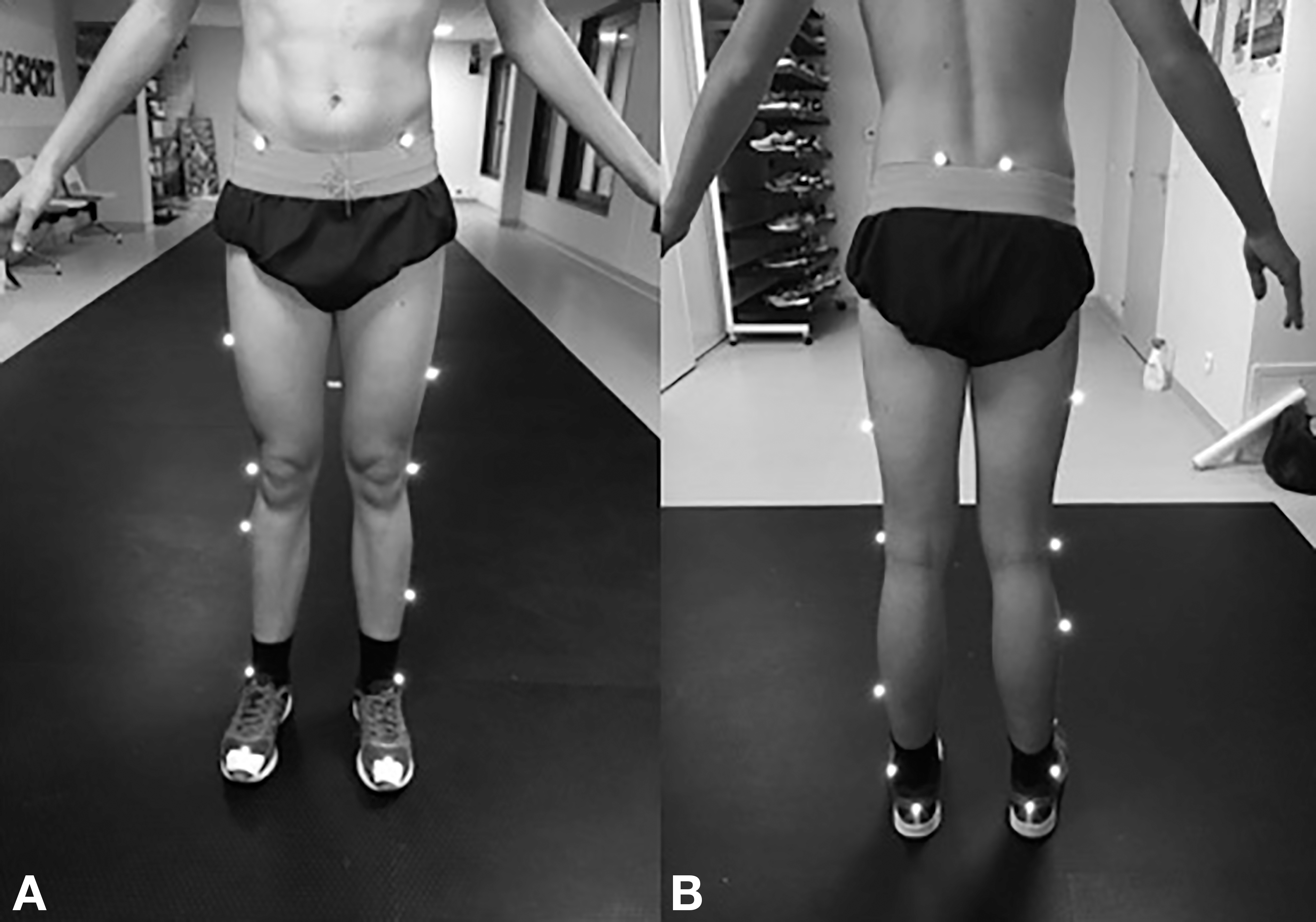
For S1, gait kinematic analysis was performed with the Vicon system (Vicon Oxford Metrics, Oxford, England) to establish the biomechanical reference values.[45] The Vicon system consists of 12 infrared video cameras set at a frequency of 240 Hz. Marker placement corresponded to the Plug-in Gait lower body model (Fig. 1),[46] with 16 reflecting passive markers placed on the skin to determine the lower-limb coordinates. The markers were carefully attached by the pedicurist-chiropodist, who had 25 years of clinical experience. Marker-attaching was repeated several times to ensure optimal repeatability and exact positioning of the markers from session to session.[46] The patients wore their own standard, commercially available shoes for this study, which was necessary because the aim was to decrease their daily pain. After 10 min of familiarization with the environment, four acquisitions over 10 m of spontaneous walking were made to obtain five gait cycles per acquisition for a total of 20 cycles per patient (the three to four first and last strikes of each acquisition were not considered in the data processing because the start and stop were located 3 m from the recording space).
Figure 1.
Marker placement: anterior (A) and posterior (B) views.
During S1, LBP was estimated using a visual analog scale (VAS) graded centimeter by centimeter from 0 (absence of pain) to 10 (maximal pain). The VAS provides a subjective assessment of pain severity.[47] The assessment was made 30 min after kinematic analysis. The instructions to the patients were to answer in a spontaneous and sincere way.
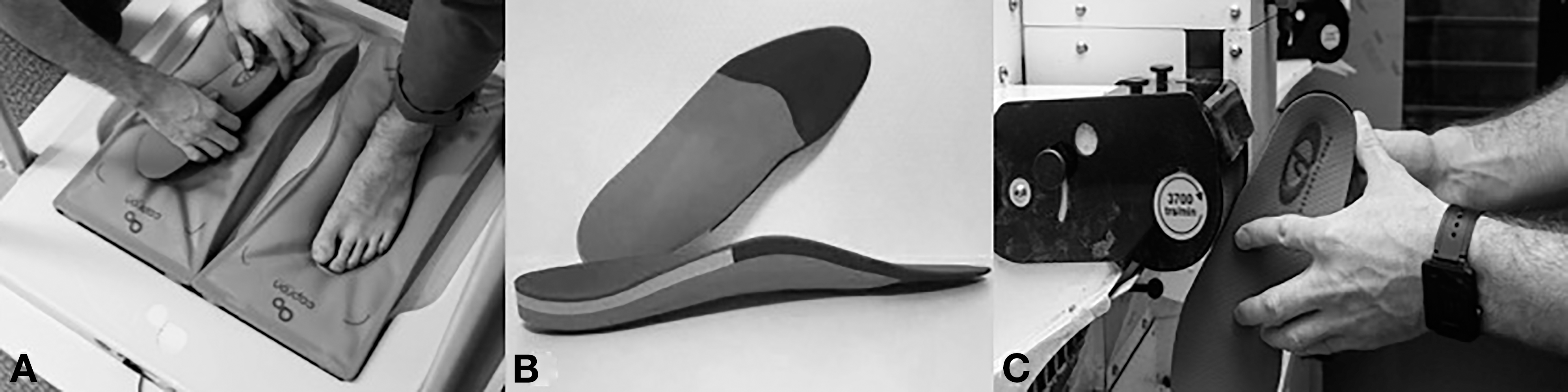
Customized bilateral OIs were made for each participant by the pedicurist-chiropodist based on the first three-dimensional gait analysis and the clinical examination. The technique was thermoforming: the OIs were first warmed before being molded under a pillow mold to obtain the footprint in the anatomical position (Fig. 2A). The components were ethyl vinyl acetate, resin, and polyethylene, and different pads were used (high density, low elasticity) for comfort, absorption, and correction (Fig. 2B). Once the OIs were molded, they were further directly shaped to effectively counteract the effects of LLD and help the patients achieve rebalance in the kinematics of walking (Fig. 2C). They were made according to the therapeutic needs of the patients, with a heel lift incorporated into the OI of the short leg. The heel lifts were partially corrective of the LLD, to 50%, and were shaped from the calcaneus to the Chopart joint. This corrective strategy is used empirically by the pedicurist-chiropodist. At the end of the process, the pedicurist-chiropodist checked the impact of the OI by examining the iliac crest position in the frontal plane while the patient was standing.
Figure 2.
A, Thermoforming of the orthotic insoles. B, Orthotic insoles in ethyl vinyl acetate, resin, and polyethylene. C, Further shaping of the orthotic insoles.
Between S1 and the final session (S2), the patients were asked to carry on their everyday activities for 3 weeks while wearing their custom OIs, as this was the standard care path of the podiatric medical center. For S2, kinematic and subjective analyses were repeated with the same shoes as in S1 but after the patients had been wearing their OIs for the 3 weeks.
Data Reduction
The kinematic parameters were assessed using Polygon software (Vicon Oxford Metrics). The gait kinematics included 1) hip flexion-extension and adduction-abduction and the rotations (the joint angle between the longitudinal axis of two adjacent segments) represented by the motion of the thigh relative to the pelvis; 2) pelvic obliquity, tilt, and rotations (the angle between a segment and the right horizontal of the distal extremity) between the pelvis and the laboratory coordinate system; 3) knee flexion-extension represented by the motion of the shank relative to the thigh; and 4) rearfoot dorsiflexion-plantarflexion between the shank end and the foot.[11] With the Plug-in Gait lower body model (Fig. 1), only two markers were used on the foot to define the measurement of ankle joint motion to flexion-extension. Furthermore, given the finite accuracy and resolution of the motion analysis system, the estimates of inversion-eversion may not have been sufficiently accurate to be of any practical use.[45]
Data Analysis
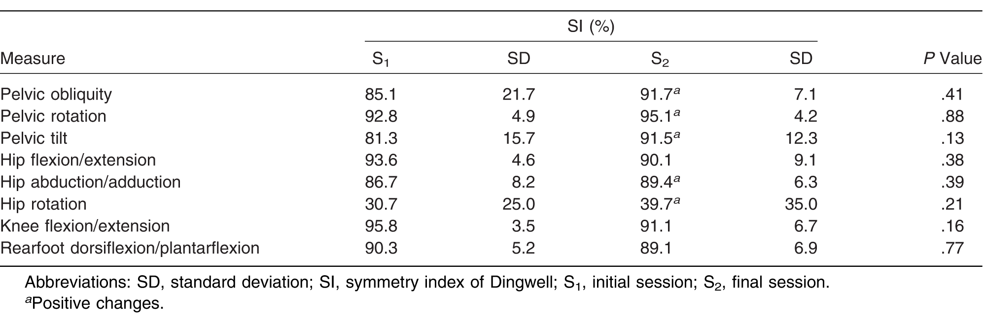
For the kinematic values, maximum and minimum peaks were assessed to identify the joint amplitudes. Then the average of the amplitudes (for the hip, knee, ankle, and pelvis) was calculated on the set of the walking cycles of each patient (n = 20 cycles) (Table 1). Furthermore, to assess the difference in joint amplitude between the lower limbs we determined the SI of Dingwell,[16] according to the following formula:
 RJ corresponds to the right joint, and LJ to the left joint. The SI supplies a percentage, which in the case of perfect symmetry gives a 100% value.
Table 1.
Values of the SI at S1 and S2
Statistical Analysis
Analysis was performed using IBM SPSS Statistics for Windows, Version 20.0 (IBM Corp, Armonk, New York). The kinematic parameters and pain scores were tested for normal distribution using the Shapiro-Wilk test, then compared using the paired Student t test (for normally distributed scores) or the Wilcoxon signed rank test (for nonnormally distributed scores).
The relationship between the changes in pain and the kinematic changes was examined with the Bravais-Pearson parametric correlation test or the Spearman nonparametric test, according to the distribution normality. Significance was set at P = .05.
Results
Comparisons of the range of motion between the short and long limbs showed no significant change (P > .05). On average we do not observe any improvement for the hip flexion/extension RoM comparison (mean S1 = 2.8 ± 1.9; mean S2 3.9 ± 3.5), knee flexion/extension (mean S1 = 2.7 ± 2.3; mean S2 = 5.4 ± 3.7), and rearfoot dosiflexion/plantarflexion (mean S1 = 2.9 ± 1.5; mean S2 = 3.5 ± 2.3).
However, some values highlight a mean decrease in RoM difference with the use of OI for hip adduction/abduction (mean S1 = 1.6° ± 1.0; mean S2 = 1.2° ± 0.9), hip rotation (mean S1 = 18. 0 ± 8.5; S2 = 15.7 ± 10.6), pelvic obliquity (mean S1 = 1.0 ± 1.5; mean S2 = 0.5 ± 0.4), pelvic tilt (mean S1 = 0.7 ± 0.7; mean S2 = 0.3 ± 0.4) and pelvic rotation (mean S1 = 0.8 ± 1.5; mean S2 = 0.4 ± 0.4). However, a complementary visual analysis of the individual results revealed considerable diversity in the kinematic evolution, underscoring the highly individual biomechanical adaptations to the OIs, with 50% partial correction of the LLD.
Range of Motion
No significant difference in the asymmetry of joint range of motion was noted between the pre-post assessments (P > .05). The intraindividual difference in range of motion (right leg –left leg [before]) / (right leg – left leg [after]) showed a nonsignificant decrease in joint asymmetry. These values highlight a decrease in the difference of range of motion with the use of OIs.
SI of Dingwell
Compared with the initial SI results, the use of OIs resulted in no significant change in any parameter observed in this study (Table 1).
Visual Analog Scale
Comparison of the VAS scores between S1 and S2 revealed that all of the patients reported less pain after 3 weeks of wearing the 50% partially corrective OIs (mean VAS score S1 = 3.3 ± 1.2; mean VAS score S2 = 1.4 ± 1.3). Correlation analysis revealed that only the improvement in ankle symmetry was significantly correlated with the decrease in LBP (P = .02; r = 0.802).
Discussion
The objective of this observational study was to analyze the effects of OIs on the parameters of gait kinematics in patients with mild LLD (≤2.0 cm) associated with changes in LBP. The case series evaluated the effect of wearing OIs for 3 weeks (partially corrective: 50% of LLD) on the biomechanics of walking and the LBP of patients with mild LLD. The results did not show a significant decrease in gait asymmetry. However, this study highlighted a significant decrease in LBP for all of the patients after 3 weeks of wearing OIs.
For decades, LLD has been recognized as a source of musculoskeletal problems,[1,4,28,42,48] yet it is a controversial issue among researchers and clinicians. Although its existence is obviously not in doubt, there is no consensus regarding the biomechanical effects of mild LLD during gait.[11-15] However, numerous authors[10,11,17,19,20,34] consider mild LLD to be at the origin of biomechanical disorders of the lower limbs.
Podiatric physicians generally consider OIs as a means to regulate the biomechanical compensations spontaneously made by patients with LLD.[36] To detect the LLD, the pedicurist-chiropodist used the direct method with a tape measure. This method measures the distance between two anatomical points while the individual is lying in a supine position.[1,2,4,40] Although imaging techniques are considered to be the most accurate method for determining LLD, they are costly and time-consuming, and, in the case of radiography and computed tomography, the patient is exposed to radiation. As a result, alternative clinical methods have been developed.[1] There are still differences of opinion in the literature as to the reliability and validity of this method. The debate can be attributed to several potential sources of error, such as the difficulty of palpating bony landmarks, anatomical bony asymmetry, bony anomalies of the anterior superior iliac spine and malleoli, excess adipose tissue due to weight gain, differences in leg circumference, and angular deformities.[4,49] However, we did not have access to radiographic or scanographic techniques to measure the LLD for this study. According to Neelly et al,[43] when clinicians need to accurately and reliably measure LLD, the use of a tape measure is appropriate. Patients with mild LLD make individual biomechanical compensations that can increase the biomechanical gait asymmetry.[17] The eight patients in this study presented mild LLD (≤2.0 cm), and the SI indicated that OIs with a partial correction of 50% of the LLD had no significant effect on the kinematic parameters. The intraindividual differences in range of motion revealed the individual character of the biomechanical adaptations.[4] For example, in one patient, the results revealed that the joint symmetry was improved for the set of variables of pelvic obliquity (S1 = 1.4°; S2 = 0.2°), pelvic tilt (S1 = 1.3°; S2 = 0.1°), pelvic rotation (S1 = 0.3°; S2= 0.2°), rearfoot (S1= 3.6°; S2 = 1.6°), hip adduction (S1 = 2.6°; S2 = 0.6°), and hip rotation (S1 = 31.1°; S2 = 26.4°). On the other hand, another patient showed improvement only for the variables of hip rotation and pelvic rotation.
In fact, the striking phenomenon was the unpredictable and highly specific effects of OIs on gait symmetry and the individual biomechanical compensations developed by the patients. According to the range-of-motion results, OI use improved joint symmetry for the hip (sagittal, frontal, transversal) in five patients and for knee flexion-extension in three patients. For the rearfoot, OIs decreased the asymmetry in three patients. For the pelvis, reductions in asymmetry were observed for four patients in the three planes. However, all of the improvements do not always concern the same patients. The findings overall agreed with those of Liu et al[18] and Seeley et al,[17] who demonstrated that the use of OIs for patients with mild LLD had no significant effect on joint symmetry, although they observed a trend toward specific improvements.
Several factors can explain the lack of improvement in some of the kinematic parameters. For example, OIs use increased the joint asymmetry in certain patients. For one patient, OIs increased the joint asymmetry except for hip rotation and pelvic rotation. It may be that the patients had to reorganize their biomechanical solutions to LLD to adapt to the modifications brought by the OIs. Indeed, the postural adjustments with mild LLD are numerous. For example, according to Resende et al[11] and Defrin et al,[28] for pelvic obliquity, the iliac crest is low on the side of the short limb and high on the side of the long limb, which may help explain the relationship between mild LLD and lower limb and lower back injuries. Resende et al[11] and Khamis and Carmeli[15] showed that during the swing phase, significantly greater knee and hip flexions were observed on the longer side. Moreover, for Blake and Ferguson,[32] ankle pronation functionally decreases the length of the longer leg. And these postural adjustments are very much specific to each patient.[50,51] It is also likely that 3 weeks between the analyses was not sufficient to allow for a regulation of the compensatory system in all of the patients, which would have improved joint symmetry. However, the literature provides no precise indication on the time needed for compensatory system regulation after correction. We, therefore, chose 3 weeks because this was the standard care path of the podiatric medical center. We, thus, assume that OIs may have disturbed the individual compensatory systems, that is, the postural mechanism that each individual had devised to counteract the effects of LLD. Indeed, Cooperstein and Lew[35] and Defrin et al[28] discussed this compensatory system before OI correction and the time for adaptation after OI use that may be needed before improvements are seen. For this pilot study, we followed the standard protocol of the center’s pedicurist-chiropodist, with a heel lift in the OI to attenuate the biomechanical disorders of LLD. Indeed, the usual conservative correction of LLD is to apply a heel lift to the shorter leg. When a heel lift was applied to patients with an LLD of more than 10 mm, it reduced their LBP and increased the range of motion of the lumbar spine.[28,33,52,53] In addition, using orthotic devices for LLD can improve gait symmetry.[28,37,54] However, Khamis and Carmeli[4] noted that a heel lift does not affect the entire stance phase of the gait cycle because during the terminal stance after the heel rise, weightbearing is solely on the forefoot. The induced lift will, therefore, be ineffective at that particular instance of gait. The corrective value of the OI (50% of the LLD) used in this study may not be optimal. However, according to Campbell et al,[7] little consensus has emerged regarding the correction strategy value. There are several strategies for correcting LLD, but in many studies they have been inadequately described.
The precise incidence and prevalence of LBP are difficult to characterize owing to the significant heterogeneity in the epidemiologic studies.[55,56] The exact mechanism by which LLD causes or augments LBP is not clear. It has been suggested that LLD causes asymmetry in the lower limb joints, spine, and pelvis, leading to stress and strain with a derangement of normal biomechanical function and functional alterations. Several authors have underlined the close relationship between LLD and LBP.[1,4,15,28,36,55] Moreover, the patients of the center were referred by their doctor for LBP linked to untreated LLD. The present results showed a significant and systematic decrease in LBP for all of the patients after 3 weeks of wearing a partially corrective OI (50% of the LLD). Furthermore, this essential finding seemed particularly associated with the kinematic change at the ankle (sagittal plane). In the present study, only the improvement in ankle symmetry was significantly correlated with the decrease in LBP (P = .02; r = 0.802). Indeed, the study by Menz et al[57] found that pronated foot function may contribute to LBP. Moreover, according to Blake and Ferguson,[32] in individuals with LLD, a significant difference between the legs regarding the position of the calcaneus can be observed during gait. This corresponds to a mechanism of shortening or compensatory extension of the lower limbs by the eversion or inversion of the foot. In the present study, OIs may have had an effect on the calcaneus by modifying its position. Thereby, OIs may have played a role in decreasing pain. However, for the ankle kinematic parameters in the frontal plane, we used the Plug-in Gait lower body model by modeling the foot as a single rigid segment.[45] As noted by Dixon et al[58] and Leardini et al,[59] the foot and ankle complex is an intricate structure composed of several independent segments. Thus, single-segment foot models are insufficient to reveal intrasegment and intersegment foot kinematic changes during gait and calculate ankle pronation (frontal plane). In a future study we will integrate a multisegment foot model, such as the Oxford Foot Model,[58] to better understand the pronation effect on LBP in patients with LLD.
The second factor that might explain the decrease in LBP was the change in pelvic kinematic parameters. To preserve skeletal integrity, muscles will compensate for shortcomings in ways that may cause LBP.[28] A good example is the psoas muscle, which attaches at the lumbar vertebrae and ensures the structural stability of the vertebral column, sometimes entailing LBP.[33] Low-back pain is one of the main reasons for consultation in podiatric medicine by people with LLD of 1.0 cm or less. Indeed, the changes in the kinematic parameters were nonsignificant despite a tendency toward improved symmetry in some of the joint amplitudes, such as the strong improvement in pelvic obliquity (S1 = 85.1%; S2 = 91.7%) and pelvic rotation (S1 = 92.8%; S2 = 95.1%). According to Rhodes and Bishop,[60] LLD can generate pelvic twisting, which can lead to LBP. The biomechanical solution often used to compensate the LLD is a lateral tilt of the pelvis and lumbar spine, which causes asymmetrical weightbearing and chronic functional changes in these joints and the lower limb joints.[36] The study by Betsch et al[61] showed that the correlation between mild LLD and pelvic tilt or pelvic twisting is significant. Indeed, in the present study, minimal and nonsignificant differences were observed for the pelvis in the frontal plane, with an average difference in range of motion of 1.0°. This confirms the study by Needham et al,[27] who reported a range-of-motion difference in pelvic obliquity of 1.4°. Differences in range of motion and patterns of movement for the pelvis were minimal in LLD conditions. These observations can be attributed to the various kinematic compensatory strategies of the lower limbs, which would require more in-depth investigation.
The biomechanical compensatory strategies related to LLD are complex, and they were not completely corrected in this study, despite significantly reduced LBP. The present study confirmed the view of White et al[23] that equalizing leg lengths should be considered even with bilateral differences less than 3.0 cm. Indeed, the slightest disparity should not be neglected because of the biomechanical changes that individuals make to compensate for the LLD effects.[11] These effects can lead to chronic pathologic conditions, such as knee and hip degenerative osteoarthritis in the short limb. At the spine, disk herniation, functional scoliosis, and LBP due to unilateral pelvic tilt are observed. These biomechanical changes can cause an overload during walking and increase the risk of injury.[11,62]
Although the sample was limited, the present results suggest new research perspectives. The OI, with a partial corrective of 50% of the LLD, might have an influence on other parameters that were not investigated in this study. Perttunen et al[63] and Gurney et al[64] suggested that LLD has an influence on kinetic, muscular, neurologic, and tendinous parameters, as well as static posture. These factors might explain the significant decrease in the sensation of pain. Perttunen et al[63] noted that electromyographic and neuromuscular measurements have rarely been used to observe the effects of LLD on the degree of asymmetry. Another method for OI design might have been considered. For example, a sole lift placed along the entire foot, from heel to toe, would have simulated an effect throughout the gait cycle. Another corrective value of the LLD might also have been considered. Complementary examinations with a larger sample should be considered in future studies to better monitor all of the mechanisms of LLD during gait, with one example being the creation of a database with all of the parameters of gait biomechanics and a comparative study of the OI, the heel lift, and the sole lift placed along the entire foot. It might be interesting to consider all of the movements in the lower limbs, such as the knee and ankle rotations in the transverse plane. For the kinematic analysis, we intend to integrate a multisegment foot model such as the Oxford Foot Model.[58] Last, we were wary of the potential bias that comes with the subjective reporting of the VAS, yet is should be noted that the patients were aware that the treatment was meant to decrease their LBP. Future trials should be blinded to reduce bias to obscure the real effect of the orthopedic treatment of LBP.
Conclusion
After patients with mild LLD of 2.0 cm or less wore OIs for 3 weeks we observed unpredictable and very individual changes in the symmetry of the joint amplitudes but significant decreases in LBP. This reduction in pain seemed to be associated with an improvement in ankle kinematics during gait. It, therefore, seems reasonable to treat mild LLD with OIs, which can reduce the postural imbalances that occur with LLD and improve well-being. Orthotic insoles are a noninvasive and inexpensive therapeutic means that can be added to the treatment of mild LLD. Optoelectronic systems (three-dimensional analyses) provide quantitative data in dynamic situations.
Financial Disclosure
This work was supported by the IRIHS “Institute of Interdisciplinary Research Human being and Society” of the Federative Research Structure of Humanities and Social Sciences (SHS) of the University of Rouen (FED 4137).
Conflicts of Interest
None reported.
References
- GurneyB:Leg length discrepancy. Gait Posture15: 195, 2002.
- WoermanALBinder-MacleodSA:Leg length discrepancy assessment: accuracy and precision in five clinical methods of evaluation*. J Orthop Sports Phys Ther5: 230, 1984.
- BaylisWJRzoncaEC:Functional and structural limb length discrepancies: evaluation and treatment. Clin Podiatr Med Surg5: 509, 1988.
- KhamisSCarmeliE:Relationship and significance of gait deviations associated with limb length discrepancy: a systematic review. Gait Posture57: 115, 2017.
- MoseleyCF:“Leg Length Discrepancy and Angular Deformity of the Lower Limbs,”inLovell and Winter’s Pediatric Orthopaedics, 4th Ed, ed byLovell,WWWinter,RBMorrissy,RTWeinstein,SLp849, Lippincott-Raven, Philadelphia, 1996.
- BradyRJDeanJBSkinnerTM: Limb length inequality: clinical implications for assessment and intervention. J Orthop Sports Phys Ther33: 221, 2003.
- CampbellTMGhaediBBTanjong GhogomuE: Shoe lifts for leg length discrepancy in adults with common painful musculoskeletal conditions: a systematic review of the literature. Arch Phys Med Rehabil99: 981, 2018.
- McCawSTBatesBT:Biomechanical implications of mild leg length inequality. Br J Sports Med25: 10, 1991.
- ReidDCSmithB:Leg length inequality: a review of etiology and management. Physiother Can36: 177, 1984.
- WalshMConnollyPJenkinsonA: Leg length discrepancy: an experimental study of compensatory changes in three dimensions using gait analysis. Gait Posture12: 156, 2000.
- ResendeRAKirkwoodRNDeluzioKJ: Biomechanical strategies implemented to compensate for mild leg length discrepancy during gait. Gait Posture46: 147, 2016.
- FribergO:Leg length asymmetry in stress fractures: a clinical and radiological study. J Sports Med Phys Fitness22: 485, 1982.
- GoelALoudonJNazareA: Joint moments in minor limb length discrepancy: a pilot study. Am J Orthop Belle Mead NJ26: 852, 1997.
- KaufmanKRMillerLSSutherlandDH:Gait asymmetry in patients with limb-length inequality. J Pediatr Orthop16: 144, 1996.
- KhamisSCarmeliE:The effect of simulated leg length discrepancy on lower limb biomechanics during gait. Gait Posture61: 73, 2018.
- DingwellJBDavisBLFrazderDM:Use of an instrumented treadmill for real-time gait symmetry evaluation and feedback in normal and trans-tibial amputee subjects. Prosthet Orthot Int20: 101, 1996.
- SeeleyMKUmbergerBRClaseyJL: The relation between mild leg-length inequality and able-bodied gait asymmetry. J Sports Sci Med9: 572, 2010.
- LiuX-CFabryGMolenaersG: Kinematic and kinetic asymmetry in patients with leg-length discrepancy. J Pediatr Orthop18: 187, 1998.
- MurrayKJAzariMF:Leg length discrepancy and osteoarthritis in the knee, hip and lumbar spine. J Can Chiropr Assoc59: 226, 2015.
- TallrothKRistolainenLManninenM:Is a long leg a risk for hip or knee osteoarthritis?Acta Orthop88: 512, 2017.
- KnutsonGA:Anatomic and functional leg-length inequality: a review and recommendation for clinical decision-making: part I. Anatomic leg-length inequality: prevalence, magnitude, effects and clinical significance. Chiropr Osteopat13: 11, 2005.
- GuichetJMSpivakJMTrouilloudP: Lower limb-length discrepancy: an epidemiologic study. Clin Orthop Relat Res272: 235, 1991.
- WhiteSCGilchristLAWilkBE:Asymmetric limb loading with true or simulated leg-length differences. Clin Orthop Relat Res421: 287, 2004.
- CoopersteinRLucenteM:Comparison of supine and prone methods of leg length inequality assessment. J Chiropr Med16: 103, 2017.
- MurrayKJMolyneuxTLe GrandeMR: Association of mild leg length discrepancy and degenerative changes in the hip joint and lumbar spine. J Manipulative Physiol Ther40: 320, 2017.
- YoungRSAndrewPDCummingsGS:Effect of simulating leg length inequality on pelvic torsion and trunk mobility. Gait Posture11: 217, 2000.
- NeedhamRChockalingamNDunningD: The effect of leg length discrepancy on pelvis and spine kinematics during gait. Stud Health Technol Inform176: 104, 2012.
- DefrinRBenyaminSBAldubiRD: Conservative correction of leg-length discrepancies of 10mm or less for the relief of chronic low back pain. Arch Phys Med Rehabil86: 2075, 2005.
- CummingsGScholzJPBarnesK:The effect of imposed leg length difference on pelvic bone symmetry. Spine18: 368, 1993.
- SpechtDLDeKB:Anatomical leg length inequality, scoliosis and lordotic curve in unselected clinic patients. J Manipulative Physiol Ther14: 368, 1991.
- PapaioannouTStokesIKenwrightJ:Scoliosis associated with limb-length inequality. J Bone Joint Surg Am64: 59, 1982.
- BlakeRLFergusonHJ:Correlation between limb length discrepancy and asymmetrical rearfoot position. JAPMA83: 625, 1993.
- FribergO:Clinical symptoms and biomechanics of lumbar spine and hip joint in leg length inequality. Spine8: 643, 1983.
- FribergO:Leg length inequality and low back pain. Lancet324: 1039, 1984.
- CoopersteinRLewM:The relationship between pelvic torsion and anatomical leg length inequality: a review of the literature. J Chiropr Med8: 107, 2009.
- KendallJCBirdARAzariMF:Foot posture, leg length discrepancy and low back pain: their relationship and clinical management using foot orthoses: an overview. Foot (Edinb)24: 75, 2014.
- BandyWDSinningWE:Kinematic effects of heel lift use to correct lower limb length differences*. J Orthop Sports Phys Ther7: 173, 1986.
- GolightlyYMTateJJBurnsCB: Changes in pain and disability secondary to shoe lift intervention in subjects with limb length inequality and chronic low back pain: a preliminary report. J Orthop Sports Phys Ther37: 380, 2007.
- da C Menezes CostaLMaherCGMcAuleyJH: Prognosis for patients with chronic low back pain: inception cohort study. BMJ339: b3829, 2009.
- JamaluddinSSulaimanARKamarul ImranM: Reliability and accuracy of the tape measurement method with a nearest reading of 5 mm in the assessment of leg length discrepancy. Singapore Med J52: 681, 2011.
- BeattiePIsaacsonKRiddleDL: Validity of derived measurements of leg-length differences obtained by use of a tape measure. Phys Ther70: 150, 1990.
- AsimHMQayyumAHashimJA:Leg length discrepancy. Prof Med J20: 995, 2013.
- NeellyKWallmannHWBackusCJ:Validity of measuring leg length with a tape measure compared to a computed tomography scan. Physiother Theory Pract29: 487, 2013.
- GogiaPPBraatzJH:Validity and reliability of leg length measurements. J Orthop Sports Phys Ther8: 185, 1986.
- KadabaMPRamakrishnanHKWoottenME:Measurement of lower extremity kinematics during level walking. J Orthop Res8: 383, 1990.
- FarahpourNJafarnezhadADamavandiM: Gait ground reaction force characteristics of low back pain patients with pronated foot and able-bodied individuals with and without foot pronation. J Biomech49: 1705, 2016.
- HayashiKIkemotoTUenoT: Regional differences of repeatability on visual analogue scale with experimental mechanical pain stimuli. Neurosci Lett585: 67, 2015.
- TiptonSCSutherlandJKSchwarzkopfR:The assessment of limb length discrepancy before total hip arthroplasty. J Arthroplasty31: 888, 2016.
- SabharwalSKumarA:Methods for assessing leg length discrepancy. Clin Orthop466: 2910, 2008.
- CarlsonMWilkersonJ:Are differences in leg length predictive of lateral patello-femoral pain?Physiother Res Int12: 29, 2007.
- EliksMOstiak-TomaszewskaWLisińskiP: Does structural leg-length discrepancy affect postural control? preliminary study. BMC Musculoskelet Disord18: 346, 2017.
- GoftonJP:Persistent low back pain and leg length disparity. J Rheumatol12: 747, 1985.
- GilesLGFTaylorJR:Low-back pain associated with leg length inequality. Spine6: 510, 1981.
- NakanowatariTSuzukamoYIzumiS-I:The effectiveness of specific exercise approach or modifiable heel lift in the treatment of functional leg length discrepancy in early post-surgery inpatients after total hip arthroplasty: a randomized controlled trial with a PROBE design. Phys Ther Res19: 39, 2016.
- O’LearyCBCahillCRRobinsonAW: A systematic review: the effects of podiatrical deviations on nonspecific chronic low back pain. J Back Musculoskelet Rehabil26: 117, 2013.
- GolobALWipfJE:Low back pain. Med Clin North Am98: 405, 2014.
- MenzHBDufourABRiskowskiJL: Foot posture, foot function and low back pain: the Framingham Foot Study. Rheumatology52: 2275, 2013.
- DixonPCBöhmHDöderleinL:Ankle and midfoot kinetics during normal gait: a multi-segment approach. J Biomech45: 1011, 2012.
- LeardiniABenedettiMGBertiL: Rear-foot, mid-foot and fore-foot motion during the stance phase of gait. Gait Posture25: 453, 2007.
- RhodesDWBishopPA:The relationship between pelvic torsion and anatomical leg length inequality: a review of the literature. J Chiropr Med9: 95, 2010.
- BetschMWildMGroßeB: The effect of simulating leg length inequality on spinal posture and pelvic position: a dynamic rasterstereographic analysis. Eur Spine21: 691, 2011.
- HarveyWYangMCookeT: Associations of leg length inequality with prevalent, incident, and progressive knee osteoarthritis: a cohort study. Ann Intern Med152: 287, 2010.
- PerttunenJRAnttilaESödergårdJ: Gait asymmetry in patients with limb length discrepancy. Scand J Med Sci Sports14: 49, 2004.
- GurneyBMermierCRobergsR: Effects of limb-length discrepancy on gait economy and lower-extremity muscle activity in older adults. J Bone Joint Surg Am83: 907, 2001.
© 2021 American Podiatric Medical Association