


Abstract
Stiff equinocavus foot deformities are challenging clinical entities that may be treated with osteotomies and extensive soft-tissue release. The most common causes of such lesions are neglected trauma and Charcot-Marie-Tooth disease; other causes include burns, neurologic diseases, and compartment leg syndrome. Conventional treatments, including extensive soft-tissue release, osteotomies, and arthrodesis combined with or without internal splinting, may result in severe complications such as neurovascular or soft-tissue damage and shortening of the foot. The Ilizarov technique may be superior to the traditional approach, because it allows surgeons to apply gradual and titrated correction of individual components of complex deformities and results in minimal surgical morbidity without shortening of the foot. This is the first case report in the literature describing the simultaneous use of Cole osteotomy, combined with external Ilizarov hinged frame fixation, soft-tissue release, and Achilles tendon lengthening for the treatment of an extreme neglected stiff equinocavus foot deformity.
Stiff equinocavus foot deformities are challenging clinical entities that may be treated with osteotomies and extensive soft-tissue release. The cavus deformity may lead to unstable gait, frequent ankle sprains, or pain along the metatarsal heads or the lateral border of the foot; in contrast, the equinus deformity is frequently associated with chronic metatarsalgia and/or forefoot plantar ulcers. The most common causes of such lesions are neglected trauma and Charcot-Marie-Tooth disease; other causes include burns, neurologic diseases, and compartment leg syndrome [1,2]. Compartment leg syndrome is a devastating condition where an osseofascial compartment pressure rises to a level that decreases perfusion, which may lead to irreversible muscle and nerve damage. The gastrocnemius muscle is the dominant calf tissue and is most vulnerable to ischemia. Thus, along with the imbalanced muscle strengths of flexor and extensor muscles caused by fibrosis and retraction of the muscles, foot and ankle deformities are inevitable [3].
Conventional treatments, including extensive soft-tissue release, osteotomies, and arthrodesis combined with or without internal splinting, may result in severe complications such as neurovascular or soft-tissue damage and shortening of the foot [4,5]. In contrast to the aforementioned, the Ilizarov technique may be superior to the traditional approach, because it allows surgeons to apply gradual and titrated correction of the individual components of complex deformities and results inminimal surgical morbidity without shortening of the foot [4,5]. This surgical procedure may be carried out with gradual distraction alone; soft-tissue release combined with gradual distraction and tendon lengthening; or osteotomy, compression, and/or distraction or arthrodesis of different joints, depending on the complexity of the case [6]. The management of such deformities aims to achieve a stable, painless, plantigrade, and cosmetically acceptable aligned foot that allows normal or nearly normal gait [7]. The Cole midfoot osteotomy is a dorsally based, transverse-wedge osteotomy through all three naviculocuneiform joints and the body of the cuboid [8], and is used to correct pes cavus foot deformity. In this case report, we aim to report a neglected stiff equinocavus deformity caused by a posterior compartment syndrome treated by Cole osteotomy combined with Ilizarov hinged fixation technique, soft-tissue release, and Achilles tendon lengthening.
Case Report
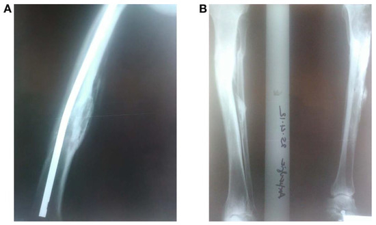
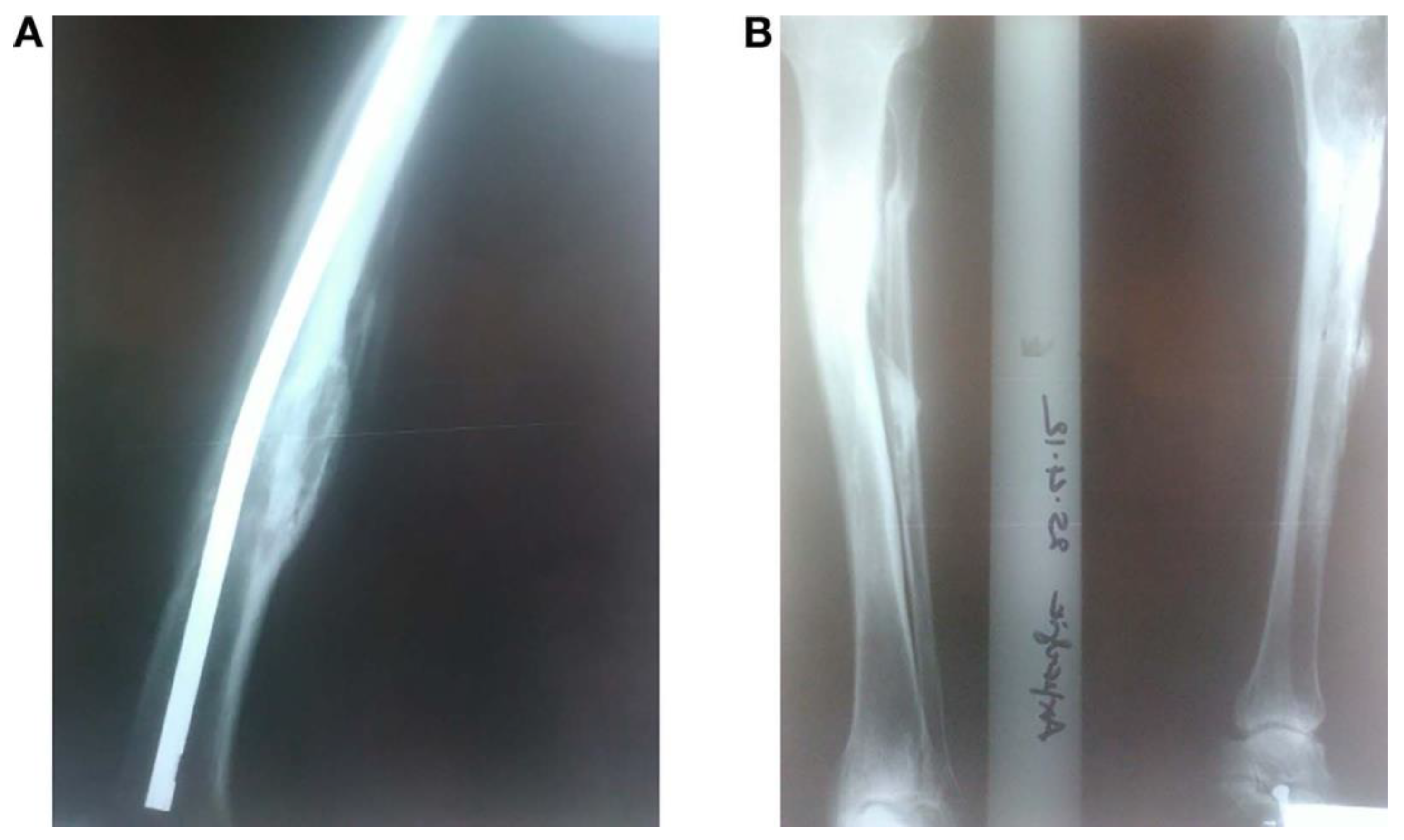
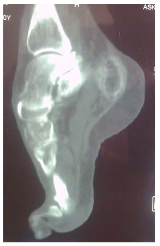
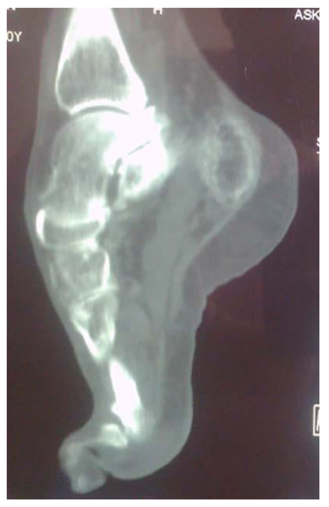
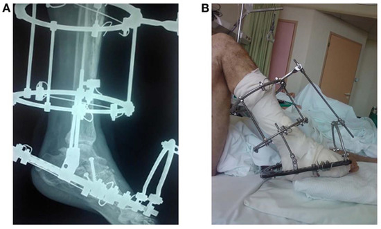
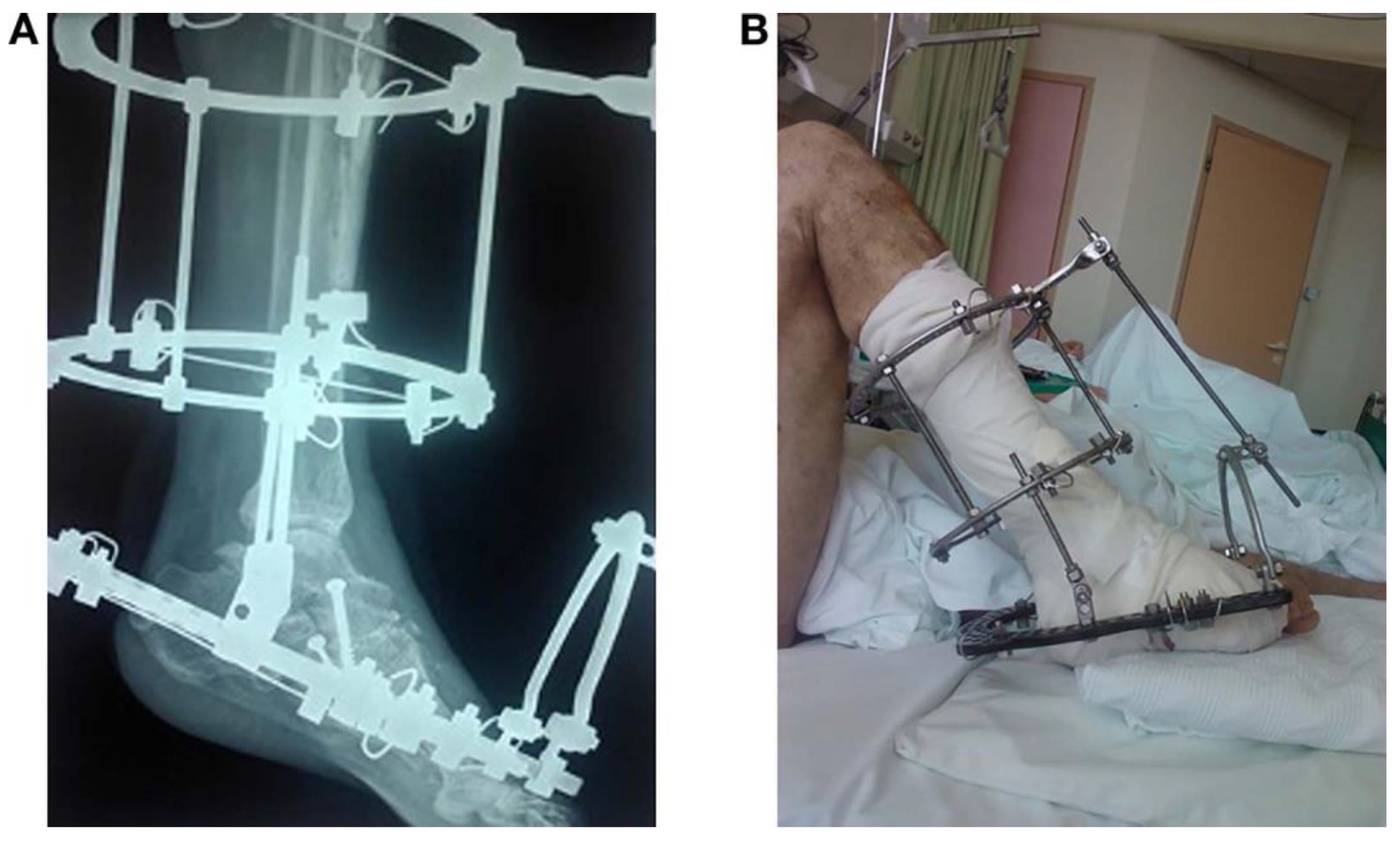
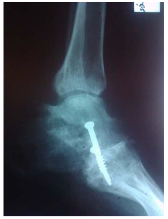
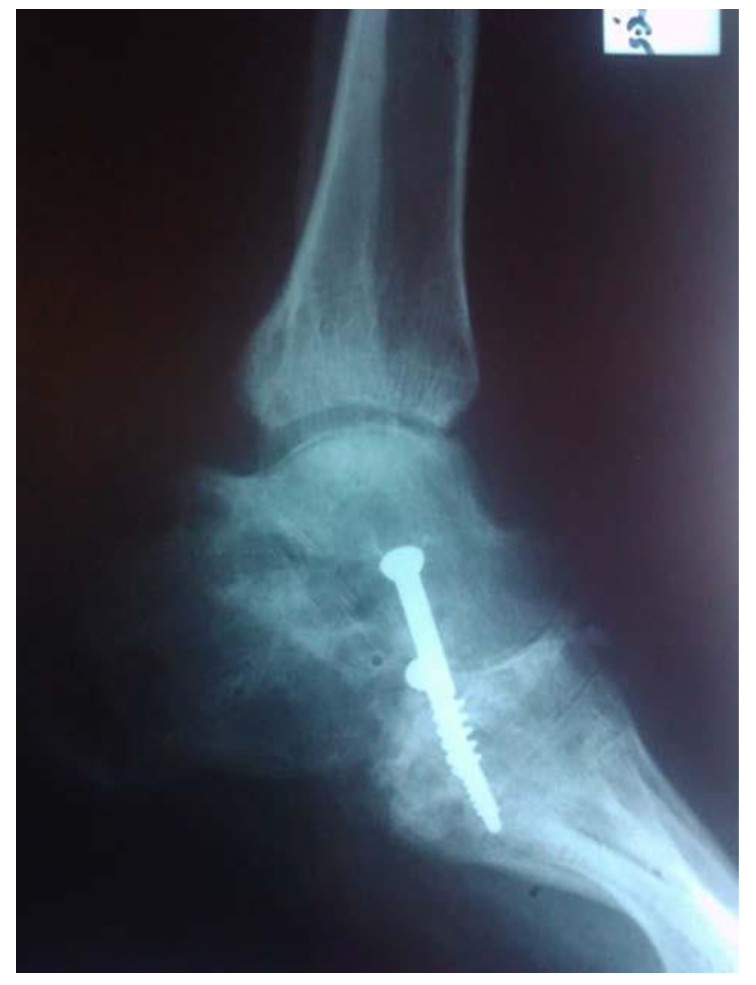
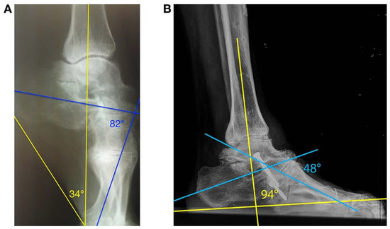
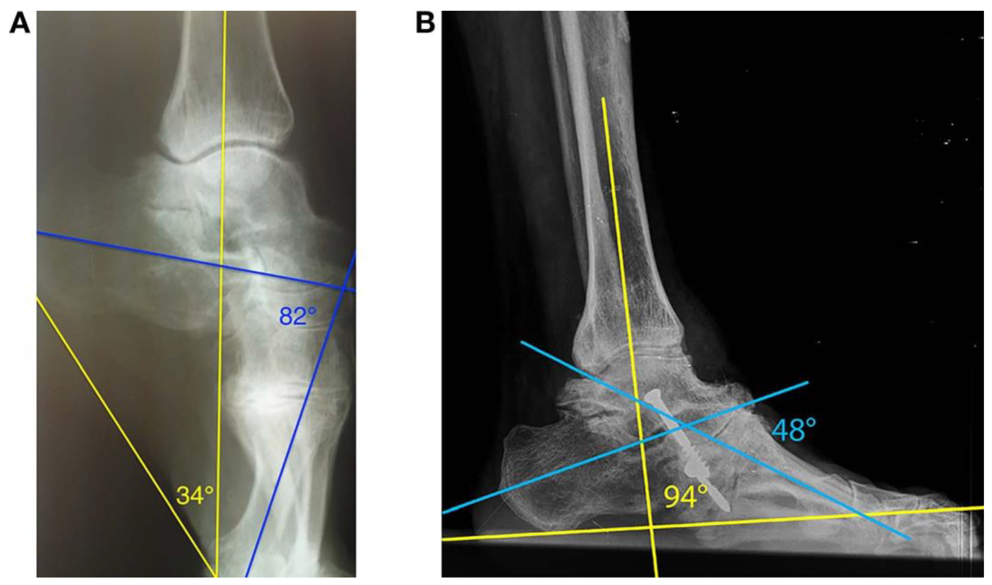
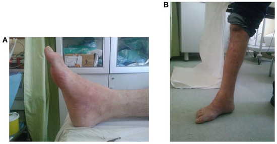
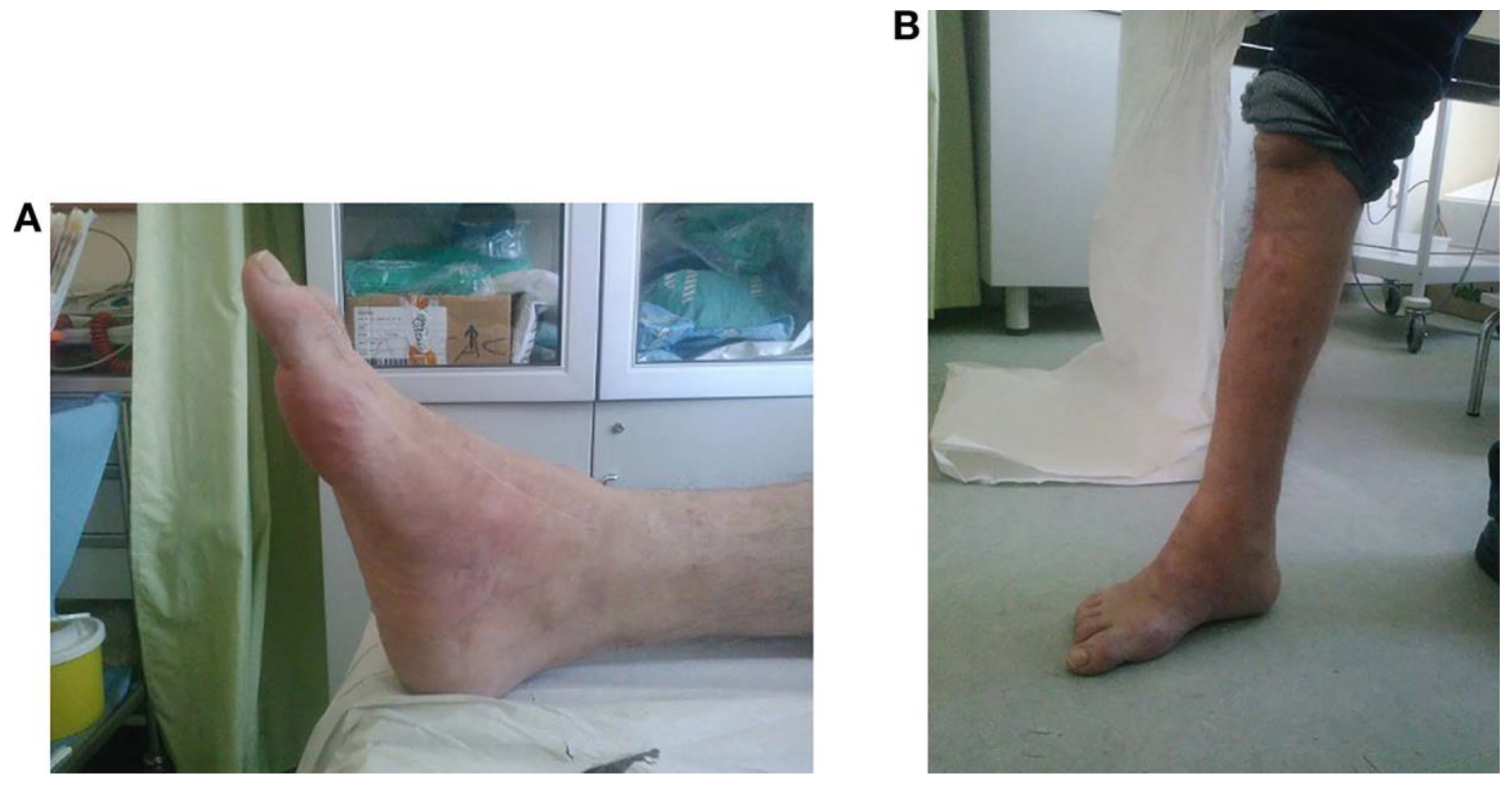
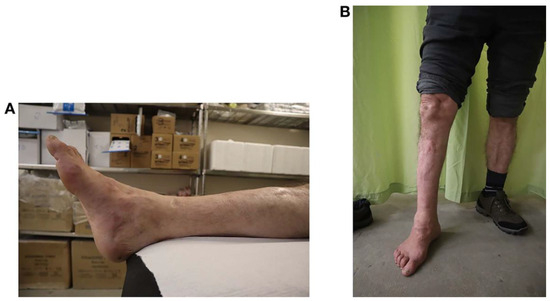
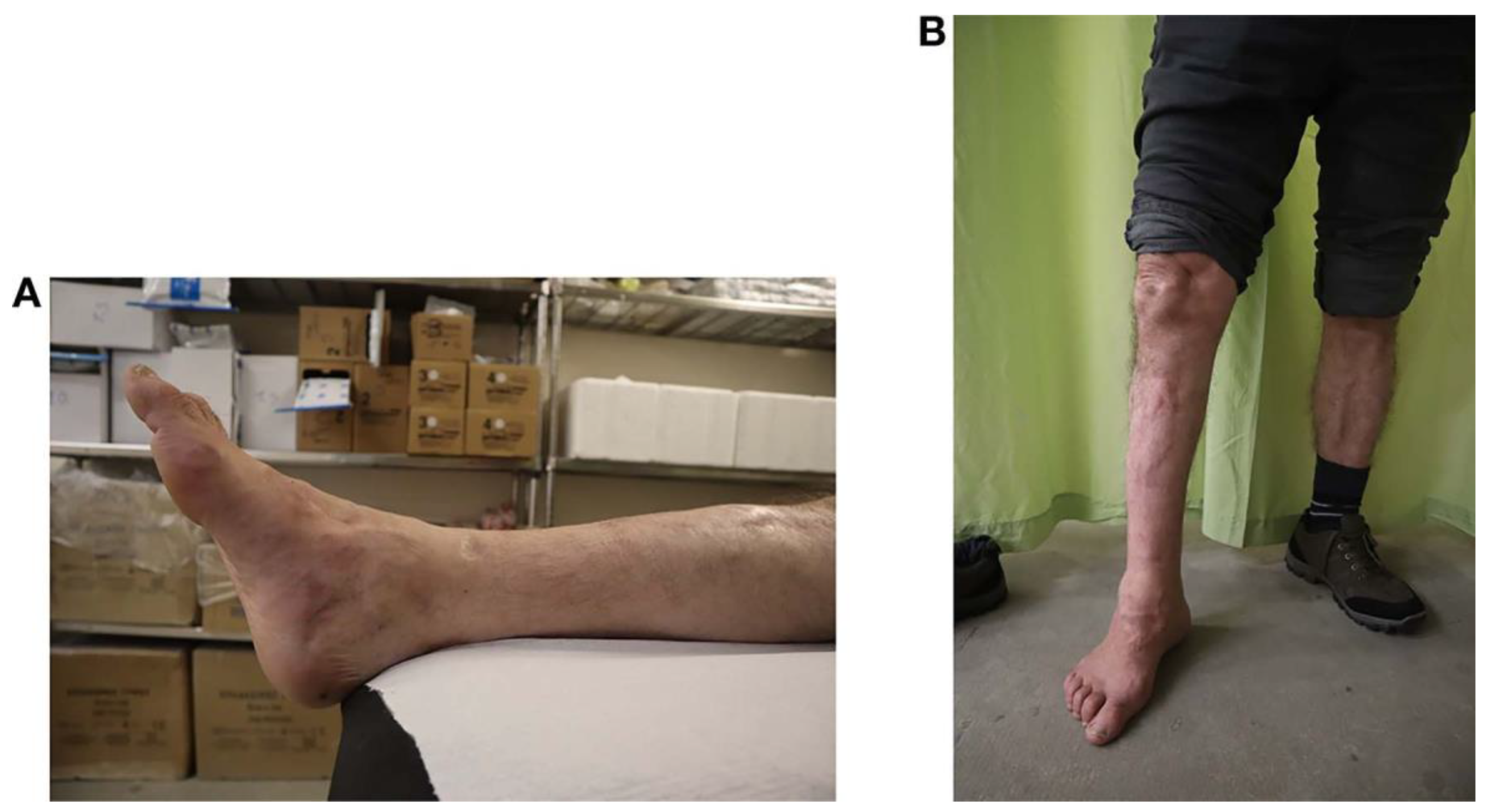
A 41-year-old man who worked as a shepherd and lived in a remote area sustained a tibial and a transverse femoral shaft fracture at the right leg (floating knee fracture) 23 years previously after a road traffic accident. The femur was treated with Kuntscher intramedullary nailing, while the tibia was treated conservatively (Figure 1). Eventually, both fractures healed, but a tibial compartment syndrome developed, leading to fibrosis and posterior compartment muscle shortening, which caused an extreme foot deformity. The patient presented at our clinic complaining of severe pain and inability to perform his daily activities. On clinical examination, he had an extremely stiff equinocavus foot deformity with associated claw toes that led to abnormal painful gait, tiptoe walking, difficulty wearing a normal shoe, and a mild knee flexion deformity of 158 (Figure 2). Mild knee flexion deformity caused leg shortening; thus, tiptoeing compensated the knee flexion, creating an abnormal and painful gait, leading to subsequent inability to perform the activities of daily living of a shepherd. A plain lateral radiograph and computed tomographic scan of the foot were obtained. The Hibbs angle and the tibial-sole angle were calculated preoperatively and after final follow-up to evaluate the degree of cavus and equinus deformity, respectively. After a diligent preoperative assessment, a lengthening of the Achilles tendon, a tarsal tunnel release, and a Cole osteotomy combined with an Ilizarov hinged frame fixation were planned in a single operative session to manage this complex deformity (Figure 3). The Cole osteotomy was stabilized with two cannulated 6.5-cm screws to the medial and lateral site to correct the cavus deformity and also contributed to the correction of claw toes because of relaxation of flexor tendons (Figure 4). Tarsal tunnel release decreased the nerve tension, and the extensive soft-tissue release contributed to the treatment. The equinus deformity was gradually corrected with the use of the hinged Ilizarov frame (0.25 mm, four times per day). The Hibbs angle was 828 preoperatively and 488 at the final follow-up (6 years), whereas the tibial-sole angle was 348 preoperatively and 948 at the final follow-up (Figure 5). The American Orthopaedic Foot and Ankle Society Midfoot Scale score was 12 preoperatively and 81 at the final follow-up. The visual analog scale score was decreased from 10 to 1 at the final follow- up. The ankle joint was corrected nearly up to normal, with 208 of residual plantarflexion, to compensate shortening caused by the lag of knee extension. The patient was given three doses of intravenous antibiotics, one intraoperatively and two postoperatively, and was taught to clean the pin sites with sterile gauzes soaked with chlorhexidine. Three months later, he revisited our department for removal of the Ilizarov device. At the last follow-up, 6 years later, the patient was satisfied with the outcome related to the correction of the deformity, which allowed him to deal with the physical needs of his daily activities (Figure 6 and Figure 7)
Figure 1.
A, The femoral fracture was treated with Kuntscher intramedullary nailing. B, The tibial fracture was treated conservatively.
Figure 2.
Computed tomographic scan of a stiff equinocavus foot deformity and associated claw toes.
Figure 3.
A, Postoperative lateral foot radiograph with Ilizarov hinged frame fixation. Cole osteotomy fixed with two 6.5-cm cannulated screws. B, Postoperative image of the patient in the clinic.
Figure 4.
Cole osteotomy stabilized with two cannulated 6.5-cm screws to the medial and lateral site.
Figure 5.
A, Preoperative tibial-sole angle of 348 (right, yellow lines) and a Hibbs angle of 828 (right, blue lines). B, Last follow up tibial-sole angle of 948 (left, yellow lines) and Hibbs angle of 488 (left, blue lines).
Figure 6.
Images obtained at 6 months postoperatively.
Figure 7.
Images obtained at 6 years postoperatively.
Discussion
In this case report, the combination of Cole osteotomy with external Ilizarov hinged frame fixation, Achilles tendon lengthening, and soft-tissue release in the setting of a neglected equinocavus deformity is found to increase functional ability and to provide satisfactory results in terms of cosmetics and no recurrence during the long-term follow-up. The Ilizarov technique was first described in 1981, allowing a three-dimensional approach for the correction of congenital or posttraumatic foot deformities, and it is considered the standard of care for the management of such lesions [4,6]. Indication for Cole osteotomy includes a cavus deformity with the apex of the deformity in the midfoot [9]. We could identify only one study in the literature concerned with equinocavus foot deformity, but with a different approach than ours. The aforementioned study was conducted by Ferreira et al [2] and included a total of 31 feet in 29 patients with stiff equinocavus foot deformity. Surgical treatment consisted, first, of percutaneous tenotomy of the Achilles and tibialis posterior tendons, and plantar fascia and short plantar muscle release; second, of transverse osteotomy of the midfoot in case of ankylosis or previous midtarsal joint fusion; and third, of a gradual realignment of the foot and ankle using a standard circular foot frame. The outcome was satisfactory in 21 feet, fair in six, and poor in four. Secondary surgery was performed in 14 feet because of symptomatic arthritis, incomplete correction of the deformity, or the need for limp amputation [2]. In our case, the Ilizarov hinged frame was applied for two reasons: first, as a supplementary stabilization of the Cole osteotomy; and second, combined with Achilles tendon lengthening, to gradually correct the equinus deformity, distract, and mobilize the ankle joint. In addition, it was a substantial way to prevent postoperative arthrodesis and arthritis to nearby supporting joints to help the patient withstand the postoperative physiotherapy.
Conclusions
In conclusion, this is the first case in the literature describing the simultaneous use of Cole osteotomy combined with external Ilizarov hinged frame fixation, soft-tissue release, and Achilles tendon lengthening for the treatment of an extreme neglected (for 23 years) stiff equinocavus foot deformity. By the use of this combination in a single operative session, we achieved additional stabilization of Cole osteotomy by the Ilizarov device, less postoperative morbidity, an immediate weightbearing of a plantigrade foot, no need for the placement of plaster in case of Cole osteotomy without the use of the Ilizarov device, and no need for extra intervention for the correction of claw toes. This case report has a principal goal to widen the operative spectrum of orthopedic and traumatology surgeons in cases of complex neglected foot deformities.
Financial Disclosure: None reported.
Conflict of Interest: None reported.
References
- ELESWARAPU, A.S.; YAMINI, B.; BIELSKI, R.J. Evaluating the cavus foot. Pediatr Ann 2016, 45, 218. [Google Scholar] [CrossRef] [Scilit] [PubMed]
- FERREIRA, R.C.; COSTA, M.T.; LOTTI, C.; et al. Minimally invasive surgery using the circular external fixator to correct neglected severe stiff equinocavus foot deformities. Foot Ankle Orthop 2018, 3, 1. [Google Scholar] [CrossRef] [Scilit]
- DU, W.; HU, X.; SHEN, Y.; et al. Surgical management of acute compartment syndrome and sequential complications. BMC Musculoskelet Disord 2019, 20, 98. [Google Scholar] [CrossRef] [Scilit] [PubMed]
- EL-MOWAFI, H. Assessment of percutaneous V osteotomy of the calcaneus with Ilizarov application for correction of complex foot deformities. Acta Orthop Belg 2004, 70, 586. [Google Scholar] [PubMed]
- SHALABY, H.; HEFNY, H. Correction of complex foot deformi-ties using the V-osteotomy and the Ilizarov technique. Strategies Trauma Limb Reconstr 2007, 2, 21. [Google Scholar] [CrossRef] [Scilit] [PubMed]
- CATAGNI, M.A.; GUERRESCHI, F.; MANZOTTI, A.; et al. Treatment of foot deformities using the Ilizarov method. Foot Ankle Surg 2000, 6, 207. [Google Scholar] [CrossRef] [Scilit]
- EL-TAYEBY, H.; EMARA, K.; GHONEEM, H.; et al. Results of using the Ilizarov method for correction of complex foot and ankle deformities in skeletally mature patients. Menoufia Med J 2017, 30, 310. [Google Scholar]
- TULLIS, B.L.; MENDICINO, R.W.; CATANZARITI, A.R.; et al. The Cole midfoot osteotomy: a retrospective review of 11 procedures in 8 patients. J Foot Ankle Surg 2004, 43, 160. [Google Scholar] [CrossRef] [Scilit] [PubMed]
- ERGUN, S.; YILDIRIM, Y. The Cole midfoot osteotomy: Clinical and radiographic retrospective review of five patients (six feet) with different etiologies. JAPMA 2019, 109, 180. [Google Scholar] [CrossRef] [Scilit] [PubMed]
© 2021 American Podiatric Medical Association