


Abstract
Tenosynovial giant cell tumor, also known as giant cell tumor of the tendon sheath (GCT-TS), is a solitary, nodular, firm, benign, soft-tissue tumor that arises from the synovial lining of a tendon. The etiology is unknown. It is a rare soft-tissue tumor, with an overall incidence of one in 50,000 individuals, and usually affects people aged between 30 and 50 years. Magnetic resonance imaging tends to be the imaging modality of choice, used for surgical preparation. Widely accepted treatment involves local excision with or without radiotherapy. The author presents a case study involving a patient with an abnormally large, longstanding GCT-TS, diagnosed with clinical examination and imaging modalities, surgically excised, and further confirmed by pathology reports. Topic of discussion includes a review of GCT-TS.
Giant cell tumor of the tendon sheath (GCT-TS) is a solitary, nodular, firm, benign, soft-tissue tumor that arises from the synovial lining of a tendon. It is the second most common benign hand tumor after a ganglion cyst [1]. Typically, patients will present with a painless mass that may cause discomfort on weightbearing or difficulty with certain footwear depending on where the mass is located. Clinically, the differential diagnosis includes ganglion cyst, lipoma, rheumatoid nodule, pigmented villonodular synovitis, synovial sarcoma, inclusion cyst, fibroma of tendon sheath, and inflammation of the synovial sheath. Plain radiology may show a soft-tissue mass or swelling that may cause splaying and erosion of the adjacent bone [2]. However, making a proper diagnosis requires advanced imaging using ultra-sound or magnetic resonance imaging. Giant cell tumor of the tendon sheath is seen with ultrasonography as a hypoechoic nodule. The mass could have either homogenous or heterogenous echogenicity. Under Doppler imaging, 71% of lesions also show substantial flow, whereas the rest show minimal flow. A finding of hypervascularity is typical of GCT-TS; however, it is not specific. This hypervascularity has led to a misdiagnosis of hemangioma. However, it will exclude ganglions, which are typically avascular or anechoic [3].
Magnetic resonance imaging is considered the most important diagnostic imaging modality. Features of GCT-TS include a low-intensity signal on T1- and T2-weighted images and homogenous enhancement on gadolinium-enhanced images. Most lesions are said to be hypointense compared with muscle. Similar low-signal images also occur in pigmented villonodular synovitis, densely mineralized tumors, and tumors with large amounts of fibrous tissue. A gradient-echo sequence is helpful for differentiating hemosiderin from fibrous tissue [4].
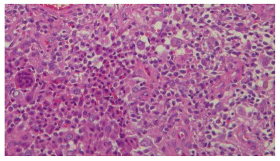
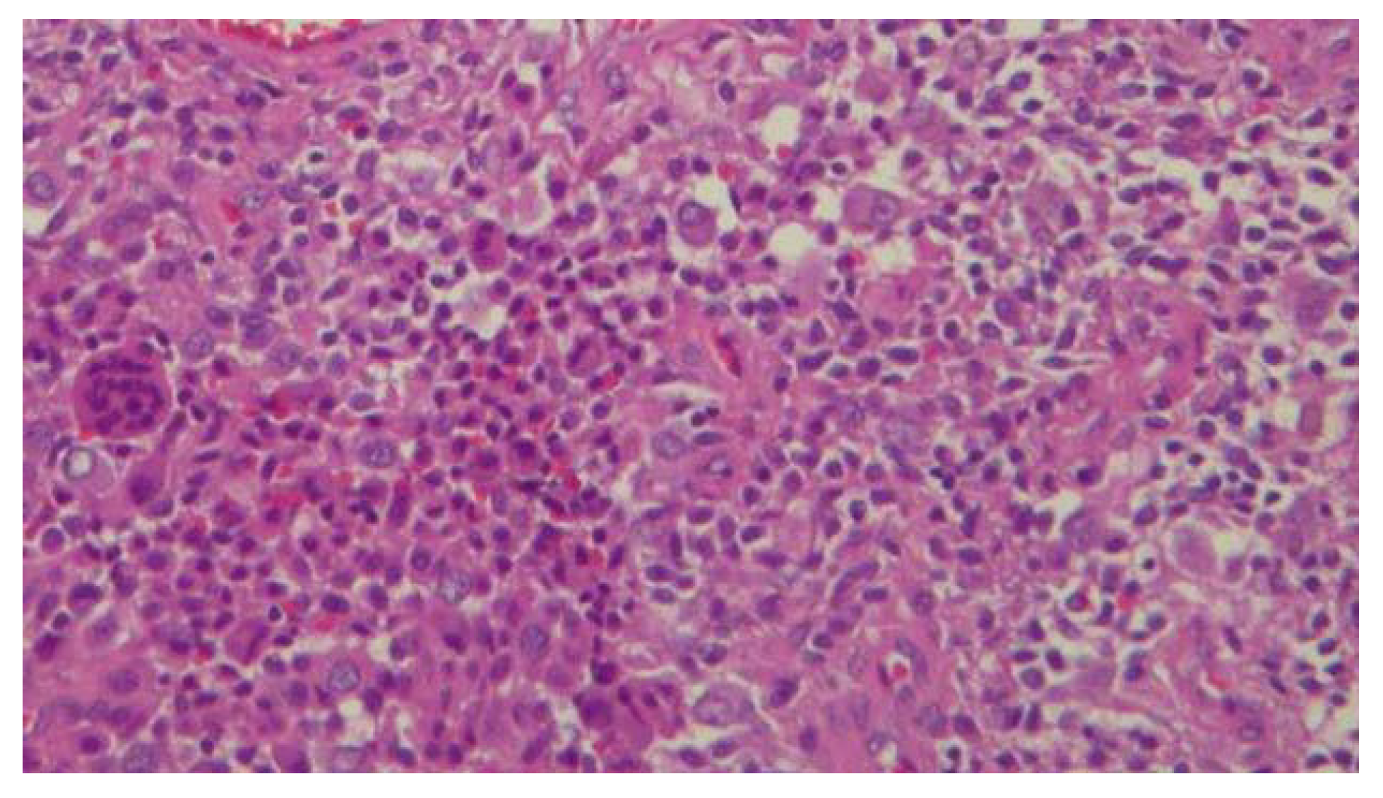
The most definitive way to acquire a diagnosis requires tissue samples and pathologic evaluation. Stromal cells can either be spindle shaped or polygonal. The nuclei of stromal cells are a source of much debate in microbiology but are now said to be of variable morphology. Giant cells that resemble osteoclasts are also seen (Figure 1). Mitoses vary considerably from zero to nine per 10 high-power fields. Lastly, xanthomatous change is present, and hemosiderin-laden macrophages can be seen [5].
Figure 1.
Histopathology.
As with many soft-tissue tumors, optimal treatment often requires marginal excision. They range in color from gray-brown to yellow-orange, depending on the hemosiderin content. Care must be taken to avoid incomplete excision, as the only cases reported with recurrence appear to be those without complete excision. Rates of recurrence in the literature vary from 0% to 44%. Recurrence is treated by marginal excision in the case of the localized forms that occur in GCT-TS of the foot [6]. Radiotherapy is used as an adjunct in other forms of the disease outside of the hand and foot [7].
Case Report
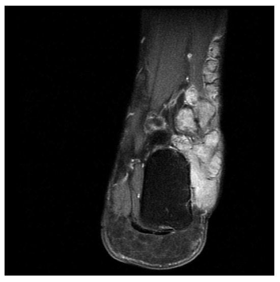
A 44-year-old man presented with an antalgic gait secondary to a slowly growing, large mass of the left lateral foot and ankle of many years’ duration. The mass was localized along the peroneal tendons and exhibited mild tenderness on palpation. Radiographs did not exhibit any bony involvement. Fine needle aspiration in the office was inconclusive because of the inability to aspirate. Magnetic resonance images revealed a prominent, lobulated, mixed signal, cystic, soft-tissue mass associated with the peroneal tendon sheath (Figure 2 and Figure 3).
Figure 2.
Coronal T1-weighted magnetic resonance imaging scan.
Figure 3.
Axial T2-weighted magnetic resonance imaging scan.
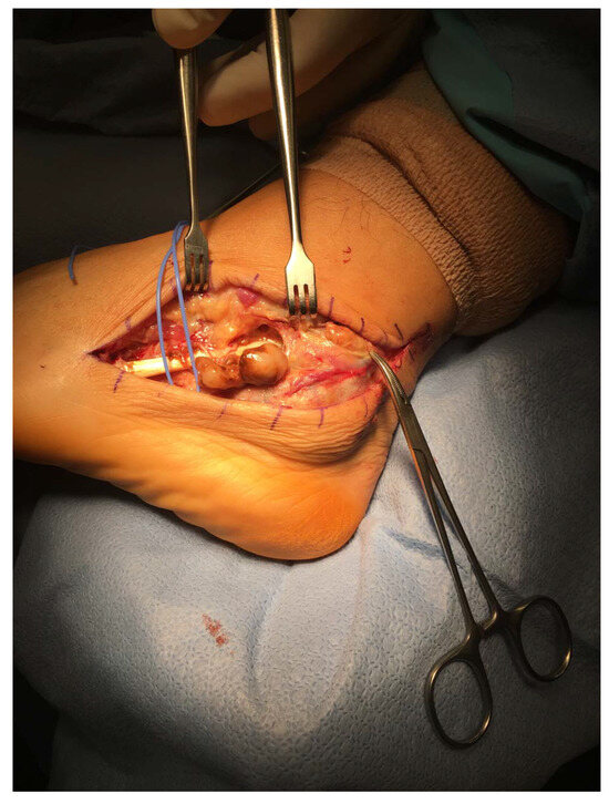
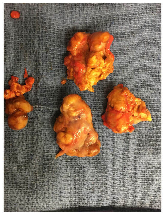
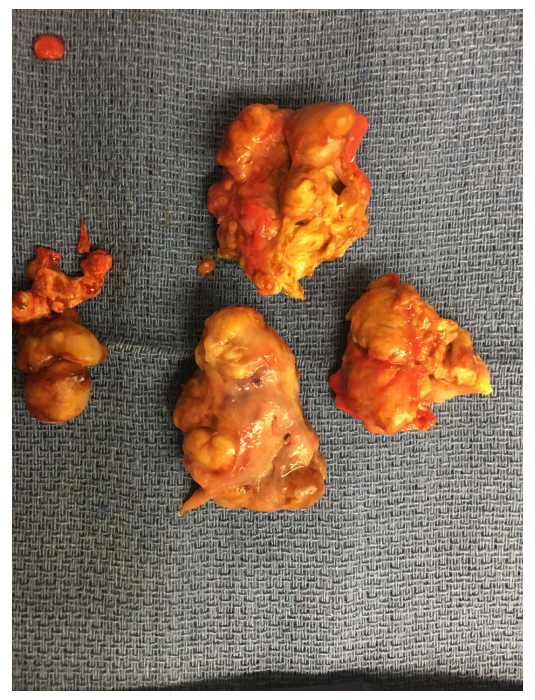
Surgical excision was the treatment of choice and was pursued. Total excision was performed and inspection of the involved peroneal tendons revealed flattening of the peroneus longus and brevis. A large, yellow-tan, rubbery-hard mass was re-moved. The mass was split into five well-encapsulated masses with different lengths, altogether measuring 15 cm in length (Figure 4).
Figure 4.
Intraoperative surgical excision.
The wound was found to have no residual traces of the mass. No tearing was appreciated. The tendons were tubularized to a more anatomical function. A pathology report later confirmed the final diagnosis of GCT-TS. The postoperative period was significant for a hematoma, which was drained, and the patient went on to full healing and recovery with no complications. The has been no evidence of recurrence over the past year.
Discussion
Giant cell tumor of the tendon sheath is a solitary, nodular, firm, benign, soft-tissue tumor that arises from the synovial lining of a tendon. It is the second most common benign hand tumor after a ganglion cyst. Although the etiology is unknown, theories have included trauma, disturbed lipid metabolism, osteoclastic proliferation, infection, vascular disturbances, immune mechanisms, inflammation, neoplasia, and metabolic disturbances [7,8,9,10,11].
The literature on GCT-TS of the foot and ankle region is based largely on a few case reports. Total reported cases of GCT-TS in the foot and ankle are 52, spanning from 1969 to 2013 [12]. This case proved especially different from other cases because of the size of the mass, being an unusually large mass (15 cm in length), and it crossed the confined anatomical spaces of the midfoot and hindfoot. The size of the mass in this case was most likely secondary to the patient’s choice to wait a long time before having the mass evaluated. His decision was based on multiple factors, including lack of symptoms and a busy lifestyle involving frequent travel for work. He decided to have an evaluation once the size of the mass had progressed to a point where it was causing pain with wearing shoes in addition to restriction in range of motion (particularly apparent with eversion). The patient went on to heal uneventfully, but did require an extensive amount of physical therapy afterward to regain strength, range of motion, and normal gait. This patient showed all the typical signs expected on clinical presentation and radiographic and histologic findings. The atypical finding here was the unusual size and its effect on foot and ankle biomechanics. Had the patient presented earlier rather than later, the mass’s effect on his biomechanics could have been prevented early on and thus led to a much shorter postoperative recovery. This is especially important to the surgeon when deciding whether to excise soft-tissue tumors or monitor them over time. The location, symptoms, and nature of the mass (benign or malignant) are often the deciding factors for surgical excision. This case gives the surgeon one more criterion to consider—the effect on biomechanics—when making that decision.
Conclusions
Giant cell tumor of the tendon sheath is an uncommon condition in the foot and ankle, with most surgeons seeing possibly only one case in a career. It presents as a mass in the foot and ankle. Making a definitive diagnosis is difficult without histologic evaluation. Clinical features are usually typical of a slow-growing mass that causes com-pressive symptoms. Symptoms vary, depending on the location of the mass. Magnetic resonance imaging is critical as a diagnostic modality and for surgical planning. Findings on magnetic resonance imaging are typically of low signal intensity on T1-and T2-weighted images. Our case report showed the typical features. However, because of the longstanding nature of the case, our patient presented with an unusually large mass, which crossed the confined anatomical spaces of the midfoot and hindfoot and affected his biomechanics.
Financial Disclosure: None reported.
Conflict of Interest: None reported.
References
- RUKAVINA, I.; ALETA, D. Giant-cell tumour of the tendon sheath: a review. OA Orthopaedics 2014, 2, 11. [Google Scholar]
- VENKATESWARAN KI; KUSUM, K.; KUSUM, V. Fine-needle aspiration cytology of giant cell tumor of tendon sheath. Diagn Cytopathol 2003, 29, 105. [Google Scholar]
- VAN DER HEIDEN, L.; Gibbons, L.; Hassan, C.L.; HASSAN, A.B.; et al. A multidisciplinary approach to giant cell tumors of tendon sheath and synovium: a critical appraisal of literature and treatment proposal. J Surg Oncol 2013, 107, 433. [Google Scholar]
- MAYET, Z.; NOBLE, T. MAGOBOTHA SKM: Giant cell tumour of the tendon sheath (GCT-TS) in the foot: a case report. SA Orthop J 2014, 13, 39. Available online: http://www.scielo.org.za/scielo.php?script=sci_arttextpid=S1681-150X2014000100007lng=ennrm=iso (accessed on 21 February 21 2018).
- WRIGHT, C.J.E. Benign giant-cell synovioma. An investiga-tion of 85 cases. Br J Surg 1951, 38, 257. [Google Scholar] [CrossRef] [Scilit] [PubMed]
- TAN, Y.W.J.; LUO, Y. The value of sonography in diagnosing giant cell tumors of the tendon sheath. J Ultrasound Med 2007, 26, 1333. [Google Scholar]
- HORVATH, R.R.; BOSTANCHE, J.; ALTMAN, M.I. Diffuse type tenosynovial giant cell tumor of the ankle. JAPMA 1993, 83, 231. [Google Scholar] [CrossRef] [Scilit] [PubMed]
- BISBINAS, I.; DE SILVA, U.; GRIMER, R.J. Pigmented villonodular synovitis of the foot and ankle: a 12 year experience from a tertiary orthopedic oncology unit. J Foot Ankle Surg 2004, 43, 407. [Google Scholar] [CrossRef] [Scilit] [PubMed]
- FRANKEL, S.L.; CHIOROS, P.G.; SIDLOW, C.J. Giant cell tumor of the plantar fascia: a case report. JAPMA 1987, 77, 557. [Google Scholar] [CrossRef] [Scilit] [PubMed]
- LO, E.P.; KETTERER, D. Giant cell tumor of tendon sheath in the toe. JAPMA 2000, 90, 270. [Google Scholar] [CrossRef] [Scilit] [PubMed]
- PAEZ, H.; VULETIN, J.C.; SOAVE, R.L.; et al. Pedal giant cell tumor of tendon sheath. JAPMA 1999, 89, 368. [Google Scholar] [CrossRef] [Scilit] [PubMed]
- VARGAONKAR, G.; SINGH, V.; ARORA, S.; et al. Giant cell tumor of the tendon sheath around the foot and ankle. JAPMA 2015, 105, 249. [Google Scholar] [CrossRef] [Scilit] [PubMed]
© 2021 American Podiatric Medical Association