


Abstract
Background: First metatarsophalangeal joint fusion is a commonly used procedure for treating many pathologic disorders of the first ray. Historically, hallux valgus deformity with severely increased intermetatarsal angle or metatarsus primus adductus indicated need for a proximal metatarsal procedure. However, the effectiveness and reliability of first metatarsophalangeal joint arthrodesis in reducing the intermetatarsal angle has been increasingly described in the literature. We compared findings at our institution with current literature for further validation of this well-accepted procedure in correcting hallux valgus deformity with high intermetatarsal angle. Methods: Weightbearing preoperative and postoperative radiographs of 43 patients, 31 women and 12 men, meeting the inclusion and exclusion criteria were identified. Two independent investigators measured the hallux abductus and intermetatarsal angles. Preoperative and postoperative measurements for each angle were compared and average reduction calculated. The data were further analyzed by grouping deformities as mild, moderate, and severe. Mean follow-up was 10 months. Results: The overall mean preoperative intermetatarsal and hallux abductus angles decreased significantly (from 13.09° to 9.33° and from 23.72° to 12.19°, respectively; both P < .01). When grouping deformities as mild, moderate, and severe, all of the categories maintained reduction of intermetatarsal and hallux abductus angles (P < .01). Furthermore, the mean reduction of the intermetatarsal and hallux abductus angles seemed to correlate with preoperative deformity severity. Conclusions: In patients undergoing correction of hallux valgus deformity, first metatarsophalangeal joint arthrodesis produced consistent reductions in the intermetatarsal and hallux abductus angles. Furthermore, these findings are consistent with those reported by other institutions.
First metatarsophalangeal joint arthrodesis is an accepted and effective procedure for many first-ray pathologic disorders. It has been widely used as surgical treatment for end-stage arthritis, hallux rigidus, and hallux valgus with associated arthritic changes and as a revision surgery. It has been postulated that hallux valgus deformity with a severely increased intermetatarsal angle indicates correction via a proximal procedure or a distal procedure with a second proximal procedure.[1,2] However, in the past several decades there has been an increasingly large amount of literature confirming the effectiveness and reproducibility of first metatarsophalangeal joint arthrodesis in reducing the intermetatarsal angle without the use of a proximal procedure in patients with metatarsus primus adductus.[3,4,5,6,7,8,9,10,11,12,13,14,15,16,17,18,19]
Most literature evaluating the intermetatarsal angle after first metatarsophalangeal joint fusion reports significant angular reduction. It has also been proposed that the reduction in intermetatarsal angle achieved is directly proportional to the preoperative intermetatarsal angle; wider preoperative intermetatarsal angles seem to correlate with greater reduction in intermetatarsal angle after first metatarsophalangeal joint fusion.[4,5,13,14] The goal of this study was to evaluate the angular correction between the first and second metatarsals and hallux abductus after first metatarsophalangeal joint arthrodesis at Ohio State University Medical Center to compare with the current literature. Furthermore, the present data were divided into groups for comparison of mild, moderate, and severe hallux valgus deformities to further evaluate the expected reduction in intermetatarsal angle based on level of hallux valgus deformity.
Successful angular correction has been documented in long-term studies as well.[10,12,16] A study by Coughlin et al[10] in 2005 of 21 patients with mean follow-up of 8.2 years found the average correction in the hallux abductus and intermetatarsal angles to be 21° and 6°, respectively. Agoropoulos et al[12] found that “first MTPJ [metatarsophalangeal joint] fusion is a consistently successful operation … and its good results remain unchanged for up to 31 years.” With long-term successful results and effective reduction of hallux abductus and metatarsus primus adductus, first metatarsophalangeal joint fusion proves to be a viable option for all levels of hallux valgus deformity.
Materials and Methods
A review was performed of patients undergoing a primary first metatarsophalangeal joint arthrodesis by one of us (I.B.) from October 2011 through October 2016. Approval from the university's institutional review board was obtained before the study. We identified 83 patients older than 18 years who had undergone a first metatarsophalangeal joint arthrodesis. Fixation constructs for fusion varied among the senior authors (E.M., S.A., and M.A.): a single plate, one screw with an overlying plate, or crossing screws. We excluded patients who had undergone previous bunion surgery or other surgery of the first ray, those without preoperative or postoperative weightbearing anteroposterior radiographs, and those with a history of hallux varus deformity. After exclusion, 49 patients met the initial criteria for inclusion. After failure of conservative treatment, the indications for arthrodesis included pain with first metatarsophalangeal joint range of motion, crepitus with first metatarsophalangeal joint range of motion, and radiographic findings consistent with degenerative joint disease.
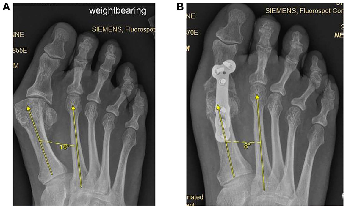
Digital weightbearing anteroposterior radiographs were used to evaluate the preoperative and postoperative hallux abductus and intermetatarsal angles, as described by Coughlin.[20] The hallux abductus angle was, therefore, defined as the intersection of the longitudinal axis of the proximal phalanx and the first metatarsal. An example of this measurement is seen in Figure 1. The intermetatarsal angle was measured as the angle formed by the intersection of the longitudinal axis of the first and second metatarsals, as seen in Figure 2. After initial radiographic evaluation, feet with a normal preoperative intermetatarsal angle, defined by Coughlin [1] as less than 9°, were excluded from the study, leaving 43 patients for evaluation. Two independent investigators (I.B. and K.C.) performed measurements for each radiographic value for all of the included patients. The mean value between the two investigators for each angle was then calculated. The feet were then grouped by mean preoperative intermetatarsal angles according to severity based on a hallux valgus classification by Coughlin [20] into mild (9°–10°), moderate (11°–16°), and severe (>16°). The differences between mean preoperative and postoperative intermetatarsal angles and the respective difference in mean preoperative and postoperative hallux abductus angles were calculated. The data were then analyzed with Microsoft Excel (Microsoft Corp, Redmond, Washington) using a paired Student t test to compare significance.
Figure 1.
Weightbearing anteroposterior radiographs depicting measurement of the hallux abductus angle before (A) and after (B) first metatarsophalangeal joint arthrodesis using plate and screw fixation.
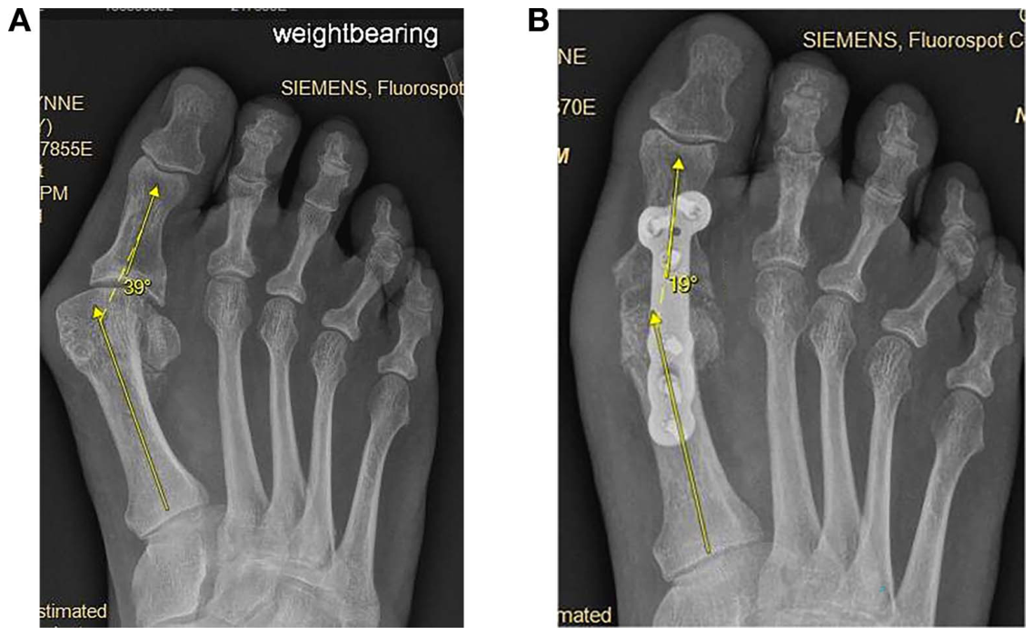
Figure 2.
Weightbearing anteroposterior radiographs depicting intermetatarsal angle measurement before (A) and after (B) first metatarsophalangeal joint arthrodesis using plate and screw fixation
Results
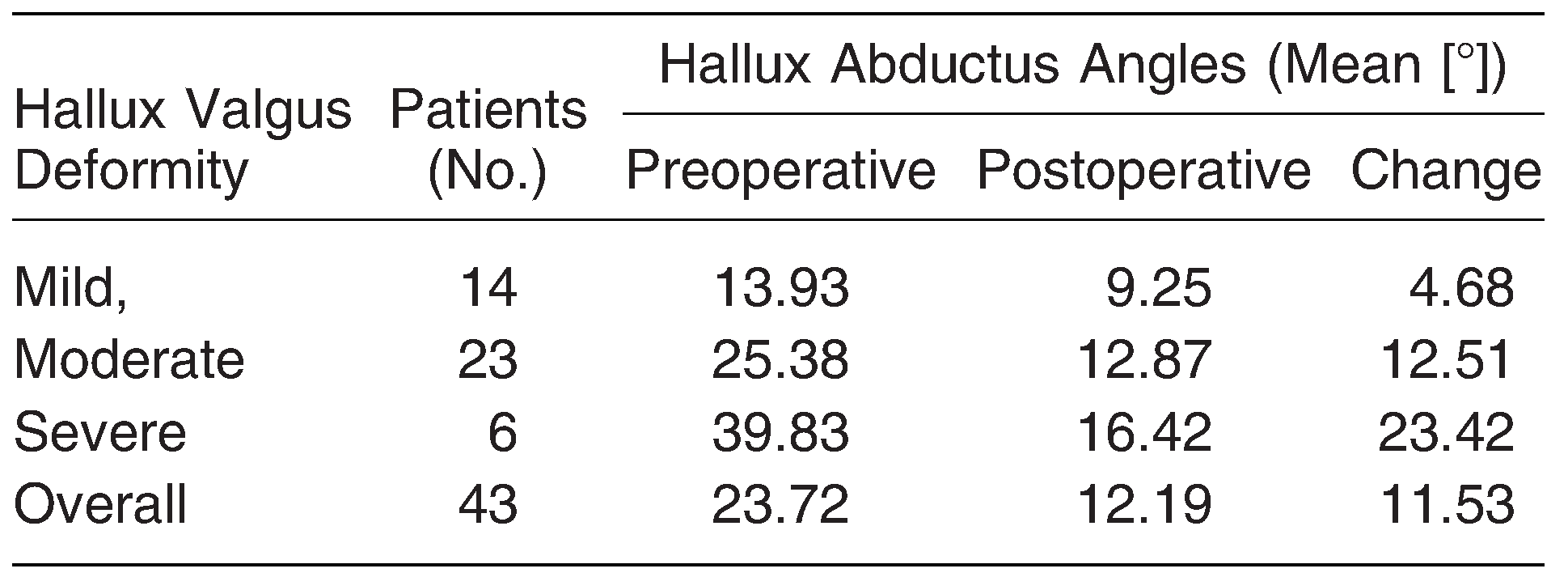
Mean follow-up in the study was 10 months. There were 31 women and 12 men. The mean participant age was 58.27 years (range, 25–75 years). The overall mean preoperative intermetatarsal angle decreased significantly from 13.09° to 9.33°, and the overall mean preoperative hallux abductus angle decreased significantly from 23.72° to 12.19° (both P < .01). When grouping deformities as mild, moderate, and severe, for mild hallux valgus, the mean measurements for intermetatarsal angle and hallux abductus angle decreased significantly from 9.39° to 8.04° and from 13.93° to 9.25°, respectively (both P < .01). The mean measurements for moderate hallux valgus deformities decreased significantly from 13.65° to 9.63° for the intermetatarsal angle and from 25.48° to 12.87° for the hallux abductus angle (both P < .01). The mean measurements for severe hallux valgus deformities decreased significantly from 19.58° to 11.17° for the intermetatarsal angle and from 39.83° to 16.42° for the hallux abductus angle (both P < .01). The mean change in intermetatarsal angle was 3.76° overall, 1.36° for mild deformities, 4.02° for moderate deformities, and 8.42° for severe deformities (Table 1). The mean change in hallux abductus angle was 11.53° overall, 4.68° for mild deformities, 12.51° for moderate deformities, and 23.42° for severe deformities (Table 2).
Table 1.
Preoperative and Postoperative Intermetatarsal Angles Grouped by Mild, Moderate, and Severe Hallux Valgus Deformities
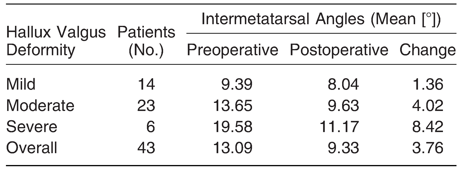
Table 2.
Preoperative and Postoperative Hallux Abductus Angles Grouped by Mild, Moderate, and Severe Hallux Valgus Deformities
Discussion
The goal of this study was to perform a retrospective radiographic review of hallux valgus correction after first metatarsophalangeal joint arthrodesis at our institution to compare with the current literature. Many previous studies have noted improvement of the intermetatarsal angle after first metatarsophalangeal joint arthrodesis. Coughlin et al[10] noted a reduction of 6.1° in the intermetatarsal angle after first metatarsophalangeal joint arthrodesis. Cronin et al[13] found that the mean change between preoperative and final follow-up intermetatarsal angles after first metatarsophalangeal joint arthrodesis was 8.22°. A large systematic review by Dayton et al[14] in 2014 overwhelmingly confirmed these findings across 701 first metatarsophalangeal joint arthrodesis procedures with an overall mean intermetatarsal angle change of 4.36°.
Mann and Katcherian[5] noted that the change in intermetatarsal angle with first metatarsophalangeal joint arthrodesis was directly proportional to the preoperative intermetatarsal angle. Cronin et al[13] found a mean intermetatarsal angle change of 8.22° and noted the correlation of increased differences with increased preoperative measurements. Similarly, Sung et al[8] found that severe hallux valgus preoperative intermetatarsal angles were reduced by a mean of 6.7° and moderate hallux valgus deformities were reduced by an average of 4.3°. This was also found by Dayton and colleagues[14] by further grouping their data into two subsets based on preoperative intermetatarsal angle less than and greater than 15°; the mean change in intermetatarsal angle of the former subset was 3.7° and of the latter was 5.42°. In the present study, mean intermetatarsal angle reductions when grouped by level of severity of hallux valgus deformity were similar to those reported in the literature. Herein, the mild hallux valgus group improved the least, with a mean change of 1.36; moderate hallux valgus improved by 4.02°; and the greatest improvement seen in the severe hallux valgus group, improving by 8.42°.
The exact mechanism of intermetatarsal angle reduction has not been determined experimentally; however, it is believed that elimination of the retrograde force of the hallux on the metatarsal allows for the decrease in the intermetatarsal angle. This may be an interesting topic for further investigation. Another area of interest may be evaluation of sesamoid position with first metatarsophalangeal joint arthrodesis, as previous investigators have not as popularly assessed frontal plane correction with this procedure. Of note, Pydah et al[7] found that the tibial sesamoid generally improved by at least one grade. Costa et al[19] also noted improvement in sesamoid position after first metatarsophalangeal joint fusion. Perhaps this is an area for further future investigation.
Conclusions
In this series of patients undergoing first metatarsophalangeal joint arthrodesis for hallux valgus, the procedure resulted in consistent reduction of the intermetatarsal and hallux abductus angles. Furthermore, the present findings are consistent with the values found at other institutions. The present results further support the current belief that first metatarsophalangeal joint arthrodesis is a reliable and effective procedure for bunion correction in reducing the hallux abductus angle as well as the intermetatarsal angle that may prevent the need for a more proximal surgical procedure in patients with severe metatarsus primus adductus.
Financial Disclosure
None reported.
Conflict of Interest
None reported.
References
- Rippstein, PF; Park, YU; Naal, FD. Combination of first metatarsophalangeal joint arthrodesis and proximal correction for severe hallux valgus deformity. Foot Ankle Int 2012, 33, 400. [Google Scholar]
- Sammarco, VJ. Surgical correction of moderate and severe hallux valgus: proximal metatarsal osteotomy with distal soft-tissue correction and arthrodesis of the metatarsophalangeal joint. Instr Course Lect 2008, 57, 415. [Google Scholar]
- McKean, RM; Bergin, PF; Watson, G; et al. Radiographic evaluation of intermetatarsal angle correction following first MTP joint arthrodesis for severe hallux valgus. Foot Ankle Int 2016, 37, 1183. [Google Scholar]
- Dayton, P; LoPiccolo, J; Kiley, J. Reduction of the intermetatarsal angle after first metatarsophalangeal joint arthrodesis in patients with moderate and severe metatarsus primus adductus. J Foot Ankle Surg 2002, 41, 316. [Google Scholar]
- Mann, RA; Katcherian, DA. Relationship of metatarsophalangeal joint fusion on the intermetatarsal angle. Foot Ankle 1989, 10, 8. [Google Scholar]
- Cronin, JJ; Limbers, JP; Kutty, S; et al. Intermetatarsal angle after first metatarsophalangeal joint arthrodesis for hallux valgus. Foot Ankle Int 2006, 27, 104. [Google Scholar]
- Pydah, SK; Toh, EM; Sirikonda, SP; et al. Intermetatarsal angular change following fusion of the first metatarsophalangeal joint. Foot Ankle Int 2009, 30, 415. [Google Scholar]
- Sung, W; Kluesner, AJ; Irrgang, J; et al. Radiographic outcomes following primary arthrodesis of the first metatarsophalangeal joint in hallux abductovalgus deformity. J Foot Ankle Surg 2010, 49, 446. [Google Scholar]
- Dalat, F; Cottalorda, F; Fessy, MH; et al. Does arthrodesis of the first metatarsophalangeal joint correct the intermetatarsal M1M2 angle? analysis of a continuous series of 208 arthrodeses fixed with plates. Orthop Traumatol Surg Res 2015, 101, 709. [Google Scholar]
- Coughlin, MJ; Grebing, BR; Jones, CP. Arthrodesis of the first metatarsophalangeal joint for idiopathic hallux valgus: intermediate results. Foot Ankle Int 2005, 26, 783. [Google Scholar]
- Coughlin, MJ. Arthrodesis of the first metatarsophalangeal joint. Orthop Rev 1990, 19, 177. [Google Scholar]
- Agoropoulos, Z; Efstathopoulos, N; Mataliotakis, J. Long term results of first metatarsophalangeal joint fusion for severe hallux valgus deformity. Foot Ankle Surg 2001, 7, 9. [Google Scholar]
- Cronin, JJ; Limbers, JP; Kutty, S; et al. Intermetatarsal angle after first metatarsophalangeal joint arthrodesis for hallux valgus. Foot Ankle Int 2006, 27, 104. [Google Scholar]
- Dayton, P; Feilmeier, M; Hunziker, B; et al. Reduction of the intermetatarsal angle following first metatarsal phalangeal arthrodesis: a systematic review. J Foot Ankle Surg 2014, 53, 620. [Google Scholar]
- Feilmeier, MJ; Dayton, PD; Wienke, JC. Reduction of the intermetatarsal angle after first MTPJ arthrodesis in patients with hallux valgus. J Foot Ankle Surg 2013, 53, 29. [Google Scholar]
- Gregory, JL; Childers, R; Higgins, KR; et al. Arthrodesis of the first metatarsophalangeal joint: a review of the literature and long-term retrospective analysis. J Foot Surg 1990, 29, 369. [Google Scholar]
- Nicholas, C; Silhanek, AD; Connolly, FG; et al. The effect of first metatarsophalangeal arthrodesis on transverse plane deviation of the second toe: a retrospective radiographic study. J Foot Ankle Surg 2005, 44, 365. [Google Scholar]
- Lombardi, CM; Silhanek, AD; Connolly, FG; et al. The effect of first metatarsophalangeal joint arthrodesis on the first ray and the medial longitudinal arch: a radiographic study. J Foot Ankle Surg 2002, 41, 96. [Google Scholar]
- Costa, MT; Neto, DL; Kojima, FH; et al. Evaluation of the intermetatarsal angle after the arthrodesis of the first metatarsophalangeal joint for treatment of the hallux valgus. Rev Bras Ortop 2015, 47, 363. [Google Scholar]
- Coughlin, MJ. Hallux valgus. J Bone Joint Surg Am 1996, 78, 932. [Google Scholar]
© 2021 American Podiatric Medical Association