


Abstract
Background: Diabetic foot ulcers combined with ischemia and infection can be difficult to treat. Few studies have quantified the level of blood supply and infection control required to treat such complex diabetic foot ulcers. We aimed to propose an index for ischemia and infection control in diabetic chronic limb-threatening ischemia (CLTI) with forefoot osteomyelitis. Methods: We retrospectively evaluated 30 patients with diabetic CLTI combined with forefoot osteomyelitis who were treated surgically from January 2009 to December 2016. After 44 surgeries, we compared patient background (age, sex, hemodialysis), infection status (preoperative and 1- and 2-week postoperative C-reactive protein [CRP] levels), surgical bone margin (with or without osteomyelitis), vascular supply (skin perfusion pressure), ulcer size (wound grade 0–3 using the Society for Vascular Surgery Wound, Ischemia, and foot Infection classification), and time to wound healing between patients with healing ulcers and those with nonhealing ulcers. Results: Preoperative CRP levels and the ratio of ulcers classified as wound grade 3 were significantly lower and skin perfusion pressure was significantly higher in the healing group than in the nonhealing group (P < .05). No other significant differences were found between groups. Conclusions: This study demonstrates that debridement should be performed first to control infection if the preoperative CRP level is greater than 40 mg/L. Skin perfusion pressure of 55 mm Hg is strongly associated with successful treatment. We believe that this research could improve the likelihood of salvaging limbs in patients with diabetes with CLTI.
When diabetic foot ulcers (DFUs) are present with concomitant ischemia and infection, treatment becomes more difficult.[1] However, few studies have investigated the necessary level of blood supply and infection control required to successfully treat such complex ulcers. In a previous study,[2] we proposed a surgical treatment strategy for diabetic forefoot osteomyelitis (DFO) based on our research[3,4] using magnetic resonance imaging (MRI) and histopathologic analysis. In this study, we aimed to propose an index for ischemia and infection control in the treatment of diabetic chronic limb-threatening ischemia (CLTI) with forefoot osteomyelitis.
Materials and Methods
We retrospectively reviewed the records of 30 patients with diabetic CLTI combined with forefoot osteomyelitis (26 men, four women; mean ± SD age, 68.4 ± 11 years; age range, 40–89 years) at our institutions from January 2009 to December 2016. The study protocol conforms to the ethical guidelines of the 1975 Declaration of Helsinki as reflected in the approval by the institution's human research review committee. Participants were provided with a detailed explanation of the study protocol and informed of their right to withdraw from the study at any time. All of the patients provided verbal informed consent. Patients with autoimmune disorders, life-threatening infections, and acute limb ischemia were excluded. Patients with another infection status, such as pneumonia or enteritis, were also excluded because such conditions may influence the laboratory data, wound healing, and clinical course. The inclusion criteria were a diagnosis of type 2 diabetes according to the classification and diagnostic criteria of the Japan Diabetes Society[5] and a diagnosis of ischemic forefoot ulcers combined with osteomyelitis. A total of 36 ulcers (26 patients with one ulcer, three patients with two ulcers, and one patient with four ulcers) were included. If the patient developed an ulcer at a different site after one ulcer healed or if the patient presented with simultaneous bilateral ulcers, we considered the patient to have two different ulcers. All of the ulcers were classified on the basis of the Society for Vascular Surgery (SVS) Wound, Ischemia, and foot Infection (WIfI) classification.[6]
Patients were suspected of having acute osteomyelitis on the basis of a positive probe-to-bone test, swollen foot, sausage toe, an unexplained high leukocyte count or inflammatory markers, and plain radiographic findings of the foot. We suspected peripheral arterial disease (PAD) if we could not detect a pulse in either the dorsalis pedis artery or the posterior tibial artery by either palpation or Doppler stethoscope, as well as the presence of an ankle-brachial index less than 0.9 and skin perfusion pressure (SPP) less than 40 mm Hg.[7] The SPP values near the ulcers were measured on the dorsal and plantar aspects of the foot, and the mean value was used for analysis. Patients with suspected PAD were evaluated using computed tomographic angiography, duplex ultrasonography, and angiography. Patients were then revascularized via distal bypass, endarterectomy, or endovascular therapy.
Patients were treated surgically. Curative surgeries[8] were performed on the basis of MRI findings, and localization of the osteomyelitis was evaluated as previously described.[3,4] However, in patients with severe infection, emergency debridement was performed. If an ulcer did not heal after the first surgery, revision surgery was performed as needed. Patients were administered parenteral antibiotic therapy preoperatively. They were first managed empirically, and then the antibiotics were modified according to culture results. The duration of antibiotic administration was determined on the basis of the severity of the infection.
We retrospectively investigated the results of surgery and categorized patients as healing or nonhealing. Healing was defined as complete epithelialization. If additional bone resection or revascularization was required, the patient was considered nonhealing. At each surgery, patient background (age, sex, and hemodialysis), infection status (preoperative and 1- and 2-week postoperative C-reactive protein [CRP] levels), surgical bone margin (with or without osteomyelitis), vascular supply (SPP), and ulcer size (SVS WIfI classification) were investigated. If the ulcer healed, the time to wound healing (ie, the number of days from surgery to healing) was also investigated. These parameters were then compared between the healing and nonhealing groups.
A definitive diagnosis of osteomyelitis was made based on the histopathologic examination of 4-μm-thick sections of formalin-fixed and paraffin-embedded bone stained with hematoxylin and eosin. Only bone marrow edema, inflammatory cell infiltration, or both were considered indicative of reactive bone marrow edema, as previously described[2,3,4]; furthermore, these two conditions, together with the presence of osteonecrosis, granulation tissue, and/or fibrosis, were considered indicative of osteomyelitis. The proximal margins of resected bones were examined histopathologically to ascertain whether osteomyelitis was completely resected (ie, negative surgical margin) or was still present (ie, positive surgical margin). Some bone margins were confirmed by culture only.
Statistical Analysis
Statistical analyses were performed using R (R Foundation for Statistical Computing, Vienna, Austria). Comparisons of the groups' characteristics were made with the Mann-Whitney U test. Statistical significance was set at P < .05 (two-tailed).
Results
Of the 36 ulcers, eight were extensive, deep ulcers or gangrene involving the forefoot, classified as wound grade 3, and 28 were limited to the digits, classified as wound grade 2.[6] All of the patients were diagnosed as having PAD and were revascularized using distal bypass, endarterectomy, or endovascular therapy. In total, there were 44 surgical interventions (19 patients had one surgery, nine patients had two surgeries, one patient had three surgeries, and one patient had four surgeries). Five surgeries were performed before or immediately after revascularization because emergency debridement was required for ulcers with severe infection (classified as foot infection grade 3). The remaining 39 were curative surgeries to treat ulcers with moderate infection that were classified as foot infection grade 2; after revascularization, three toe amputations, 14 ray amputations, ten metatarsophalangeal joint disarticulations, seven modified transmetatarsal amputations,[9] two Lisfranc joint disarticulations, and three Chopart joint disarticulations were performed.
Figure 1 shows the summary of patient outcomes. After 44 surgeries, 28 ulcers had healed and 16 had not healed. The mean ± SD preoperative CRP level was significantly lower in the healing group (15 ± 17 mg/L) than in the nonhealing group (43 ± 32 mg/L) (P < .05) (Table 1; examination 1-1). The ratio of ulcers classified as wound grade 3 in the healing group was significantly lower than that in the nonhealing group (P < .05) (Table 2; examination 1-2). The mean ± SD SPP was significantly higher in the healing group than in the nonhealing group (54.2 ± 14.1 mm Hg versus 36.4 ± 14.8 mm Hg; P < .05) (Table 1; examination 1-1). There were no significant differences in age, sex, 1- and 2-week postoperative CRP levels, and number of patients with or without hemodialysis between groups. Of the five patients who underwent emergency debridement, all had positive surgical margins, and no ulcers healed. Although the difference was not statistically significant, emergency debridement was associated with decreased CRP level (P = .095) (Fig. 2). In addition to the five emergency debridement surgeries, osteomyelitis was not completely resected in three curative surgeries, despite the performance of MRI. After these eight surgeries, the ulcers did not heal, and additional bone resection was required. In the remaining 36 surgeries, osteomyelitis was completely resected; finally, 28 ulcers healed and eight did not heal. The same parameters were again compared between the healing group (n = 28) and the nonhealing group (n = 8) (Table 3; examination 2-1 and Table 4; examination 2-2). Only mean ± SD SPP was significantly different between the healing and nonhealing groups (54.2 ± 14.1 mm Hg versus 40.6 ± 15.8 mm Hg; P < .05). Furthermore, we focused on healing time in the healing group after complete resection of osteomyelitis (n = 28). The mean ± SD healing time was 59.5 ± 48.5 days (range, 14–232 days). We compared the differences between ulcers with a healing time less than 1 month (n = 8) and those with a healing time of 1 month and longer (n = 20) (Table 5; Examination 3-1 and Table 6; Examination 3-2). The SPP did not differ significantly between ulcers with a healing time less than 1 month versus 1 month and longer (57.1 ± 12.5 mm Hg versus; 53.1 ± 14.8 mm Hg; P = .525). However, age (76.88 ± 7.36 years versus 65.50 ± 12.49 years), preoperative CRP level (6.2 ± 4.7 mg/L versus 19 ± 18 mg/L), and the ratio of ulcers classified as wound grade 3 (three versus 17) were significantly associated with healing time (P < .05).
Figure 1.
Summary of patient outcomes.
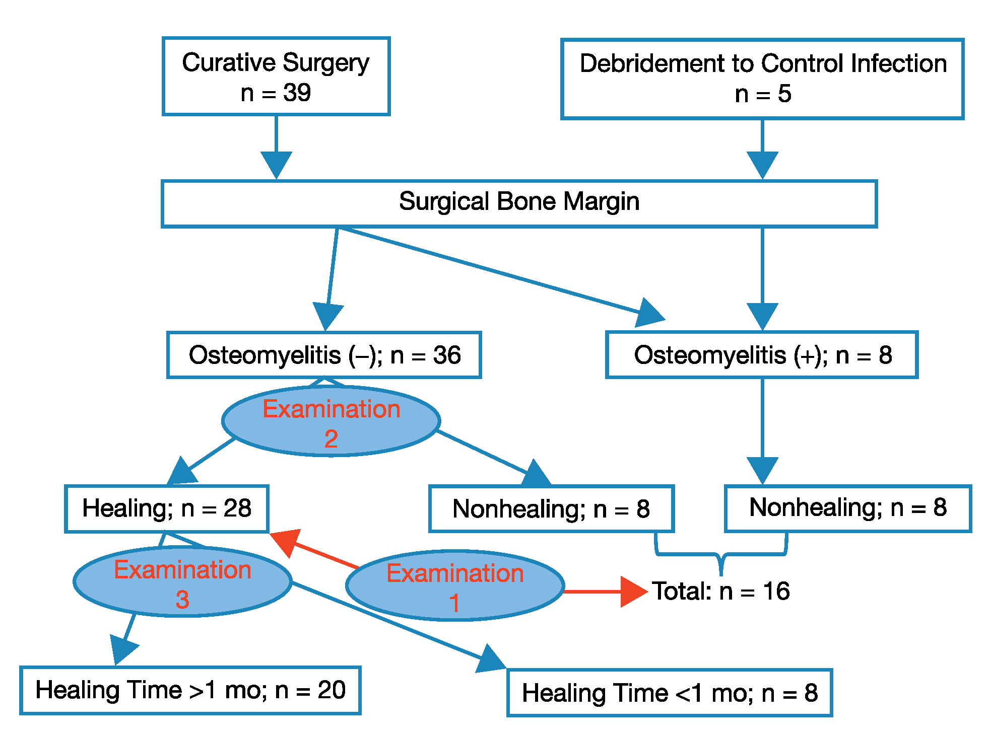
Table 1.
Examination 1-1
Table 2.
Examination 1-2
Figure 2.
C-reactive protein (CRP) level before debridement and after emergency debridement. P = .095.
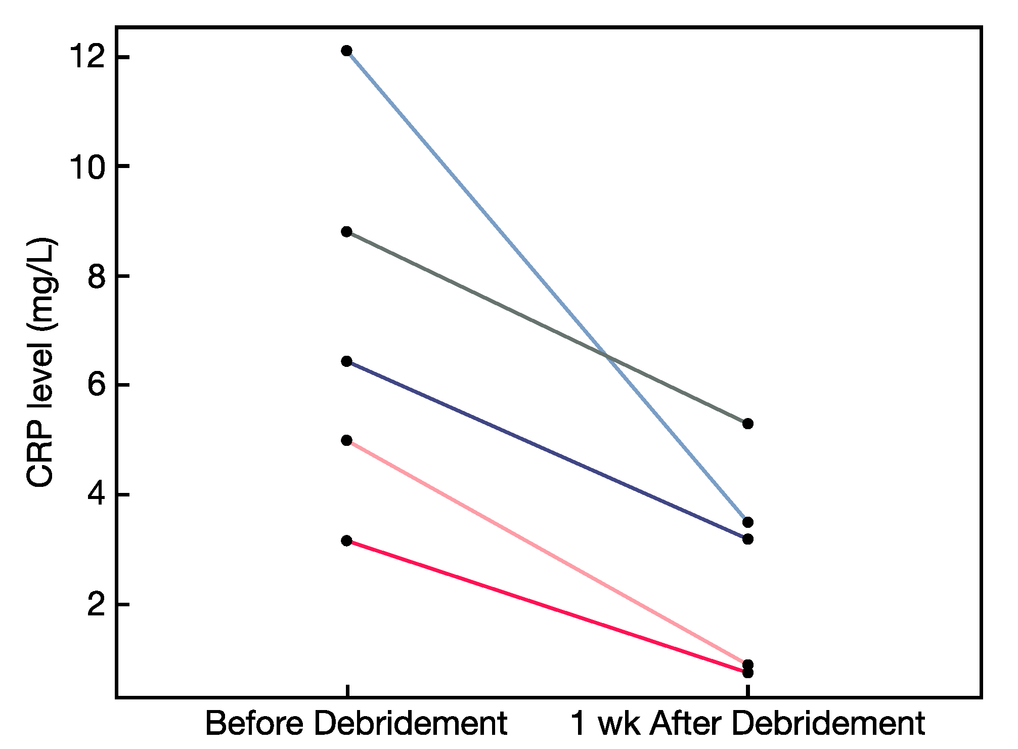
Table 3.
Examination 2-1
Table 4.
Examination 2-2
Table 5.
Examination 3-1
Table 6.
Examination 3-2
Discussion
These results suggest that for the successful treatment of diabetic CLTI with forefoot osteomyelitis, infection has to be controlled at a preoperative CRP level greater than 40 mg/L, and the necessary level of blood supply is represented by an SPP of approximately 55 mm Hg. If the preoperative CRP level is greater than 40 mg/L, debridement should be performed to control infection instead of curative surgery. Although debridement should be performed after revascularization on the basis of MRI findings, many patients with high CRP levels require emergency debridement before or immediately after revascularization; thus, MRI is not effective due to insufficient blood supply.[2,3] In emergency debridement, only obviously necrotic and infected tissue should be resected. Other ambiguous tissue should be preserved because these tissues may become viable after revascularization and antibiotic administration.
Although the normal SPP value is between 80 and 90 mm Hg, it is generally accepted that a value of at least 40 mm Hg is a prerequisite for successful wound healing in patients with CLTI.[7] However, the present results (examinations 1-1 and 2-1) indicate that an SPP of 40 mm Hg is insufficient for the treatment of diabetic CLTI with osteomyelitis, and an SPP of approximately 55 mm Hg is needed for successful treatment, even after complete resection of osteomyelitis. Moreover, examination 3-1 shows that SPP does not have a significant effect on healing time. Ulcer size was statistically related to wound healing and healing time (examination 3-2).
In previous studies,[3,4] we demonstrated the efficacy of MRI in the diagnosis of osteomyelitis. Diabetic forefoot osteomyelitis can be reliably distinguished from reactive bone marrow edema, even in the presence of severe soft-tissue infection. However, ischemic DFO cannot be reliably distinguished because of insufficient interstitial fluid; MRI should, thus, be performed after revascularization. In another study by our group,[2] we proposed an appropriate surgical treatment strategy for DFO involving ischemia or moderate-to-severe soft-tissue infection. We showed that osteomyelitis should be resected completely, and the appropriate surgical margin for DFO surgery should be defined as the area of bone marrow edema on the basis of MRI findings. Furthermore, the present study demonstrated that after osteomyelitis is completely resected, the time to wound healing for ischemic DFO is significantly longer than that for nonischemic DFO. Lower preoperative and postoperative CRP levels were associated with early healing of DFO, supporting the importance of controlling soft-tissue infection after complete resection of osteomyelitis. Furthermore, Rutherford class 6 ulcers required longer than 3 months to heal after complete resection of osteomyelitis, demonstrating that vascular bypass is the preferred revascularization method because it provides more durable patency than endovascular therapy, as previously described.[10,11] The healing rates were 100% for nonischemic DFO and 88.5% for ischemic DFO, which was better than those for conservative surgery.[12,13,14] In the present evaluation of mean ± SD long-term follow-up (1,157.1 ± 571 days for nonischemic DFO and 699 ± 637 days for ischemic DFO), all of the patients could walk with customized footwear, and no recurrence was observed.
The outcome of DFUs is influenced by many factors. Most notably, infection and ischemia are associated with a greater risk of amputation.[15] A previous report[16] showed that PAD was associated with an approximately 2-fold increased risk of foot infection, and another report[17] showed that patients with infection and ischemia were nearly 90 times more likely to receive a midfoot or higher amputation compared with patients with less advanced wound stages (76.5 versus 3.5%; P < .001). The EURODIALE (European Study Group on Diabetes and the Lower Extremity) study,[1] one of the few large, prospective, multinational studies on outcomes and determinations of outcomes in diabetic foot disease, confirmed that the combination of PAD and infection has a major effect on healing rates. The authors concluded that the treatment of PAD and non-PAD DFU should be defined as two separate disease entities because these two distinct diagnoses have significant differences in clinical characteristics, outcomes, and predictors of outcome. However, the current literature on the treatment of DFU has largely not included patients with DFU involving PAD or severe infection. Recently, on the basis of the widespread need for a system that focuses not only on wound healing but also on vascular disease, the SVS Lower Extremity Guidelines Committee created a new threatened limb classification system.[6] This system established a risk stratification on the basis of WIfI. Several studies[18,19,20] demonstrated the effectiveness of this classification system for the correlation of important clinical outcomes for limb salvage and wound healing. This is an iterative process with the goal of more precisely stratifying patients according to their initial disease burden, but it is not aimed at dictating therapy or clinical decision making. The present study findings propose an actual index of blood supply and infection control required for the successful treatment of diabetic critical ischemia with osteomyelitis.
This study has several limitations. First, it was limited to 30 patients. However, because we were able to demonstrate statistically significant differences between patient groups, we believe that the results are relevant for the treatment of diabetic CLTI with osteomyelitis. We hope that these results can serve as baseline data for the performance of a randomized controlled study in the future. Second, we used only CRP level as an infection marker because CRP is an acute-phase protein that is widely used in clinical settings and has high sensitivity and specificity for diagnosing bacterial infections.[21] Third, our definitive diagnosis of osteomyelitis was primarily based on histopathologic examination and not on microbiologic assessment. This is because we wanted to confirm the location of osteomyelitis in the resected bone. Fourth, we did not use the SVS WIfI system completely. We used only the wound and infection grades because we wanted to demonstrate the actual blood supply level as an SPP value. Fifth, we used SPP to evaluate vascular supply rather than toe pressure or transcutaneous oxygen pressure, which are widely accepted methods. All of the patients in this study were Japanese individuals with CLTI, a population with a higher prevalence of renal failure and vascular calcifications compared with other ethnic groups. Skin perfusion pressure is a reliable method for assessing the severity of PAD and for predicting wound-healing ability in such patients.[21,22] Because this trend is rapidly increasing worldwide, we think that the present data provide clinically relevant information.
In conclusion, to treat diabetic CLTI with foot osteomyelitis, osteomyelitis should be resected completely, and, if the preoperative CRP level is greater than 40 mg/L, debridement should be performed first to control infection before curative surgery. An SPP of 55 mm Hg is necessary for successful treatment, even after complete resection of osteomyelitis; however, it has no effect on healing time. We believe that the present research could improve the likelihood of limb salvage in patients with diabetes with CLTI.
Acknowledgments
The patients and staff of the radiology and histopathology departments at Kitaharima Medical Center.
Financial Disclosure
This study is partially supported by National Institutes of Health, National Institute of Diabetes and Digestive and Kidney Diseases Award Number 1R01124789-01A1.
Conflict of Interest
None reported.
References
- Prompers, L; Schaper, N; Apelqvist, J; et al. Prediction of outcome in individuals with diabetic foot ulcers: focus on the differences between individuals with and without peripheral arterial disease: the EURODIALE Study. Diabetologia 2008, 51, 747. [Google Scholar]
- Fujii, M; Terashi, H; Yokono, K:. Surgical treatment strategy for diabetic forefoot osteomyelitis. Wound Repair Regen 2016, 24, 447. [Google Scholar]
- Fujii, M; Armstrong, DG; Terashi, H:. Efficacy of magnetic resonance imaging in diagnosing diabetic foot osteomyelitis in the presence of ischemia. J Foot Ankle Surg 2013, 52, 717. [Google Scholar]
- Fujii, M; Terashi, H; Tahara, S:. Efficacy of magnetic resonance imaging in diagnosing osteomyelitis in diabetic foot ulcers. JAPMA 2014, 104, 24. [Google Scholar]
- Seino, Y; Nanjo, K; Tajima, N; et al. Committee of the Japan Diabetes Society on the Diagnostic Criteria of Diabetes Mellitus; Report of the committee on the classification and diagnostic criteria of diabetes mellitus. J Diabetes Investig 2010, 19, 212. [Google Scholar]
- Mills, JL, Sr,; Conte, MS; Armstrong, DG; et al. The Society for Vascular Surgery Lower Extremity Threatened Limb Classification System: risk stratification based on wound, ischemia, and foot infection (WIfI). J Vasc Surg 2014, 59, 220. [Google Scholar]
- Castronuovo, JJ, Jr,; Adera, HM; Smiell, JM; et al. Skin perfusion pressure measurement is valuable in the diagnosis of critical limb ischemia. J Vasc Surg 1997, 26, 629. [Google Scholar]
- Armstrong, DG; Lavery, LA; Frykberg, RG; et al. Validation of a diabetic foot surgery classification. Int Wound J 2006, 3, 240. [Google Scholar]
- Terashi, H; Kitano, I; Tsuji, Y; et al. A modified transmetatarsal amputation. J Foot Ankle Surg 2011, 50, 441. [Google Scholar]
- Conte, MS:. Understanding objective performance goals for critical limb ischemia trials. Semin Vasc Surg 2010, 23, 129. [Google Scholar]
- Khan, MU; Lall, P; Harris, LM; et al. Predictors of limb loss despite a patent endovascular-treated arterial segment. J Vasc Surg 2009, 49, 1440. [Google Scholar]
- Lázaro-Martínez, JL; Aragón-Sánchez, J; García-Morales, E:. Antibiotics versus conservative surgery for treating diabetic foot osteomyelitis: a randomized comparative trial. Diabetes Care 2014, 37, 789. [Google Scholar]
- Aragón-Sánchez, J; Lázaro-Martínez, JL; Hernández-Herrero, C; et al. Does osteomyelitis in the feet of patients with diabetes really recur after surgical treatment? natural history of a surgical series. Diabet Med 2012, 29, 813. [Google Scholar]
- Ha Van, G; Siney, H; Danan, JP; et al. Treatment of osteomyelitis in the diabetic foot: contribution of conservative surgery. Diabetes Care 1996, 19, 1257. [Google Scholar]
- Armstrong, DG; Boulton, AJM; Bus, SA:. Diabetic foot ulcers and their recurrence. N Engl J Med 2017, 376, 2367. [Google Scholar]
- Lavery, LA; Armstrong, DG; Wunderlich, RP; et al. Risk factors for foot infections in individuals with diabetes. Diabetes Care 2006, 29, 1288. [Google Scholar]
- Armstrong, DG; Lavery, LA; Harkless, LB:. Validation of a diabetic wound classification system: the contribution of depth, infection, and ischemia to risk of amputation. Diabetes Care 1998, 21, 855. [Google Scholar]
- Cull, DL; Manos, G; Hartley, M; et al. SS12 Prospective analysis of wound characteristics and degree of ischemia on time to wound healing and limb salvage: an early validation of the Society for Vascular Surgery Lower Extremity Threatened Limb Classification System. J Vasc Surg 2014, 59, 28S. [Google Scholar]
- Zhan, LX; Branco, BC; Armstrong, DG; et al. The Society for Vascular Surgery Lower Extremity Threatened Limb Classification System based on Wound, Ischemia, and foot Infection (WIfI) correlates with risk of major amputation and time to wound healing. J Vasc Surg 2014, 61, 939. [Google Scholar]
- Causey, MW; Ahmed, A; Wu, B; et al. Society for Vascular Surgery limb stage and patient risk correlate with outcomes in an amputation prevention program. J Vasc Surg 2016, 63, 1563. [Google Scholar]
- Mölkänen, T; Ruotsalainen, E; Rintala, EM; et al. Predictive value of C-reactive protein (CRP) in identifying fatal outcome and deep infections in Staphylococcus aureus bacteremia. PLoS One 2016, 11, e0155644. [Google Scholar]
- Yamada, T; Ohta, T; Ishibashi, H; et al. Clinical reliability and utility of skin perfusion pressure measurement in ischemic limbs: comparison with other noninvasive diagnostic methods. J Vasc Surg 2008, 47, 318. [Google Scholar]
© 2021 American Podiatric Medical Association