


Abstract
This case study presents a 72-year-old male patient who presented with multiple schwannomas. The patient underwent several soft-tissue mass excisions performed by general surgery and podiatry. The physical examination displayed multiple nodules on the left foot, ankle, and previously in the groin. Magnetic resonance imaging displayed high–T2-weighted signal masses in the left plantar heel and ankle. These masses were excised and, through pathologic analysis, were diagnosed as schwannomas. Immuno- histochemistry displayed a mosaic pattern of INI1 expression, a gene associated with tumor suppression. The patient demonstrated findings consistent with multiple schwannomas (eg, schwannomatosis or neurofibromatosis type 2). Because of the patient’s medical history, neurofibromatosis type 2 was more probable.
Schwannomas are one of the most common benign peripheral nerve tumors, which develop from Schwann cells. Schwannomas are found throughout the body, and they clinically resemble various neoplasms. Gross examination of Schwannomas typically demonstrate well-circumscribed masses with degenerative changes; variable admixture of compact spindled areas; and hypocellular, micro- cystic areas rich in macrophages and collagen fibers. The annual incidence of schwannomatosis is 0.58 case per 1 million individuals.[1] The incidence of neurofibromatosis type 2 is one in 25,000.[2] Radiologic or histologic evaluation, alone, has proven ineffective at diagnosing schwannomas. More advanced imaging such as magnetic resonance imaging (MRI) has been useful in determining the involvement of the surrounding tissue.[3] However, MRI alone does not provide a definitive diagnosis and is often inconclusive.[4] Previous studies have shown that one of the most effective ways to differentiate these masses from other abnormalities is with the use of immunohistochemistry. S100 protein is a marker for Schwann cells in normal peripheral nervous tissue. The S100 protein can be detected by using anti–S protein antibodies. Cor- rectly diagnosing schwannomas is important for providing the best treatments for patients. The treatment of choice for schwannomas is surgical excision, which has a good prognosis and a low rate of recurrence.[5]
The presence of multiple schwannomas is associated with neurofibromatosis. There are three types of neurofibromatosis: type 1, type 2, and schwannomatosis.
Neurofibromatosis type 2 (NF2) is an inherited disorder characterized by multiple benign tumors of the central nervous system.[6] The presence of vestibular schwannomas is diagnostic for NF2.[7] Vestibular schwannomas often cause tinnitus, hearing loss, and balance dysfunction.[8] Other common findings of NF2 include meningiomas, ependymomas, neurofibromas, and spinal and peripheral nerve schwannomas.[9] No mutation to the INI1 tumor suppressor gene is seen in NF2.[10] An MRI scan of the brain is required to identify vestibular schwannomas and make the diagnosis of NF2.
Schwannomatosis is a form of neurofibromatosis characterized by multiple schwannomas of the peripheral nervous system in the absence of vestibular schwannomas.[7] The presence of a mosaic pattern of INI1 expression is characteristic of familial schwannomatosis.[7] Schwannomatosis may be sporadic or familial, which accounts for 20% of patients.[7] These patients generally present in adulthood with pain that may or may not involve the schwannoma.[7]
Case Report
A 72-year-old male presented to the clinic with the chief concern of a palpable mass to the plantar aspect of his left foot. He had noticed the mass several months previously. He denied any previous injuries or punctures to the foot. He complained of discomfort from the mass when walking. Changes in shoe gear had provided no relief. The patient’s medical history included hemorrhoids, hypertension, gastroesophageal reflux disease, and difficulty hearing. The patient’s medical history was unremarkable for any other soft-tissue masses. The patient previously had surgery to his left hand and left inguinal mass excision.
A physical examination was performed to evaluate the chief complaint. Vascular and neurologic examinations were within normal limits. The Mulder sign was negative bilaterally. The dermato- logic examination revealed a palpable, superficial soft-tissue mass on the plantar rearfoot, measuring 3.0 3 3.0 cm. The mass was soft and immobile, free from underlying tendons and plantar fascia, and tender with palpation. An antalgic gait was noted.
The initial radiographs were negative for periosteal reaction, cortical changes, and soft-tissue calcifications. The preoperative diagnosis was ganglionic cyst.
Treatment options were discussed and the patient elected to undergo an excisional biopsy of the lesion. The case was scheduled as an outpatient operation under local anesthesia with monitored anesthesia care. A 5-cm lazy S–type incision was made over the soft-tissue mass on the left plantar rearfoot. Sharp dissection was carried out to free the mass from its attachments. The stalk was identified near the distal aspect of the calcaneus. The incision site was further searched for any remaining cystic material. All remaining material was excisionally debrided to the level of deep fascia and removed from the operative site. The specimen was then submitted for pathologic evaluation after being removed in total. The pathology report indicated a 4.5 3 3.8 3 3.0-cm, gray-tan, rubbery mass with lobulations. Cut sections revealed a beige, glistening, well-circumscribed nodule partially surrounded by gray-tan tissue. The specimen was diagnosed as plantar fibromatosis.
One year previously, this patient had undergone an excision of a left inguinal groin mass. The preoperative diagnosis made by the general surgeon was lymphadenopathy. The specimen was sent to the pathology department for evaluation. The initial report was negative for immunophenotypic evidence of a lymphoproliferative disorder; a second opinion on the pathology report was obtained from an outpatient pathologist. The final diagnosis was schwannoma, and the report displayed tumor cells that were diffusely positive for S100 and CD34 and negative for B-catenin and anaplastic lymphoma kinase.
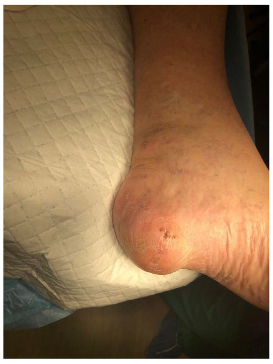
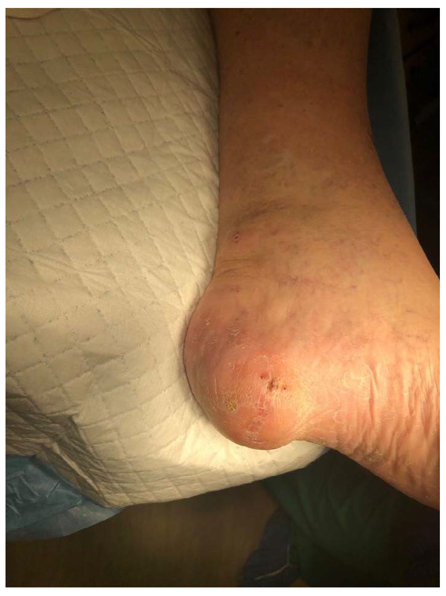
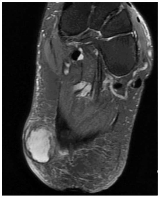
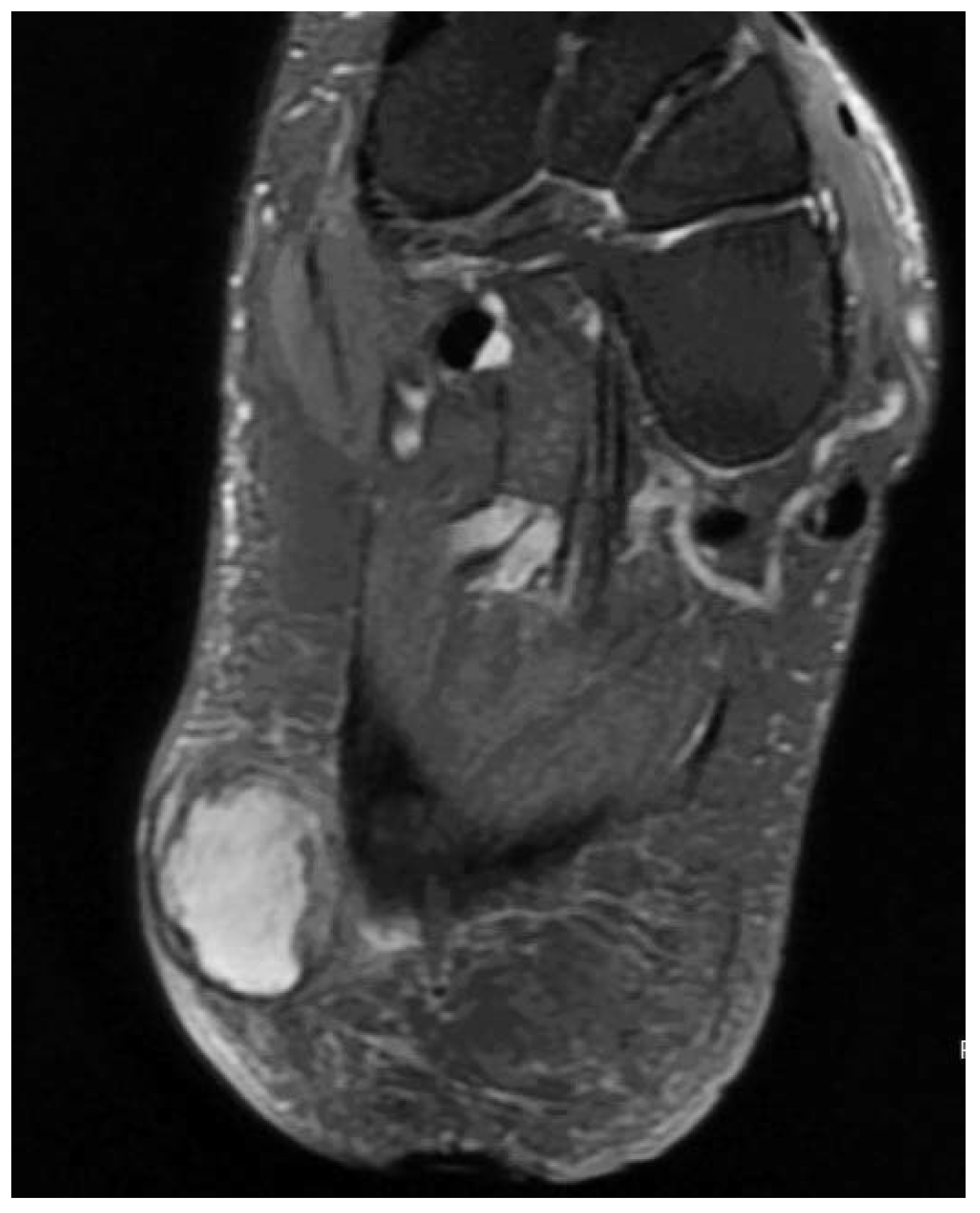
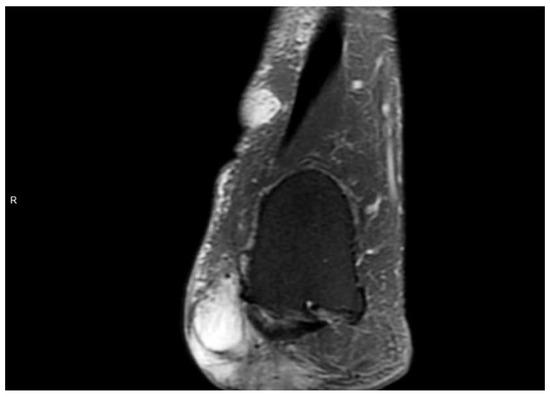
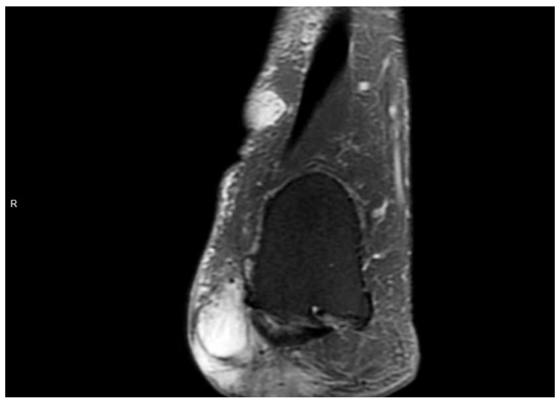
This patient then returned to the clinic approximately 5 months later with a similar complaint of soft-tissue masses to the posteromedial left ankle and plantar foot (Figure 1). An MRI scan without contrast was taken of the left ankle and foot, which displayed a large multilobulated and multiseptated heterogeneous subcutaneous structure, in the plan- tar heel, with high T2- and low T1-weighted signal intensity (Figure 2 and Figure 3). This mass measured 4.9 3 2.7 3 1.7 cm. This mass was suggested to represent a complex fluid collection; however, further characterization was limited because of the lack of intravenous gadolinium. There was severe, extensive soft-tissue edema in the medial aspect of the ankle. The MRI scan was negative for any signs of osteomyelitis.
Figure 1.
Clinical photograph of left heel soft-tissue mass.
Figure 2.
Magnetic resonance imaging coronal view of plantar heel displaying high signal mass.
Figure 3.
Magnetic resonance imaging axial view of left plantar heel displaying high signal intensity mass.
Punch biopsy specimens of both soft-tissue masses were obtained in the outpatient setting. The medial ankle and plantar foot masses were diagnosed as neurofibroma and plantar fibroma with invasion into the level of the dermis, respectively.
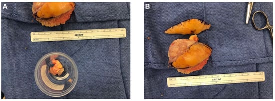
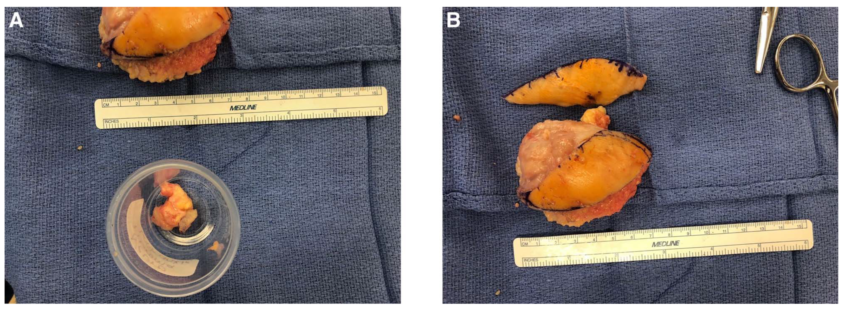
The patient elected to undergo surgical excision of the left lower-extremity masses. Attention was directed to the medial ankle soft-tissue mass, where a 7-cm curvilinear incision was made over the palpable mass. Incision was deepened through the subcutaneous tissue, and care was taken to identify posterior medial neurovascular structures and ensure that they were retracted out of the way. The mass was fully resected from its underlying soft-tissue attachments. There was no identifiable stalk noted (Figure 4A). The mass was solid in nature. Attention was then directed to the plantar heel mass. An 8-cm S-shaped incision was made directly overlying the mass. The mass was dissected from the underlying soft-tissue attachments down to the level of deep fascia. It was noted that a branch of the medial calcaneal nerve led directly to the soft- tissue mass, and a portion of this nerve was resected along with the mass. This mass also was noted to be firm in nature (Figure 4B). Because of the void remaining, negative-pressure wound therapy was used to aid in closure.
Figure 4.
A, Excised soft-tissue mass from the left ankle. B, Excised mass from the left heel.
Both specimens were sent for outside pathologic evaluation. The final report demonstrated schwannoma to the left heel and benign nerve sheath tumor, with hybrid schwannoma and neurofibroma features to the left ankle. Multiple schwannoma and mosaic pattern of INI1 on immunohistochemistry were discussed on the pathology report, which raised the suspicion for various multiple schwannoma syndromes (eg, sporadic schwannomatosis, familial schwannomatosis, and NF2).
Discussion
Schwannomas are benign peripheral nerve sheath tumors that may indicate an underlying neurologic disorder. This case describes a patient with recurring, painful, soft-tissue masses of the lower extremities. The initial soft-tissue mass was diagnosed as plantar fibromatosis. The ensuing mass resections by the general surgeon and podiatrist revealed schwannomas. The immunohistochemistry studies were suggestive of schwannomatosis and NF2. A definitive diagnosis was unable to be made because of the patient not undergoing MRI of the brain to identify vestibular schwannomas. This patient’s medical history did reveal hearing impairment. Because of his physical findings and immunohistology, a diagnosis of NF2 was most probable.
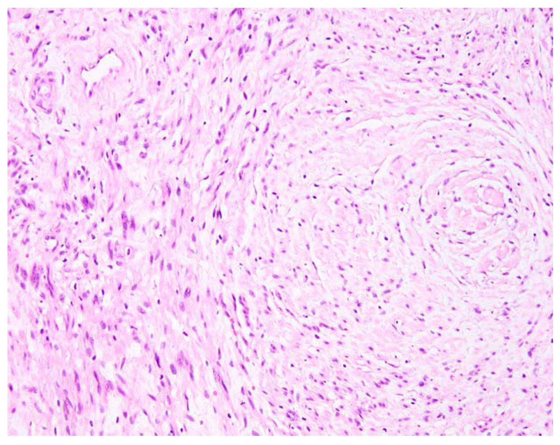
Figure 5.
Pathology slide demonstrating cell histology consistent with schwannoma.
Financial Disclosure
None reported.
Conflicts of Interest
None reported.
References
- MERKER, V.L.; ESPARZA, S.; SMITH, M.J. ET AL: Clinical features of schwannomatosis: a retrospective analysis of 87 pa- tients. Oncologist 2012, 17, 1317. [Google Scholar] [CrossRef] [Scilit] [PubMed]
- EVANS, D.G.; MORAN, A.; KING, A.; ET, A.L. Incidence of vestibular schwannoma and neurofibromatosis 2 in the North West of England over a 10-year period: higher incidence than previously thought. Otol Neurotol 2005, 26, 93. [Google Scholar] [CrossRef] [Scilit] [PubMed]
- SOTO, R.; ALDUNCE, M.J.; WORTSMAN, X. ET AL: Subungual schwannoma with clinical, sonographic, and histologic correlation. JAPMA 2014, 104, 302. [Google Scholar] [CrossRef] [Scilit] [PubMed]
- PASTERNACK, W.A.; WINTER-REIKEN, D.J. Unusually large cellular schwannoma of the foot. JAPMA 2005, 95, 157. [Google Scholar] [CrossRef] [Scilit] [PubMed]
- MOTT, R.C.; DELLON, A.L. Multiple schwannomas of the foot. Case report and strategy for treatment. JAPMA 2003, 93, 51. [Google Scholar] [CrossRef] [Scilit] [PubMed]
- EVANS DG: Neurofibromatosis type 2 (NF2): a clinical and molecular review. Orphanet J Rare Dis 2009, 4, 16. [CrossRef] [Scilit] [PubMed]
- HULSEBOS, T.J.; PLOMP, A.S.; WOLTERMAN, R.A. ET AL: Germline mutation of INI1/SMARCB1 in familial schwannomato- sis. Am J Hum Genet 2007, 80, 805. [Google Scholar] [CrossRef] [Scilit] [PubMed]
- EVANS DGR: Neurofibromatosis 2 [bilateral acoustic neurofibromatosis, central neurofibromatosis, NF2, neurofibromatosis type II]. Genet Med 2009, 11, 599. [CrossRef] [Scilit] [PubMed]
- LU-EMERSON, C.; PLOTKIN, S.R. The neurofibromatoses. Part 2. NF2 and schwannomatosis. Rev Neurol Dis 2009, 6, E81. [Google Scholar] [PubMed]
- MACCOLLIN, M.; CHIOCCA, E.A.; EVANS, D.G. ET AL: Diagnostic criteria for schwannomatosis. Neurology 2005, 64, 1838. [Google Scholar] [CrossRef] [Scilit] [PubMed]
© 2020 American Podiatric Medical Association