


Abstract
Background The purpose of this study was to compare the clinical and radiographic outcomes of stabilization of the lateral ligament combined with joint debridement in patients with ligamentous moderate neutral ankle osteoarthritis with those achieved for patients with varus ankle osteoarthritis. Methods We reviewed integrated data from 40 patients (40 ankles) with ligamentous moderate ankle osteoarthritis. Matched for age, gender, and follow-up duration, they were divided into two groups by preoperative coronal plane hindfoot moment arm values (HMAV): neutral (20 ankles, ≤15 mm) and varus (20 ankles, >15 mm) deformity. Stabilization of lateral ligament combined with joint debridement was performed. American Orthopaedic Foot and Ankle Society (AOFAS) ankle-hind foot score, hindfood moment arm values, and classification of Takakura were used to compare clinical and radiographic outcomes after a mean follow-up period of 64.2 months (range, 60-84 months). Results Mean post-operative AOFAS was 86.0 and 72.6 in the neutral and varus groups, respectively. The post-operative AOFAS of both groups improved significantly, although the outcome improvement of the neutral group was better than that of the varus group (P = 0.0006). There was obvious improvement in HMAV of the neutral group (P = 0.0469) and less improvement in HMAV of the varus group (P = 0.8509). The mean postoperative HMAV was 4.60 mm (0–10 mm) and 17.85 mm (8–23 mm) in the neutral and varus groups, respectively. The radiographic classification of Takakura was unchanged in the neutral group, whereas four cases in the varus group had a worse classification. Conclusions Stabilization of the lateral ligament combined with joint debridement for ligamentous moderate ankle osteoarthritis showed better clinical and radiographic outcomes in patients with neutral alignment than that achieved for patients with varus malalignment.
Osteoarthritis (OA) is significantly less common in the ankle than in the knee or hip. However, Glazebrook et al [1]. have demonstrated that patients with end-stage ankle OA have mental and physical disabilities comparable with those of patients with end-stage hip OA, and the clinical value of treatment of ankle OA should not be underestimated. There are several causes of ankle arthritis; posttraumatic osteoarthritis, considered to be the most common type, accounts for more than 70% of ankle arthritis cases [2]. Trauma is the main cause of ankle OA and primary OA is rare [3-4]. Lateral ligament lesions are the most frequent ankle injuries sustained in sports and recreational activities, and the resulting ankle arthritis is known as ligamentous arthritis [56]. Approximately 15% of the world's adult population is affected by joint pain and disability resulting from OA [7]. and approximately 1% have OA of the ankle [8].
Arthrodesis is often considered the gold standard for advanced stage painful ankle OA, but limited ankle motion caused by arthrodesis increases the possibility of adjacent joint arthritis [9]. The aim of ankle arthrodesis is to provide a pain-free plantigrade foot during weightbearing activities [10]. The effort made to prevent moderate ankle OA from deteriorating to end-stage ankle OA is of great significance. Several studies have documented clinical outcomes of stabilization of the lateral ligament for the treatment of ligamentous ankle osteoarthritis [211]. However, the different outcomes between patients with neutral alignment and with varus malalignment have not been concerned. In this study, we compared the clinical and radiographic outcomes of stabilization of lateral ligament combined with joint debridement in patients with ligamentous moderate neutral ankle osteoarthritis with those achieved for patients with varus ankles osteoarthritis.
Materials and Methods
Patients
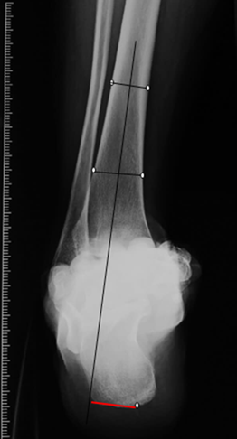
This study was performed in the Department of Orthopedic Surgery. Between May 2007 and March 2011, 65 patients with ligamentous moderate ankle OA were treated by a single, experienced team, and the integrated data of 40 patients (40 ankles) were obtained. Matched for age, gender, and follow-up duration, participants were divided into two groups (20 per group) according to preoperative coronal plane hindfoot moment arm values (HMAV): neutral (20 ankles, ≤15 mm) and varus (20 ankles, >15 mm) deformity [12]. The HMAV was represented by the perpendicular distance from the most inferior point on the calcaneus and midlongitudinal axis line of the tibia (Fig. 1). The moderate stage of ankle OA was quantified and evaluated based on the Takakura et al [13]. classification, including: stage 2, narrowing of the medial joint space; stage 3, obliteration of this space with subchondral bone contact; stage 3a, obliteration of the joint space limited to the medial malleolus; and stage 3b, obliteration extended to the roof of the dome of the talus. This study was undertaken in conformity with the Declaration of Helsinki after approval of our institutional review board. Written informed consent was obtained from all participants.
Figure 1.
Hindfood alignment view. Hindfood moment arm values (HMAV) are determined by measuring the distance between the lowest point on the calcaneus to the longitudinal axis of the tibia with weightbearing.
Operative procedure
Joint debridement, medial release, and lateral ligament stabilization were performed in all patients. Combined open cheilectomy and arthroscopic joint debridement were performed in 22 patients. Arthroscopic joint debridement alone was performed in eight patients and open cheilectomy alone in 10 patients. Bony spurs on the anterior distal tibia or dorsal talus (or both) obstructing the normal dorsiflexion of the ankle were resected completely no matter what procedure was applied, and holes were drilled at approximately 2-mm intervals at a depth of approximately 10 mm at the region of degenerative cartilage [14]. Full passive range of activity was obtained in all the ankles during surgery. Contracted deltoid ligament release was performed in most cases (80%).
Following joint debridement, a procedure to reconstruct the lateral ligaments of the ankle was performed. By use of a lateral approach, lateral ligaments of 25 patients were augmented via a modified Brostrom operation (MBO) [15]. and the remaining 15 patients were reconstructed via a modified Chrisman-Snook technique [16]. In the latter, the upper part of the peroneous brevis tendon was cut down and transferred through the hole drilled in the lateral malleolus to reinforce the lateral collateral ligaments, including the anterior talofibular ligament and the calcaneofibular.
Postoperative management
A splint was maintained in all patients until suture removal at 2 weeks postoperative. Subsequently, the ankle was immobilized in a soft ankleorthosis for 4 weeks. Active range of motion and partial weightbearing were allowed at 4 weeks after surgery; full weightbearing was allowed at 8 weeks after surgery. Patients were evaluated at 6 weeks, 3 months, 6 months, 1 year, 2 years, and 5 years postoperative for range of motion and evaluation of pain and function.
Clinical and radiological analysis
The American Orthopaedic Foot and Ankle Society (AOFAS) clinical ankle-hindfoot scale was used to evaluate the function of the ankle preoperatively and at the last follow-up.
Radiologic evaluation was performed using weightbearing anteroposterior, lateral, and Saltzman views [12]. before surgery and at the most recent follow-up. On the anteroposterior view, the classification described by Takakura et al [13]. was used to identify verities of the ankle arthrodesis. On the Saltzman view, according to Saltzman CL [12]. the hindfoot alignment was represented by the coronal plane hindfoot moment arm values (HMAV), which referred to the distance between the point of heel-floor contact to the longitudinal axis of the tibia (Fig. 1). Since the 95% confidence interval was 15 mm, the ankle with HMAV exceeding 15 mm was identified as the varus ankle.
Statistical analysis
Statistical analysis was performed with statistical software (SPSS, Chicago, IL,). The AOFAS score and HMAV on the coronal plane before surgery and at the most recent follow-up were compared by means of the independent samples t-test for continuous variables and chi-square tests for dichotomous values. The AOFAS score and HMAV at the most recent follow-up were compared between the two groups by the same method. A P value <0.05 was considered to be significant.
Results
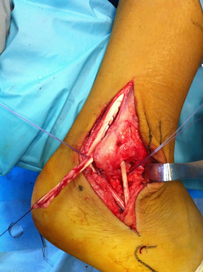
All 40 patients (40 ankles; 17 men, 23 women), with a mean age of 54.95 years (range, 32–75 years), met the criteria of having sustained one or more lateral ligamentous lesions and a history of chronic lateral instability of the ankle (LIA), but no other injury (such as fracture) to the same ankle, and were included in the study. Talartilt and anterior drawer tests were conducted before and after surgery, but the stabilization of the ankle was difficult to assess clinically because of osteophytosis around the ankle preoperatively. All patients had normal knee alignment and there was no leg-length discrepancy. None of the patients had undergone any other ankle surgery before our therapy. Of the 40 patients, there were 25 patients with stage 2 and 15 patients with stage 3a osteoarthritis; 16 patients in the neutral group had stage 2 ankle and four had stage 3a, and these values were 7 and 13 in the varus group, respectively (Fig. 2). There were no significant differences between the two groups in the AOFAS between surgery (P = 0.6663; Table 1). One or two suture anchors were implanted to reconstruct the attachment point of the ligaments (Fig. 3). The improvement of the medial space ratio indicated that the talus was in a better position postoperatively due to lateral stabilization.
Figure 2.
Preoperative (A) and postoperative 62 months (B) weightbearing anteroposterior views of the ankle of a 42-year-old male. Note the widened medial clear space and narrowed lateral clear space after surgery. Joint debridement and modified Brostrom procedure were applied (C), which resulted in substantial postoperative pain relief, functional improvement, and slowing of the degeneration process (D).
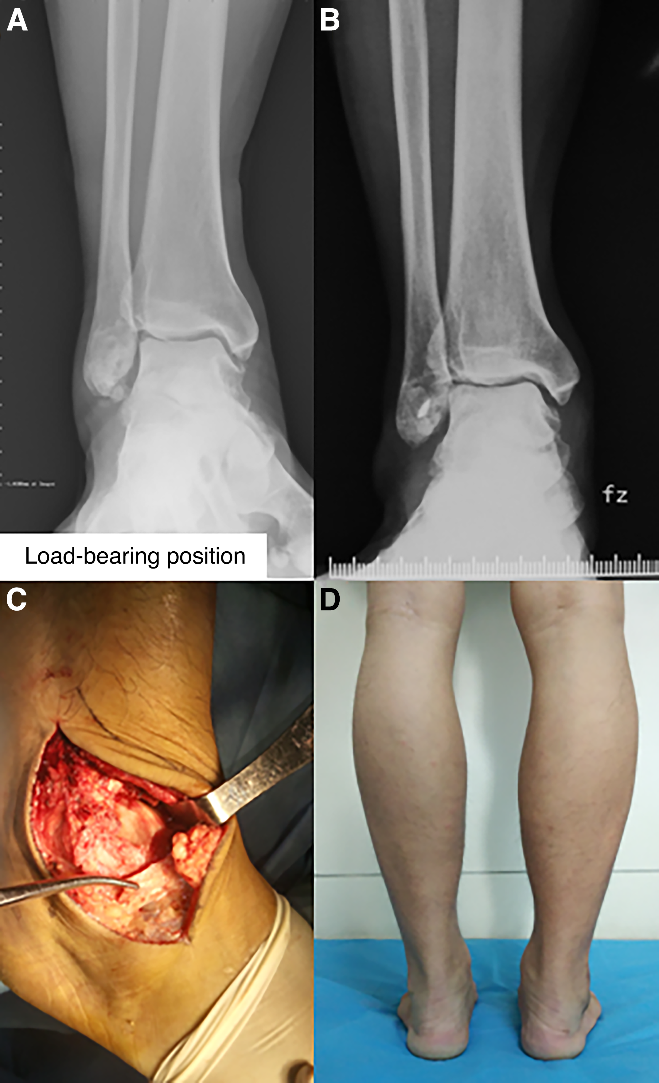
Figure 3.
Modified Chrisman-Snook technique. The transferred semi-peroneousbrevis tendon was fixed by 1 or 2 suture anchors.
The mean AOFAS scores and mean HMAV in the neutral and varus groups before surgery and at the most recent follow-up are shown in Table 2. There were significant differences between the AOFAS score before surgery and that at the most recent follow-up in the neutral group (42.45 ± 7.15, 86 ± 6.25, P < 0.001) and varus group (41.5 ± 6.67, 72.6 ± 14.02, P < 0.001). There were significant differences between the most recent follow-up AOFAS score in the neutral group (86 ± 6.25) and varus group (72.6 ± 14.02, P = 0.0006). These findings showed that the function of the neutral group improved more significantly than that of the varus group.
There were significant differences between the mean HMAV before surgery and at the most recent follow-up in the neutral group (6.45 ± 3.19, 4.60 ± 2.46, P = 0.0469) and no significant differences in the varus group (17.65 ± 2.08, 17.85 ± 4.23, P = 0.0851); the scores of nine patients in the varus group got worse postoperatively. The mean postoperative HMAV was 4.60 mm (0–10 mm) and 17.85 mm (8–23 mm) in the neutral and varus groups, respectively.
At the most recent follow-up, 16 patients of the neutral group had stage 2 ankle OA and four had stage 3a (just as many as before surgery), while four cases got worse in the varus group, three cases at stage 2 deteriorated to stage 3a, and one at stage 3a deteriorated to stage 3b.
Discussion
Ankle OA is mostly caused by chronic lateral instability of the ankle (LIA) after later alligamentous injury [17]. The ligament system plays a fundamental role in the ankle's stability and includes a talocrural complex and a subtalar complex that are functionally related [18]. Because of the functional and biomechanical factors involved, the lateral ligament complex is the most often injured structure of all the stabilizing components of the ankle joint, contributing to about 85% of all chronic LIA and the ankle arthritis resulting from chronic LIA is known as ligamentous arthritis [5-6]. The mechanism may involve concomitant peritalar instability, which allows the talus to translate and rotate on the calcaneus. Beside maintaining lateral ankle stability, the lateral ankle ligaments have been shown to play a significant role in providing rotational ankle stability and it is especially true for the anterior talofibular ligament [19]. This internal rotated talus with medially displaced heel can injure the articular cartilage on the medial side rather than lateral side of the ankle joint [17]. Newer studies have shown that, in up to 60% of affected ankles with progression of osteoarthritic process, the talus did showed a varus or valgus tilt within the ankle mortise. Therefore, with the increasing HMAV, the heel progresses into varus displacement gradually and ligamentous varus ankle osteoarthritisis emerges. In the neutral type of instability, without obvious malalignment, the talus has not changed its position in the coronal plane, but it has translational movement on the calcaneus in the sagittal and horizontal plane latently [20]. Notably, this would be more serious in patients with varus hindfoot alignment.
The goals of treating moderate ligamentous ankle OA are to preserve partial joint function, prevent arthrosis development, or halt additional progression and to avoid ankle arthrodesis or arthroplasty [21]. The most important aspect of preoperative planning is assessment of the origin of the deformity and the understanding of deformation forces. For this purpose, several realignment procedures, such as supramalleolar osteotomies [22-23]. inframalleolar osteotomies [24]. calcaneal osteotomies, tendon transfers [20]. and ligament reconstructions [17]. have been recommended for joint-preserving treatments and the indication of the surgery type is taken mainly based on the ankle osteoarthritis staging. Realignment procedures may restore the normal biomechanics of the ankle joint, resulting in substantial postoperative pain relief, functional improvement, and slowing of the degeneration process. Bony realignment procedures are adopted to correct obvious ankle malalignment, which would otherwise lead to greater damage than isolated soft-tissue surgery.
Although it remains controversial whether the ligaments have to be reconstructed, the isolated mortise plasty osteotomy without ligament reconstruction described by Kobayashi et al [21]. provided good clinical and radiographical outcomes for patients with varus ankle osteoarthritis and instability, and Al-Nammari and Myerson [24]. recommended any associated instability and multilevel deformity must be recognized and addressed. We propose the pathomechanism mentioned above. There is still another important but neglected reason for stabilization of lateral ligament; in most cases, with the tilted talus, the calcaneus goes in the opposite direction and results in a so-called Z-shaped deformity, which leads to decreased hindfoot height and subsequent loss of ligaments [20].
The aim of ligament reconstructions is to stabilize the talus in a correct position within the ankle mortise. Of the many surgical options available, anatomical repair of the anterior talofibular and calcaneofibular ligaments (Brostrom procedure) is recommended most widely when the intensity of the ruptured ligaments permits, whereas anatomical or nonanatomical reconstruction with autograft or allograft should be performed when the ruptured ligaments are attenuated. Although Brostrom reported that end-to-end repair of the ruptured anterior talofibular ligament can restore good stability of the ankle even after a long delay between injury and repair, Lee et al [17]. found the Brostrom and modified Brostrom procedure had limitations in stabilizing a severely tilted talus. In the 40 ankles we assessed, 25 ankles with a strong ligament scar were repaired by the Brostrom procedure, while the remaining 15 ankles with slim scars were stabilized by the Chrisman-Snook procedure, which was considered to be a more reliable method to restore ankle stability compared with the other reconstruction procedure [25].
It should be noted that the differences in results were based mainly on the initial Takakura Stage. Although no improvement of the Takakura stage was seen in our 40 cases, AOFAS score, hindfood alignment ratio, and clear space ratio were improved after surgery, especially in the neutral group. In our study, there were significant differences between the mean HMAV before surgery and at the most recent follow-up in the neutral group and no significant differences in the varus group; this was consistent with the improvement of postoperative AOFA scores. This finding shows that isolated procedure of stabilization of lateral ligament without bony realignment surgery cannot improve hindfoot alignment sufficiently in the ligamentous moderate ankle osteoarthritis with obvious varus malalignment. Osteotomies surrounding the ankle should be performed to provide the advantage of a stabilization effect for lateral instability as well as correct an obvious varus displaced heel. There were limitations in the study, such as the relatively small sample size, the multivariate analysis between the two groups, and follow up of the exact effect of these methods.
Conclusions
Stabilization of the lateral ligament combined with joint debridement for ligamentous moderate ankle osteoarthritis may partially restore normal biomechanics of the ankle joint, resulting in substantial postoperative pain relief, functional improvement, and slowing of the degeneration process, and it shows better clinical and radiographical outcomes in patients with neutral alignment than that achieved for patients with varusmal alignment.
Acknowledgments
Financial Disclosure: This work was supported by key research and development plan of Yantai city science and technology bureau (2016WS033), Key research and development plan of Shandong Province (2016GSF201112), and the medical science and technology development plan project of Shandong Province (2016WS0696).
Conflict of interest: None reported.
References
- Glazebrook, M.; Daniels, T.; Younger, A.; et al. Comparison of health-related quality of life between patients with end-stage ankle and hip arthrosis. J Bone Joint Surg Am 2008, 90, 499. [Google Scholar]
- Valderrabano, V.; Hintermann, B.; Horisberger, M.; et al. Ligamentous posttraumatic ankle osteoarthritis. Am J Sports Med 2006, 34, 612. [Google Scholar]
- Barg, A.; Pagenstert, GI.; Hugle, T.; et al. Ankle osteoarthritis: etiology, diagnostics, and classification. Foot Ankle Clin 2013, 18, 411. [Google Scholar]
- Valderrabano, V.; Horisberger, M.; Russell, I.; et al. Etiology of ankle osteoarthritis. Clin Orthop Relat Res 2009, 467, 1800. [Google Scholar]
- Elgafy, H.; Ebraheim, NA.; Tile, M.; et al. Fractures of the talus: experience of two level 1 trauma centers. Foot Ankle Int 2000, 21, 1023. [Google Scholar]
- Hirose, K.; Murakami, G.; Minowa, T.; et al. Lateral ligament injury of the ankle and associated articular cartilage degeneration in the talocrural joint: anatomic study using elderly cadavers. J Orthop Sci 2004, 9, 37. [Google Scholar]
- Corti, MC.; Rigon, C. Epidemiology of osteoarthritis: prevalence, risk factors and functional impact. Aging Clin Exp Res 2003, 15, 359. [Google Scholar]
- Bruyere, O.; Cooper, C.; Arden, N.; et al. Can we identify patients with high risk of osteoarthritis progression who will respond to treatment? A focus on epidemiology and phenotype of osteoarthritis. Drugs Aging 2015, 32, 179. [Google Scholar]
- DeHeer, PA.; Catoire, SM.; Taulman, J.; et al. Ankle arthrodesis: a literature review. Clin Podiatr Med Surg 2012, 29, 509. [Google Scholar]
- Nihal, A.; Gellman, RE.; Embil, JM.; et al. Ankle arthrodesis. Foot Ankle Surg 2008, 14, 1. [Google Scholar]
- Takao, M.; Komatsu, F.; Naito, K.; et al. Reconstruction of lateral ligament with arthroscopic drilling for treatment of early-stage osteoarthritis in unstable ankles. Arthroscopy 2006, 22, 1119. [Google Scholar]
- Saltzman, CL.; el-Khoury, GY. The hindfoot alignment view. Foot Ankle Int 1995, 16, 572. [Google Scholar]
- Takakura, Y.; Tanaka, Y.; Kumai, T.; et al. Low tibial osteotomy for osteoarthritis of the ankle. Results of a new operation in 18 patients. J Bone Joint Surg Br 1995, 77, 50. [Google Scholar]
- Choi, JI.; Lee, KB. Comparison of clinical outcomes between arthroscopic subchondral drilling and microfracture for osteochondral lesions of the talus. Knee Surg Sports Traumatol Arthrosc 2016, 24, 2140. [Google Scholar]
- Gould, N.; Seligson, D.; Gassman, J. Early and late repair of lateral ligament of the ankle. Foot Ankle 1980, 1, 84. [Google Scholar]
- Acevedo, JI.; Myerson, MS. Modification of the Chrisman-Snook technique. Foot Ankle Int 2000, 21, 154. [Google Scholar]
- Lee, HS.; Wapner, KL.; Park, SS.; et al. Ligament reconstruction and calcaneal osteotomy for osteoarthritis of the ankle. Foot Ankle Int 2009, 30, 475. [Google Scholar]
- Bonnel, F.; Toullec, E.; Mabit, C.; et al. Chronic ankle instability: biomechanics and pathomechanics of ligaments injury and associated lesions. Orthop Traumatol Surg Res 2010, 96, 424. [Google Scholar]
- Hintermann, B. Biomechanics of the ligaments of the unstable ankle joint. Sportverletz Sportschaden 1996, 10, 48. [Google Scholar]
- Hintermann, B.; Knupp, M.; Barg, A. Joint-preserving surgery of asymmetric ankle osteoarthritis with peritalar instability. Foot Ankle Clin 2013, 18, 503. [Google Scholar]
- Kobayashi, H.; Kageyama, Y.; Shido, Y. Treatment of Varus Ankle Osteoarthritis and Instability With a Novel Mortise-Plasty Osteotomy Procedure. J Foot Ankle Surg 2016, 55, 60. [Google Scholar]
- Knupp, M.; Barg, A.; Bolliger, L.; Hintermann, B. Reconstructive surgery for overcorrected clubfoot in adults. J Bone Joint Surg Am 2012, 94, e1101. [Google Scholar]
- Myerson, MS.; Zide, JR. Management of varus ankle osteoarthritis with joint-preserving osteotomy. Foot Ankle Clin 2013, 18, 471. [Google Scholar]
- Al-Nammari, SS.; Myerson, MS. The Use of Tibial Osteotomy (Ankle Plafondplasty) for Joint Preservation of Ankle Deformity and Early Arthritis. Foot Ankle Clin 2016, 21, 15. [Google Scholar]
- Hollis, JM.; Blasier, RD.; Flahiff, CM.; et al. Biomechanical comparison of reconstruction techniques in simulated lateral ankle ligament injury. Am J Sports Med 1995, 23, 678. [Google Scholar]
© 2020 American Podiatric Medical Association