


Abstract
Background Hallux abducto valgus (HAV) is a frequently seen abnormality of the first metatarsophalangeal joint. Limited conservative treatment options exist, making surgery the only definitive treatment option for a mild to moderate deformity. Since initially published in 2008, treatment of HAV with botulinum toxin injection has evolved as a potentially effective modality as shown in several subsequent independent studies. Methods Botulinum injection of two intrinsic foot muscles (extensor halluces brevis and flexor hallucis brevis) in addition to adductor hallucis under electrical stimulation is presented as an improvement to the original method. Results The additional muscle injections of botulinum resulted in an further reduction of the HAV deformity and associated pain. Conclusions A significant improvement to the injection paradigm developed the author may prove to be more effective in reducing the HAV deformity and its associated pain.
Botulinum toxin is a protein produced by the anaerobic bacterium Clostridium botulinum Seven serotypes (A–G) of botulinum neurotoxin exist; type A was the first to be approved by the US Food and Drug Administration and is the most frequently used [1]. In small amounts, botulinum toxin type A causes muscle paralysis by blocking presynaptic release of the neurotransmitter acetylcholine. Acetylcholine plays a vital role in sending signals from the nerve to the muscle, causing movement. Botulinum toxin type A blocks the synaptic transmission and causes the muscle to which the nerve is attached to become temporarily paralyzed [2]. The clinical applications of botulinum toxin type A have been expanding since its first use in the 1980s for strabismus, or misalignment of the eyes [3]. The scope of treatment in the lower extremity has broadened, suggesting its use not only for a spastic foot or ankle, as seen in patients with cerebral palsy, but also for spastic toes [4], plantar hyperhidrosis [5], plantar fasciitis [6], and hallux abducto valgus (HAV) [7,8,9,10].
Botulinum toxin type A use in pain control studies [11,12]. have demonstrated that the pain relief associated with botulinum toxin type A therapy is often seen because of botulinum toxin type A receptors also found on autonomic nerve terminals. It has been shown that botulinum A toxin type blocks peripheral sensitization and indirectly reduces central sensitization by means of its antinociceptive effect [13].
Hallux abducto valgus (commonly known as “bunions”) is one of the most frequently seen abnormalities of the first metatarsophalangeal joint. In a patient suffering from HAV, the proximal phalanx of the hallux (the great toe) points toward the second toe. This results in a lateral deviation of the great toe (tilting of the great toe away from the midline of the body and a valgus rotation of the toe in the frontal plane) and a widening of the angle between the first and second metatarsals. With intrinsic muscle imbalances to the first metatarsophalangeal joint, one of the greatest deforming forces in the development of HAV is the adductor hallucis (AdH) muscle. This muscle has two muscle bellies, a transverse and an oblique. In a patient with HAV, the AdH muscle gains mechanical advantage, pulling the hallux laterally and forcing (retrograde) the metatarsal head medially [14,15]. The severity of HAV deformities has traditionally been quantified based on a variety of measurements from radiographs. One common measurement is the intermetatarsal (IM) angle between the line of the first and second metatarsals. Normally, this angle is less than 9°, with mild deformity less than 11°. Moderate HAV deformity is between 11° and 16° and with an IM angle with severe abnormalities measuring greater than 16° [16]. Another common measurement is the hallux abductus (HA) angle, which is the angle between the longitudinal axes of the first metatarsal and the great toe. Normally, this angle can average from approximately 10° to approximately 15° [13]. In HAV, the HA angle is increased, with severe cases measuring greater than 40° [13]. A third common measurement is the tibial sesamoid position (TSP). With HAV, the first metatarsal everts and deviates medially off the sesamoids, causing apparent lateral dislocation. The position of the tibial sesamoid in relation to a line drawn through the mid–longitudinal axis of the first metatarsal determines the TSP.
Several studies have been published since the original publication in this journal in 2008 that have shown efficacy in reduction of HAV deformity and its associated pain with botulinum toxin injections [8,9,10]. However, there was some variation from the original injection method in the study by Wu et al [8]. (ie, including injecting the extensor hallucis longus and both arms of the flexor hallucis brevis [FHB]). This is likely problematic because of the actions of both, and the location and volume of the extensor hallucis longus muscle and its limited contribution to mild or moderate HAV, not to mention its proximity to the anterior tibial artery, which increases the risk for inadvertent systemic infiltration of toxin. One might also expect a risk of “drop toe” adversely affecting gait.
In the following case studies involving two patients with similar bilateral HAV deformities to compare, one can appreciate the improvement in deformity reduction by additionally injecting the lateral head of the FHB and the extensor hallucis brevis (EHB) in addition to the original method if injecting both arms of AdH. This improvement is logical in that both these additional target muscles for paralysis are commonly released in HAV surgery. The lateral head of the FHB inserts onto the fibular sesamoid and is a contributing force in progression of the HAV deformity, as is in many instances the EHB because of its oblique insertion into the dorsolateral aspect of the base of the hallux proximal phalanx. This shows that the completed improved injection paradigm of temporarily paralyzing all four distinct muscles when compared to the original injection of only the two arms of the AdH may have improved efficacy in reducing the HAV deformity.
Materials and Methods
Materials used are a nerve/muscle stimulator (DigiStim III; Neuro Technology Inc, Kerrville, Texas); a lead wire with a gel electrode; a needle electrode (Neuro-line Inoject; Ambu, Ballerup, Denmark), 35 × 0.40 (1.4 inches × 27 gauge), with a 30-cm lead wire; 150 U of botulinum toxin type A solubilized in 0.9% sterile saline without preservative; and a 3-mL sterile syringe with a draw needle. Before treatment, patients gave consent both verbally and in writing and were made aware of the off-label use of botulinum toxin in this study.
The patient is placed supine, with the low-voltage lead (gel electrode) placed on the distal posterior leg. Lead wires are attached to the DigiStim jacks. The Inoject needle attached to the DigiStim is placed from the dorsal middle first interspace toward the first and second metatarsophalangeal joints.
The DigiStim is turned on and the 2-Hz button is depressed, allowing for a frequency of two pulses per second to be introduced into the needle. Before each injection, an aspiration retraction of the syringe is performed. The needle is gently advanced plantarly toward the proximal second metatarsophalangeal joint area in the direction of the transverse AdH muscle belly until an abduction motor response is noted. A low frequency is used to confirm the motor response of the AdH. At this point, 25 U of toxin is administered. The needle is then partially retracted and redirected toward the oblique arm of the AdH muscle, more proximal laterally. Care is taken in obtaining the motor response from the correct muscle by avoiding stimulation of the muscles in the surrounding area by using a high frequency. The needle is gently advanced until abduction of the hallux is noted again. Seventy-five units of toxin is then administered. The needle is then partially retracted and directed adjacent to the lateral aspect of the first metatarsal shaft plantar and slightly medial to the lateral head of the FHB, eliciting a slight plantar flexion/abduction of the hallux. Twenty-five units is then injected into the lateral head of the FHB. The needle is then removed and placed into the EHB muscle belly at the dorsum of the foot and, after aspiration, injected with 25 U of the toxin. The dose selected was based on the use of botulinum toxin type A in other disorders, and the muscle size, the innervation ratio, and the end-plate density were noted to be variables [17].
Case Reports
As discussed in further detail below, it has been discovered that intramuscular injection of toxin into the EHB muscle alone can significantly reduce abnormalities of HAV. Surprisingly, combination treatment with intramuscular toxin injection into other intrinsic muscles of the foot, including the AdH more commonly associated with HAV, and in combination with intramuscular injection into the FHB muscle (lateral head) of the affected foot, can result in synergistic effects.
In the original injection paradigm, a female patient suffering from HAV was treated with 100 U of botulinum toxin type A by direct injection of the toxin into the AdH muscle [7]. Determination of the injection sites was performed by placing the INOJECT needle from the dorsal mid first interspace of the foot proximal to the first and second metatarsophalangeal joints and delivering a 2-Hz pulse while advancing the needle in the direction of the transverse AdH muscle belly. Once a motor response was elicited (pulsating abduction of the hallux), 25 U of the toxin was injected into the transverse belly of the AdH muscle. The needle was then partially retracted and redirected upward toward the oblique arm of the AdH muscle and advanced plantarly until a motor response was elicited (abduction of the hallux). At this point, 75 U of the toxin was injected into the oblique AdH muscle. Within 1 week, the symptoms of HAV were markedly reduced. The patient was followed for 41 days after injection, with measurements of radiographs obtained both before and after the procedure to monitor the patient's progress. Before the procedure, the patient exhibited an IM angle of 14°, a TSP of 4, and an HA angle of 20°. Twenty-four days after injection, the patient's IM angle was reduced to 10°, her TSP was 3, and her HA angle was 10°. Forty-one days after injection, the patient's IM angle was further reduced to 9°, her TSP was 2, and her HA angle was 7°. Through day 41, the patient has reported none of her previous symptoms associated with HAV.
Improvements to the angular reduction of HAV can be obtained by using certain additional injection techniques. It has been discovered that injecting the EHB muscle either alone or in combination with other sites can provide more effective reduction of HAV deformity. In many cases, this muscle is a contributing adductor of the hallux, relative to the second metatarsal. This study discovered that injecting the lateral head of the FHB muscle either alone or in combination with other sites may provide more effective reduction of HAV deformity.
Case 1
A radiographic comparison study of the HA angles, first IM angles, and TSP was performed on the same patient with almost identical bilateral HAV deformities to assess the contribution to reduction of the HAV deformity with the EHB injection (injection B) as an isolated procedure and as an improvement/adjunct to the original isolated procedure (injection A). Radiographs of the right and left feet of the patient were taken before injection, 21 days after injection, and 42 days after injection. All radiographs were positioned and taken by the same technician with the patient in their natural weightbearing angle and base of gait. Comparisons for this study were anteroposterior views at 13 degrees from vertical taken before treatment and then at 3-week intervals two times after each injection. The first injection series was as follows (immediately after the initial radiographs): right foot, 25 U of botulinum toxin type A into the EHB (injection B); left foot, 75 U into the oblique and 25 U into the transverse heads of the AdH (injection A). The second injection series was as follows (immediately after 3 weeks following the initial injection radiographs): right foot, 75 U into the oblique and 25 U into the transverse heads of the AdH (injection A); left foot, 25 U of botulinum toxin A into the EHB (injection B).
As reported in Table 1 and when comparing radiographs, following injection A into the oblique and transverse heads of the AdH of the left foot, the patient exhibited a reduction in IM angle, HA angle, and TSP. In a radiograph of the left foot taken before the procedure, the patient's left foot exhibited an IM angle of 14°, an HA angle of 20°, and an TSP of 4. In a radiograph taken at 21 days after the injection A into the patient's left foot, the patient's IM angle was reduced to 10°, her HA angle was 10°, and her TSP was 3.
Table 1.
Left Foot Radiographic Angles
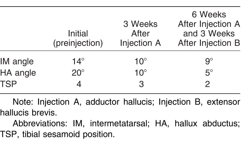
The patient's left foot was then injected with injection B into the EHB muscle after injection A into the oblique and transverse heads of the AdH. Following injection B into the EHB muscle, the patient exhibited an unexpected further reduction in IM angle, HA angle, and TSP of the left foot when compared with injection A alone. As reported in Table 1 on a radiograph of the left foot taken at 42 days after injection A and 21 days after injection B, the patient's IM angle was further reduced to 9°, her HA angle was 5°, and her TSP was 2. That is, after injection B in addition to injection A into the patient's left foot, the patient exhibited further reduction in the deformity than when compared to injection A alone.
Injection B into the EHB alone also provided an unexpected reduction in IM angle, HA angle, and TSP of the great toe. As reported in Table 2 in a radiograph of the right foot obtained before the procedure, the patient's right foot exhibited an IM angle of 13°, an HA angle of 20°, and a TSP of 3. When compared with a radiograph of the right foot obtained 21 days after injection B into the EHB of the patient's right foot, the patient's IM angle in the right foot was reduced to 11°, her HA angle was 12°, and her TSP was 2. That is, following injection B into the EHB alone into the patient's right foot, the patient exhibited reduction in the deformity.
Table 2.
Right Foot Radiographic Angles
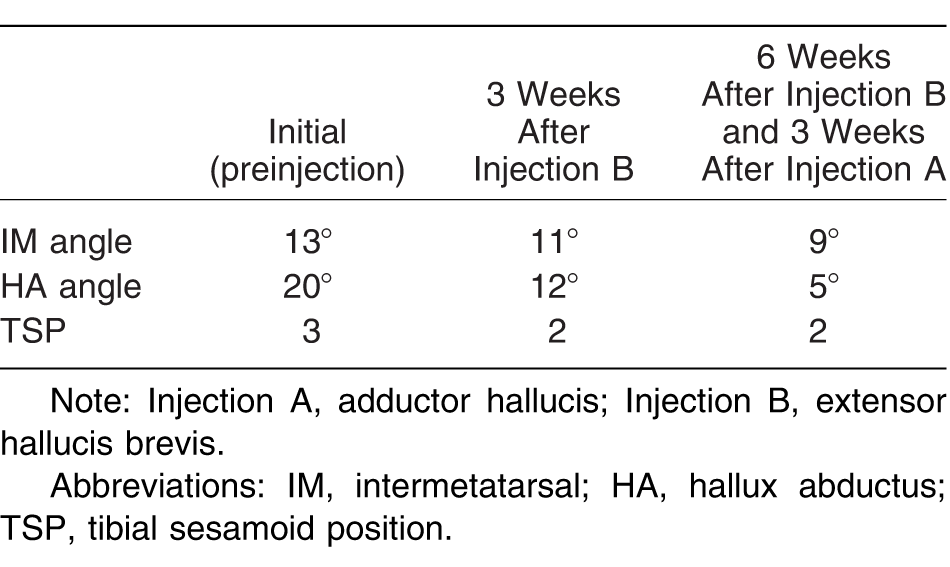
The patient's right foot was then injected with injection A into the oblique and transverse heads of the adductor halluces 3 weeks after injection B into the EHB. Following injection A into the oblique and transverse heads of the AdH, the patient exhibited an unexpected further reduction in IM angle, HA angle, and TSP of the left foot when compared to injection B alone. As reported in Table 2 which lists results for the right foot at 42 days after injection B into the patient's right foot and 21 days after injection A into the patient's right foot, the patient's IM angle was further reduced to 9°, her HA angle was reduced to 5°, and her TSP decreased to 2. That is, after injection A following injection B, the patient exhibited further reduction in the deformity than when compared to injection B alone. The above data in Tables 1 and 2 and Figures 1 through 3 show that injection B is an improvement to the application of injection A in the reduction of the two main radiographic measurement angles and TSP in evaluating severity of the HAV deformity. Of note is that, at 6 weeks, both bunions were pain free as described by the patient, and there was a reduction of the actual valgus rotation of the great toe (paralleling of the great toe nail to the transverse surface plane) noted by the clinician.
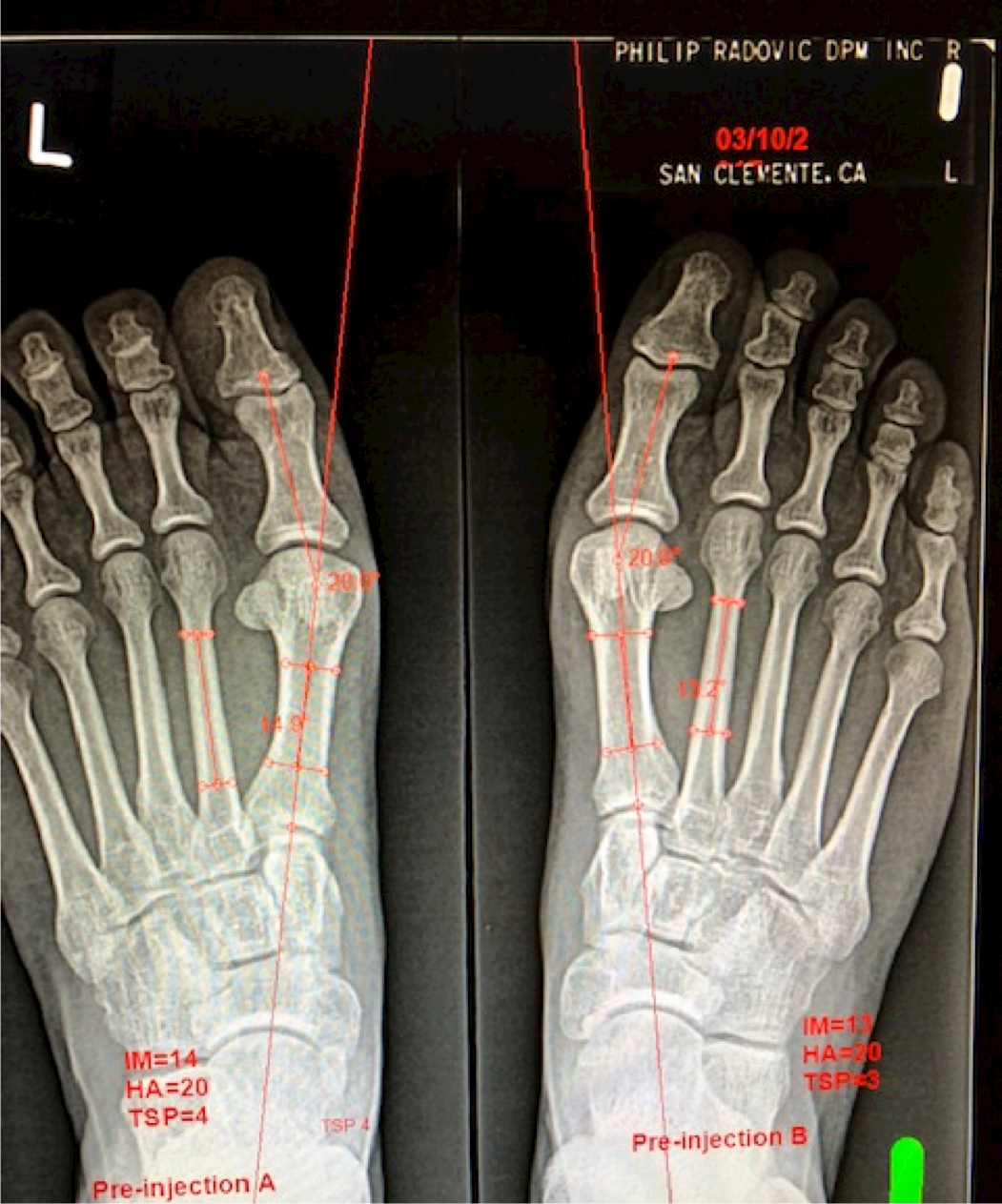
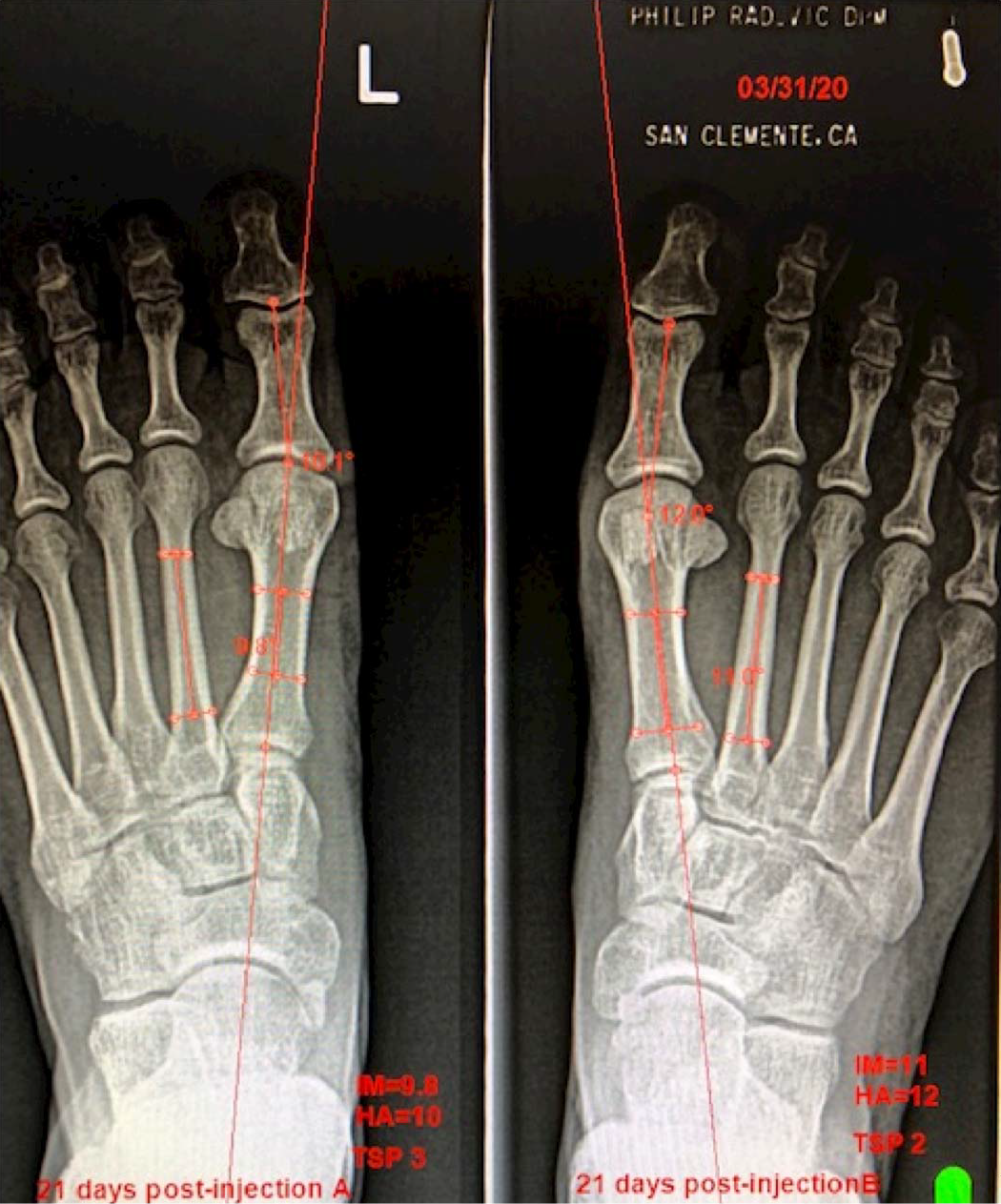
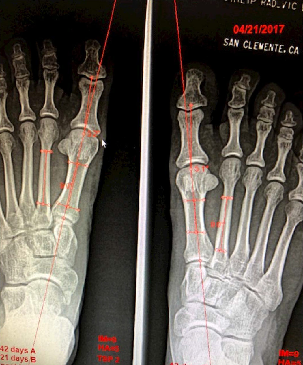
Figure 4.
Before injection A (Table 3).
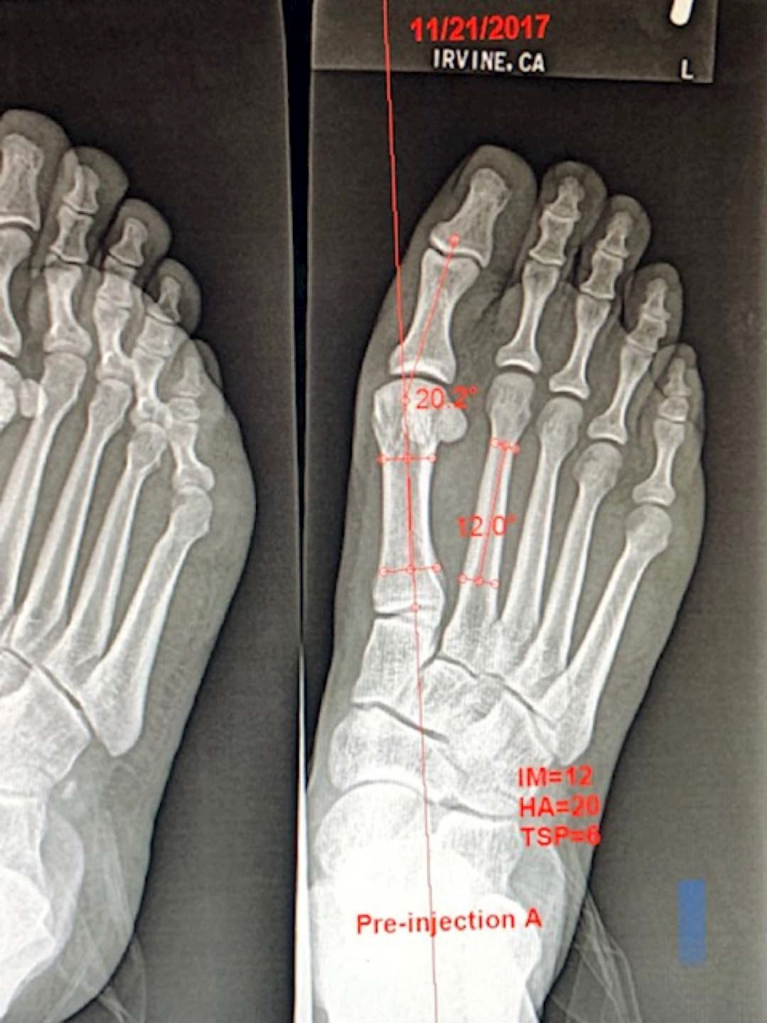
Figure 5.
Twenty-seven days after injection A (Table 3).
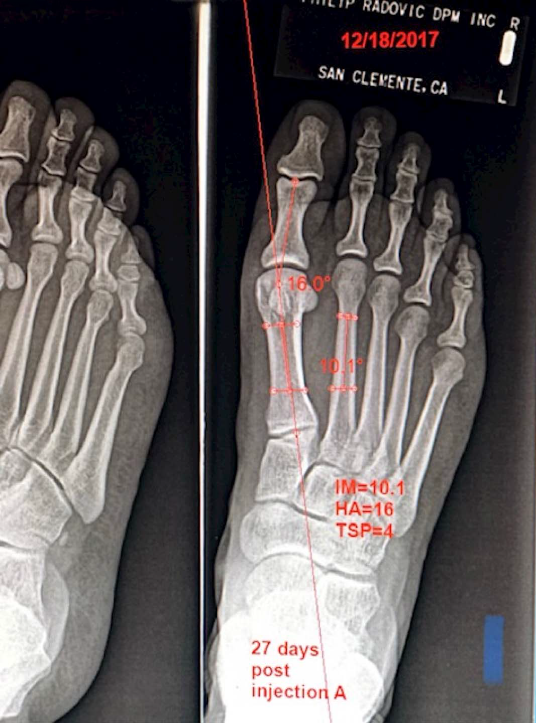
Figure 6.
Forty-three days after injection A (Table 3).
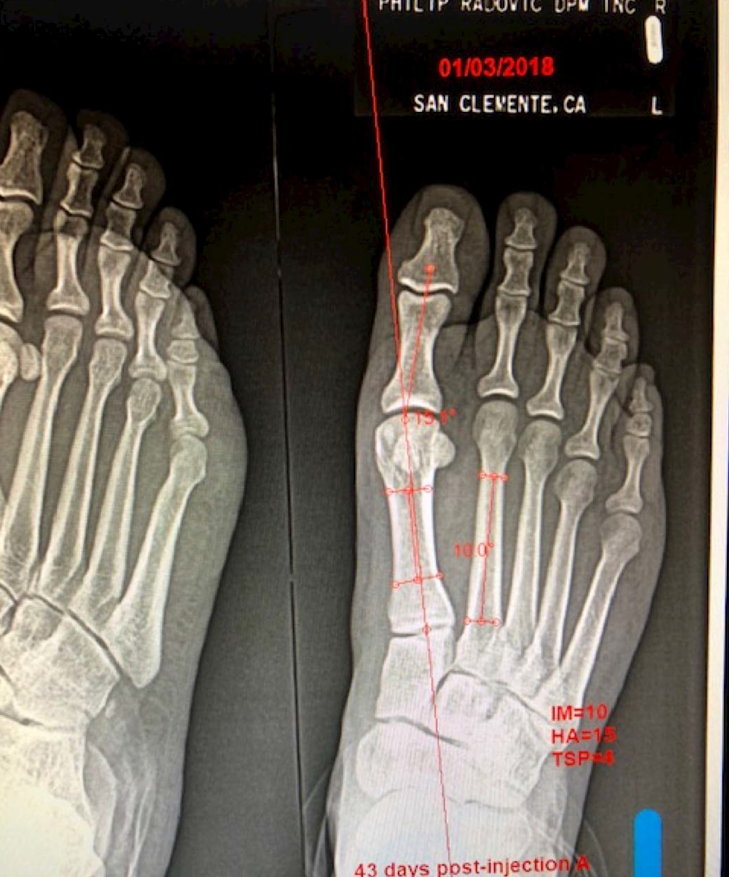
Figure 7.
Nine weeks after injection A (Table 3).
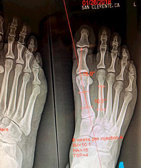
Figure 8.
Before injections A, B, and C (Table 4).
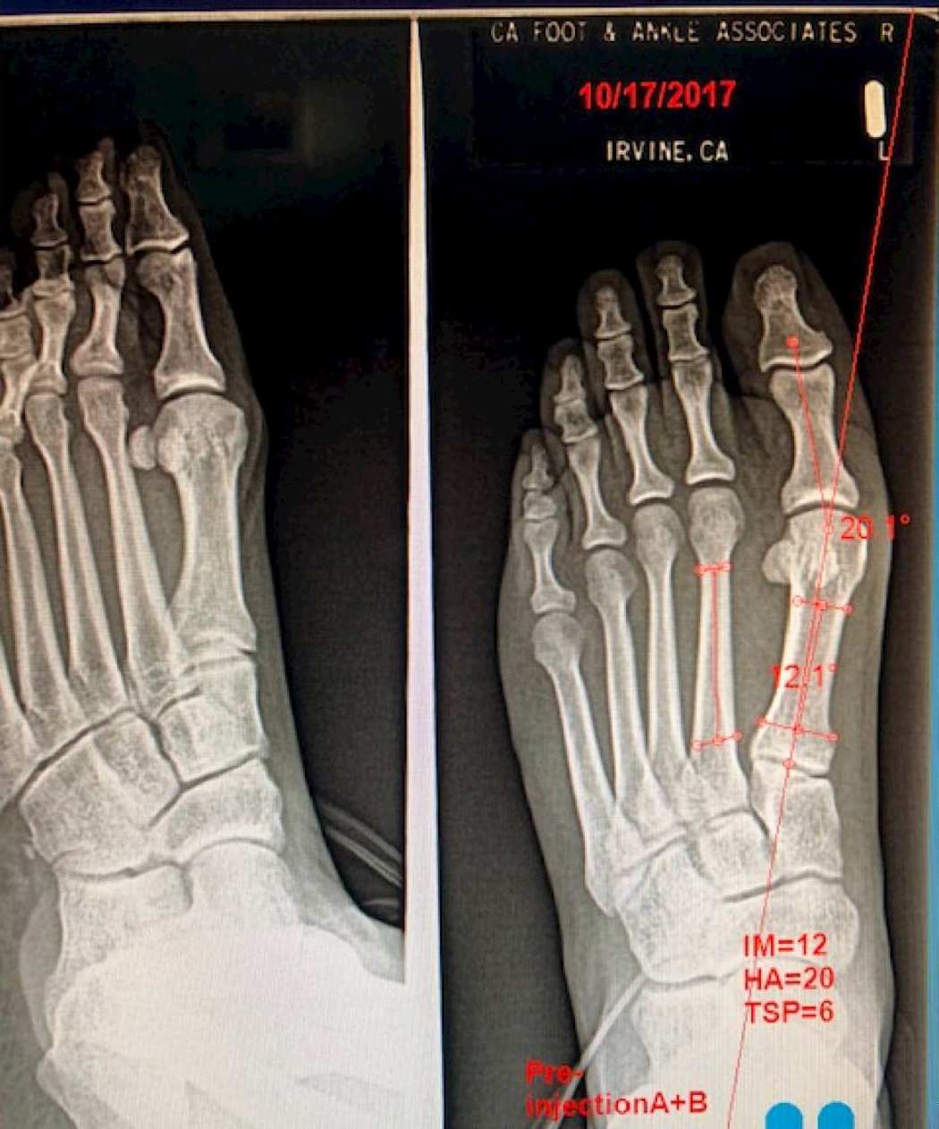
Figure 9.
Twenty-one days after injections A, B, and C (Table 4).
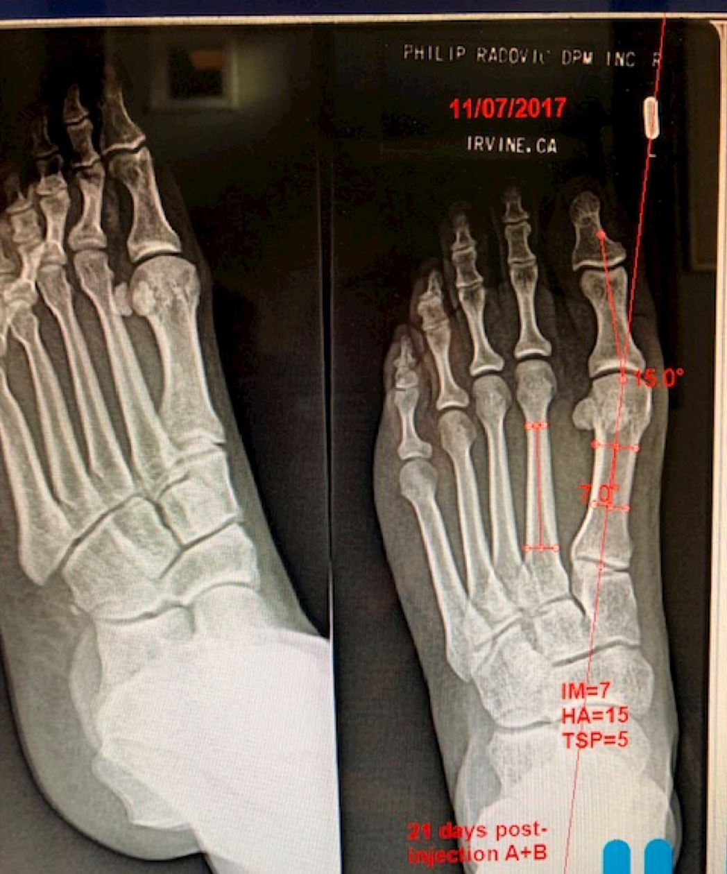
Figure 10.
Thirty-five days after injections A, B, and C (Table 4).
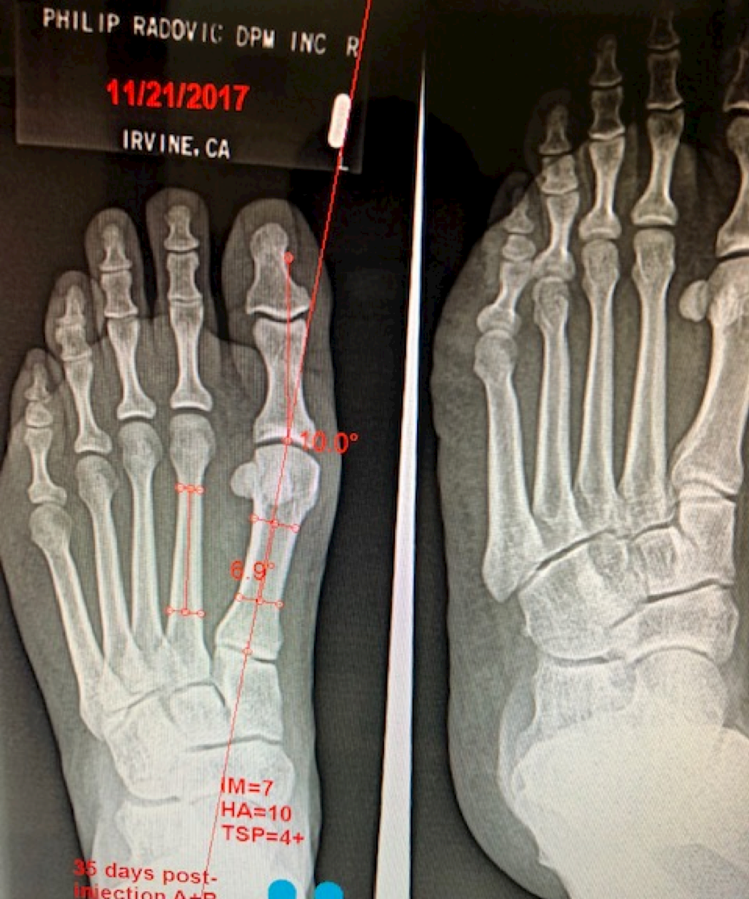
Figure 11.
Sixty-three days after injections A, B, and C (Table 4).
Case 2
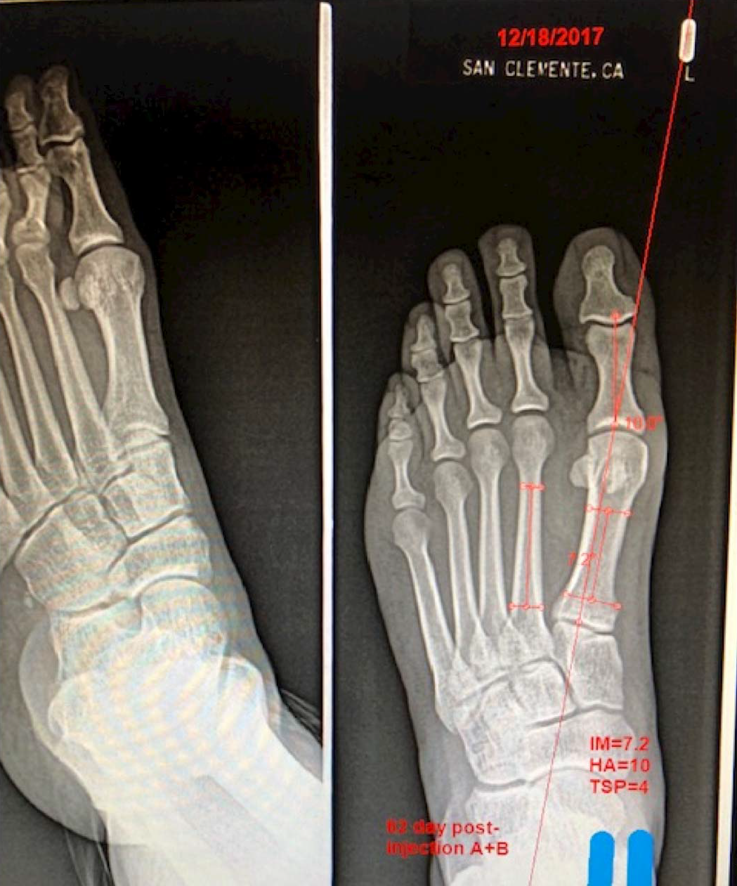
A radiographic comparison study of the HA angles, first IM angles, and TSP was performed on the patient with almost identical bilateral HAV deformities to assess the contribution to reduction of the HAV deformity with EHB injection (injection B) and with injection of the FHB lateral head (injection C) in combination with injection A into the left foot as an improvement/adjunct to the original isolated procedure (injection A) into the right foot. Radiographs of the right foot of the patient were taken before injection, 27 days after injection, 43 days after injection, and 63 days after injection. Radiographs of the left foot of the patient were taken before injection, 21 days after injection, 35 days after injection, and 63 days after injection. All radiographs were positioned and taken by the same technician with the patient in their natural weightbearing angle and base of gait. Comparisons for this study were anteroposterior views at 13° from vertical taken before treatment and then at 2- to 4-week intervals, three times, after each injection. The injections were as follows (immediately after initial radiographs): left foot, 25 U of botulinum toxin type A into the EHB (injection B), 25 U into the lateral head of the FHB (injection C), and 75 U into the oblique and 25 U into the transverse heads of the AdH (injection A); right foot, 75 U into the oblique and 25 U into the transverse heads of the AdH (injection A).
When comparing the reduction of the deformity following injection A into the oblique and transverse heads of the AdH, injection B into the EHB, and injection C into the lateral head of the FHB, the patient exhibited an unexpected reduction in IM angle, HA angle, and TSP when compared to injection A into the oblique and transverse heads of the AdHs alone.
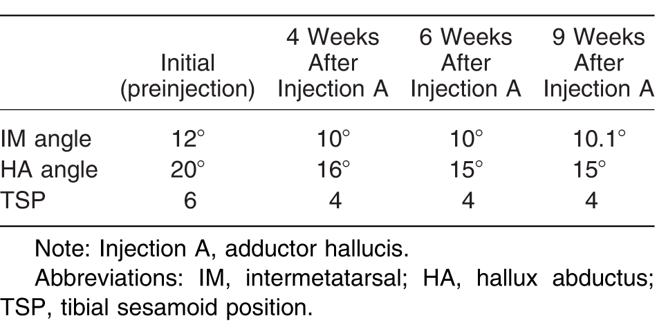
As reported in Table 3 and in a radiograph of the patient's right foot taken before injection A, the patient's initial IM angle was 12°, her HA angle was 20°, and her TSP was 6. At 4 weeks after injection A, the patient exhibited an initial reduction in the deformity. As reported in Table 3 the patient's right foot exhibited an IM angle of 10°, an HA angle of 16°, and a TSP of 4. At 6 weeks after injection A, the patient exhibited further but slight reduction in the deformity. As reported in Table 3 the patient's right foot exhibited an IM angle of 10°, an HA angle of 15°, and a TSP of 4. At 9 weeks after injection A, the patient exhibited little to no further reduction in the deformity. As reported in Table 3 the patient's right foot exhibited an IM angle of 10.1°, an HA angle of 15°, and a TSP of 4. In total, the patient exhibited an IM angle reduction of approximately 2°, an HA angle reduction of 5°, and a reduction of TSP of 2.
Table 3.
Right Foot Radiographic Angles (Injection A Only)
In contrast, the patient exhibited greater deformity reduction of the great toe following injection A into the oblique and transverse heads of the AdH, injection B into the EHB, and injection C into the lateral head of the FHB. As reported in Table 4 in a radiograph of the patient's right foot taken before injections A, B, and C, the patient's initial IM angle was 12°, her HA angle was 20°, and her TSP was 6. At 3 weeks after injections A, B, and C, the patient exhibited an initial reduction in the deformity that was greater than the initial reduction in the deformity of just injection A alone. As reported in Table 4 the patient's right foot exhibited an IM angle of 7 degrees, an HA angle of 15 degrees, and a TSP of 5. When compared to the deformity reduction at 4 weeks after just injection A alone, injections A, B, and C provided an additional reduction of IM angle of 3° and HA angle of 1°. At 5 weeks after injections A, B, and C, the patient exhibited further reduction in the deformity when compared to the results after 3 weeks. As reported in Table 4 the patient's right foot exhibited an IM angle of 7°, an HA angle of 10°, and a TSP of 4+. When compared to the deformity reduction at 6 weeks after just injection A alone, injections A, B, and C provided an additional reduction of IM angle of 3° and HA angle of 5°. Nine weeks after injections A, B, and C, the patient exhibited slight reduction in the deformity when compared to the results after 5 weeks.
Table 4.
Left Foot Radiographic Angles (Injections A, B, and C)
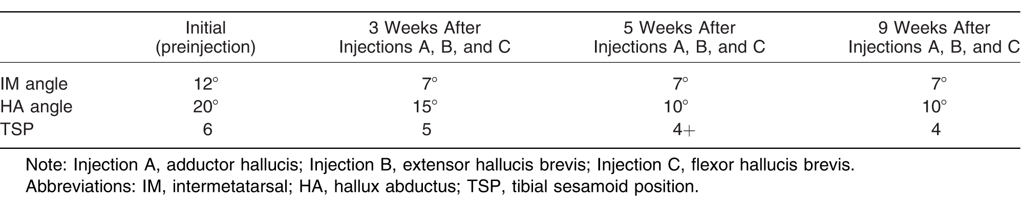
As reported in Table 4 the patient's right foot exhibited an IM angle of 7°, an HA angle of 10°, and a TSP of 4. When compared to the deformity reduction at 9 weeks after injection A alone, injections A, B, and C provided a further reduction of IM angle of 3.1° and HA angle of 5°.
The above data show that injections B and C are an initial and sustainable improvement at 9 weeks to the application of injection A in the reduction of the two main radiographic measurement angles (IM and HA) when compared to injection A alone. Furthermore, injecting the lateral head of the FHB appears to be an improvement to the injection paradigm for treatment of hallux valgus.
Discussion
It is commonly known that certain intrinsic muscle imbalances that act on the first metatarsophalangeal joint can contribute to HAV deformity. It has previously been shown that temporary paralysis of the AdH muscle with botulinum toxin can reduce HAV and its associated pain [7,8]. Modification to the original injection paradigm by including injection paralysis of additional intrinsic muscles that act on the first metatarsophalangeal joint (EHB and FHB lateral head) may show, as in this study, further synergistic reduction of HAV deformity. It is likely that each muscle can have a variable deforming contribution to HAV depending on the individual deformity. It therefore is logical that there would be a synergistic effect when paralysis is induced by means of botulinum toxin injection of those muscles that insert into the lateral aspect of the first metatarsophalangeal joint apparatus. These synergistic effects can be such that the one or more effects of the combination injection are greater than the one or more effects of each injection alone at a comparable dosing level, or they can be greater than the predicted sum of the effects of all of the injections at a comparable dosing level, assuming that each injection acts independently. However, further studies with a larger patient population with well-defined inclusion/exclusion criteria clearly need to be performed, and one must consider factors not limited to degree of deformity, flexibility, reducibility, congruity of the joint, arthritic changes, proximal articular set angle, and protrusion to be of significant influence on the amenability of intrinsic muscle paralysis to reduce HAV and its associated pain. Although paralytic efficacy of botulinum toxin is commonly known to be approximately 3 to 6 months, its use for HAV treatment may be valuable as a periodic or serial application. Clearly, a higher level study is necessary to confirm the viability of this interesting treatment for the general population on this very common foot deformity.
Conclusions
Treatment of HAV with botulinum toxin injection has evolved as a potentially effective modality, as shown in several subsequent international studies in addition to the original description published in this journal in 2008. A potential improvement to the injection paradigm developed by this author has been shown to possibly be more effective in reducing the HAV deformity and its associated pain.
Financial Disclosure:
None reported.
Conflict of Interest:
None reported.
References
- Hambleton, P.; Moore, AP. “Botulinum Neurotoxins: Origin, Structure, Molecular Actions and Antibodies,”. in. Handbook of Botulinum Toxin Treatment edited by. 1995. [Google Scholar]
- Simpson, LL. “Current Concepts on the Mechanism of Action of Clostridial Neurotoxins,”. in. Botulinum and Tetanus Neurotoxins edited by. 1993. [Google Scholar]
- Biglan, AW.; Burnstine, RA.; Rogers, GL.; et al. Management of strabismus with botulinum toxin A. Ophthalmology 1989, 96, 935. [Google Scholar]
- Suputtitada, A. Local botulinum toxin type A injections in the treatment of spastic toes. Am J Phys Med Rehabil 2002, 81, 770. [Google Scholar]
- Tamura, BM.; Cuce, LC.; Souza, RL.; et al. Plantar hyperhidrosis and pitted keratolysis treated with botulinum toxin injection. Dermatol Surg 2004, 30, 1510. [Google Scholar]
- Babcock, MS.; Foster, L.; Pasquina, P.; et al. Treatment of pain attributed to plantar fasciitis with botulinum toxin a: a short-term, randomized, placebo-controlled, double-blind study. Am J Phys Med Rehabil 2005, 84, 649. [Google Scholar]
- Radovic, P.; Shah, E. Nonsurgical treatment for hallux abducto valgus with botulinum toxin. JAPMA 2008, 98, A1. [Google Scholar]
- Wu, KP.; Chen, CK.; Lin, SC.; et al. Botulinum toxin type A injections for patients with painful hallux valgus: a double-blind, randomized controlled study. Clin Neurol Neurosurg 2015, 129((suppl 1)), S58. [Google Scholar]
- Tang; Chen, JT.; Chen, CK.; et al. Effective conservative treatment for managing painful hallux valgus. Med Res Arch 2020. 4: October 2016. Available at https://journals.ke-i.org/mra/article/view/685 Accessed March 10,.
- Moghtaderi, A.; Dehghan, F.; Mousavizadeh, A.; et al. Evaluation of the therapeutic effect of botulinum toxin A on hallux valgus deformity and pain. Int J Phys Med Rehabil 2017, 5, 438. [Google Scholar]
- Bhidayasiri, R.; Truong, DD. Expanding use of botulinum toxin. J Neurol Sci 2005, 235, 1. [Google Scholar]
- Placzek, R.; Sohling, M.; Gessler, M.; et al. Botulinum toxin A in orthopedic pain therapy. Schmerz 2004, 18, 498. [Google Scholar]
- Aoki, KR. Evidence for antinociceptive activity for botulinum toxin type A in pain management. Headache 2003, 43((suppl 1)), S9. [Google Scholar]
- Arinci, I.; Genc, H.; Erdem, HR.; et al. Muscle imbalance in hallux valgus: an electromyographic study. Am J Phys Med Rehabil 2003, 82, 345. [Google Scholar]
- Hoffmeyer, P.; Cox, JN.; Blanc, Y.; et al. Muscle in hallux valgus. Clin Orthop Relat Res 1988, 232, 112. [Google Scholar]
- Coughlin, MJ.; Mann, RA.; Saltzman, CL. Surgery of the Foot and Ankle 8th Ed, Mosby Elsevier, Philadelphia, PA,. 2007.
- Sherman, A.; Willick, S.; Cardenas, D. Management of focal dystonia of extensor hallucis longus muscle with botulinum toxin injection: a case report. Arch Phys Med Rehabil 1998, 79, 1303. [Google Scholar]
© 2020 American Podiatric Medical Association