


Abstract
Background: In the presence of a large gap where end-to-end repair of the torn Achilles tendon is difficult and V-Y advancement would likely be insufficient, augmentation is sometimes required. At our institute we have used primarily the hamstring autograft augmentation technique for the past two decades. The aim of this study was to analyze the complications after surgical treatment of Achilles tendon rupture with semitendinous tendon augmentation. Methods: We retrospectively analyzed 58 consecutive patients treated with semitendinous tendon autograft augmentation at the Helsinki University Hospital between January 1, 2006, and January 1, 2016. Results: During the study period, 58 patients were operated on by six different surgeons. Of 14 observed complications (24%), seven were major and seven were minor. Most of the complications were infections (n = 10 [71%]) The infections were noted within a mean of 62 days postoperatively (range, 22–180 days). Seven patients with a complication underwent repeated operation because of skin edge necrosis and deep infection (five patients), hematoma formation (one patient), and a repeated rupture (one patient). Conclusions: In light of the experience we have had with autologous semitendinous tendon graft augmentation, we cannot recommend this technique, and, hence, we should abandon reconstruction of Achilles tendon ruptures with autologous semitendinous tendon grafts at our institute. Instead, other augmentation techniques, such as flexor hallucis longus tendon transfer, should be used.
In the presence of a large gap, where end-to-end repair of the torn Achilles tendon is difficult and V-Y advancement would likely be insufficient, augmentation is sometimes required. However, augmentation has quite often seemed to be associated with postoperative complications, especially surgical site infections. Therefore, we decided to analyze further complications associated with semitendinous tendon autograft augmentation. The technique is very similar to that described by Maffulli[1] in which a semitendinous tendon graft is used to bridge the ruptured ends of the Achilles tendon.
Several other augmentation techniques and grafts have been presented in the literature. The defects can be reconstructed using allografts, fascial augmentation, or flexor hallucis longus (FHL), flexor digitorum longus, or peroneus brevis tendon transfer[2-7] with the common aims of restoring the Achilles tendon continuity, allowing the tendon to heal in physiologic position, and restoring muscle function. None of these techniques have shown clear superiority; however, during the past decade, flexor hallucis longus tendon grafts have become widespread in ankle surgery.[7]
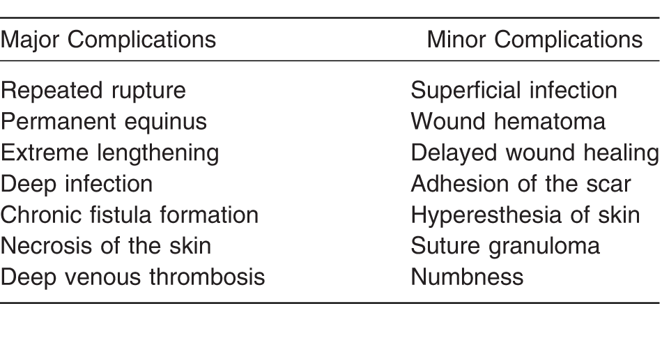
The complications after primary suturation of a complete Achilles tendon rupture are well described in the literature and have been divided into major and minor categories by Paavola et al[8] and Cetti et al.[9] The complication rate after primary suturation of Achilles tendon rupture has varied from 0% to 18%.[8-12] There are only a few studies of complications of augmented Achilles repairs.[13-18] Furthermore, the complication rate after Achilles tendon rupture repair with semitendinous tendon grafts is unknown. To our knowledge, the use of an autologous semitendinous tendon has been reported in only three studies.[10,19,20] The aim of the present study was to provide more clarity on the effects of surgically treating Achilles tendon ruptures with semitendinous augmentation by analyzing the complication rate in 58 consecutive patients.
Materials and Methods
All of the patients treated with semitendinous tendon autograft augmentation at Helsinki University Hospital (Helsinki, Finland) between January 1, 2006, and January 1, 2016, were analyzed. Patient identification was performed from full medical record reviews of all patients treated for Achilles tendon rupture or Achilles tendinosis (International Classification of Diseases, 10th Revision diagnosis codes S86.0 and M76.6) and suture or reinsertion of Achilles tendon (NOMESCO [Nordic Medico-Statistical Committee] Classification of Surgical Procedures code NHL10) at the Helsinki University Hospital's orthopedic unit. Data concerning surgical technique were obtained by reviewing the surgery reports. The presented surgical method was used for 58 patients during the 10-year analysis period. Hospital records of these 58 patients were retrospectively reviewed to register demographic data, comorbidities, length of the defect, previous local corticosteroid injections, heparin injections, postoperative regimen, and possible complications.
The patients were divided in five groups according to the diagnosis leading to semitendinous augmentation: primary rupture, repeated rupture after operative treatment, neglected/misdiagnosed rupture, failed conservative treatment, and chronic rupture. In the present study, neglected/misdiagnosed rupture was defined as a diagnosis occurring 2 or more weeks after the injury. Failed conservative treatment was defined as extreme lengthening of the Achilles tendon or repeated rupture after conservative treatment. Chronic injury was defined as magnetic resonance imaging–diagnosed total rupture without injury and with chronic Achilles tendinitis. Complications were classified as major and minor (Table 1).
Table 1.
Complications After Augmentation and Suturation of a Complete Achilles Tendon Rupture.
The study was approved by Helsinki University's ethics committee.
Surgical Technique
A standardized operative and postoperative protocol was used during the study period. The Helsinki University Hospital has obliged at least 3 months between the last cortisone injection and surgical treatment with the idea of preventing postoperative infection. All of the patients received perioperative antibiotics and postoperative anticoagulant treatment. All of the patients underwent preoperative magnetic resonance imaging, where the gap size was estimated.
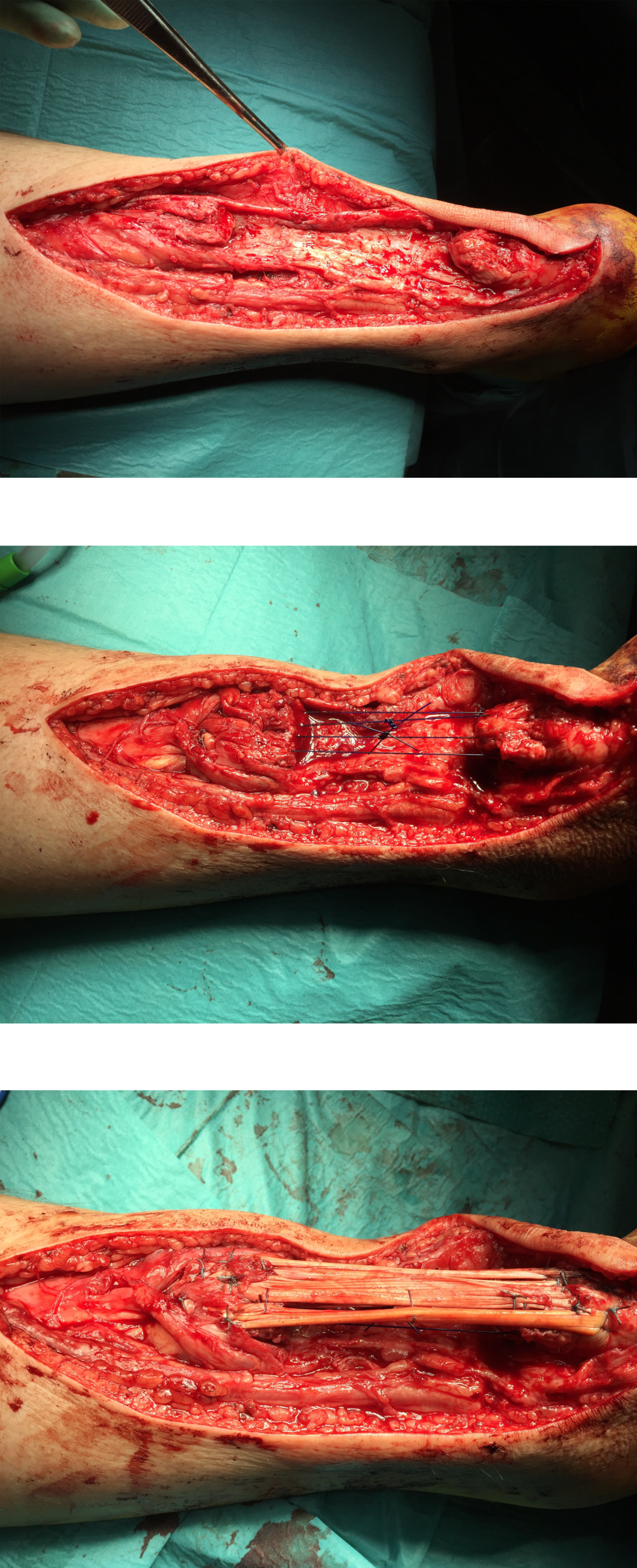
With the patient prone, a posteromedial longitudinal lazy-S–shaped incision is made in the posterior area of the ankle joint. The length of the incision is approximately 8 to 10 cm, depending on the exposure required for retracted tendon ends. In the case of tendinopathy, tendon ends are debrided, thereby creating the final defect. After thorough tendon debridement, excessive mobilization of tendon ends by soft-tissue dissection is avoided. Traction on the proximal stump is performed manually by two 1-0 Kessler loops (PDS; Ethicon, Somerville, New Jersey) (Fig. 1).
Figure 1.
The surgical technique. A, Tendon ends are debrided, thereby creating the final defect. B, Traction on the proximal stump is performed manually by two 1-0 PDS Kessler loops. C, The harvested semitendinous tendon is passed through the revised Achilles tendon ends, creating a three-time loop.
With the knee flexed in the prone position, the semitendinous tendon, and the gracilis tendon if needed, is harvested through a 3-cm incision medial to the tibial tuberosity. The length of the graft obtained is in the range of 20 to 30 cm. The semitendinous tendon is prepared by shaving off the muscular part from the tendon. Whip sutures are placed on a length of approximately 3 cm of the tendon on either side.
The harvested semitendinous tendon is passed through the revised Achilles tendon ends, creating a three-time loop, if graft length allows. The graft is sutured firmly onto the original tendon with Ti-Cron (Medtronic, Minneapolis, Minnesota) or PDS sutures. Subsequently, to maximize the fixation, the graft is sutured with additional inverted 3-0 PDS sutures.
The paratenon is closed with 3-0 PDS sutures, subcutaneous tissue is closed with 2-0 Vicryl sutures (Ethicon Inc), and skin closure is performed with 3-0 Ethilon sutures (Ethicon Inc).
Postoperative full weightbearing is not allowed. After 2 weeks, the skin sutures are removed, and the equinus cast is removed and changed to a Walker boot with a heel raise. Physiotherapy is commenced with limited dorsiflexion of the ankle, focusing on proprioception, plantarflexion of the ankle, inversion, and eversion. During this period of rehabilitation, the patient is permitted to bear half of his or her weight as comfort allows with a Walker boot. After 6 weeks the heel raise is removed and full weightbearing is allowed. The Walker boot is removed 12 weeks after surgery.
Statistical Analysis
The results are presented as medians and means ± SD for continuous nonskewed variables. The frequency distribution of the categorical variables is compared between groups with the χ2 test. Statistical significance is set as P < .05. Correlations between continuous parameters were assessed by using Pearson correlation analysis. Binary logistic regression analysis was performed to determine which parameter (age, length of the defect, use of local corticosteroids, and other comorbidities) was of greatest and independent significance for the prediction of complication. A statistical software program (IBM SPSS Statistics for Windows, Version 22.0; IBM Corp, Armonk, New York) was used for the analyses.
Results
During the study period, 58 patients (21 women and 37 men) were operated on by six surgeons (one orthopedic fellow and five consultants). Median (range) patient demographic characteristics are as follows: age, 47 years (33–72 years); body mass index (the weight in kilograms divided by the square of the height in meters), 25 (21–40); defect size, 3.9 cm (2–9 cm); and comorbidities, 8 (14%) (colitis ulcerosa, myeloma, atherosclerosis of the lower extremities, systemic lupus erythematosus, paroxysmal atrial fibrillation, Parkinson disease, asthma, and ankylosing spondylitis). The median number of operations performed by a single surgeon was four (range, 1–27). Overall, patients were considered healthy, with 50 (86%) reporting no underlying diseases. The remaining eight patients (14%) reported an underlying health concern that could be a contributing factor to the overall outcome. Only one patient reported a history of smoking. None of the patients reported current smoking. Before the diagnosed rupture, 22 patients (38%) had received local cortisone. These patients had a mean of 4.5 months (range, 3–13 months) between the last cortisone injection and surgery. Six patients had systemic cortisone treatment.
The most common reason for operation was chronic total rupture (n = 23, 39%), followed by neglected/misdiagnosed rupture (n = 15, 26%), failed conservative treatment (n = 11, 19%), repeated rupture (n = 8, 14%), and a single case of primary rupture (2%).
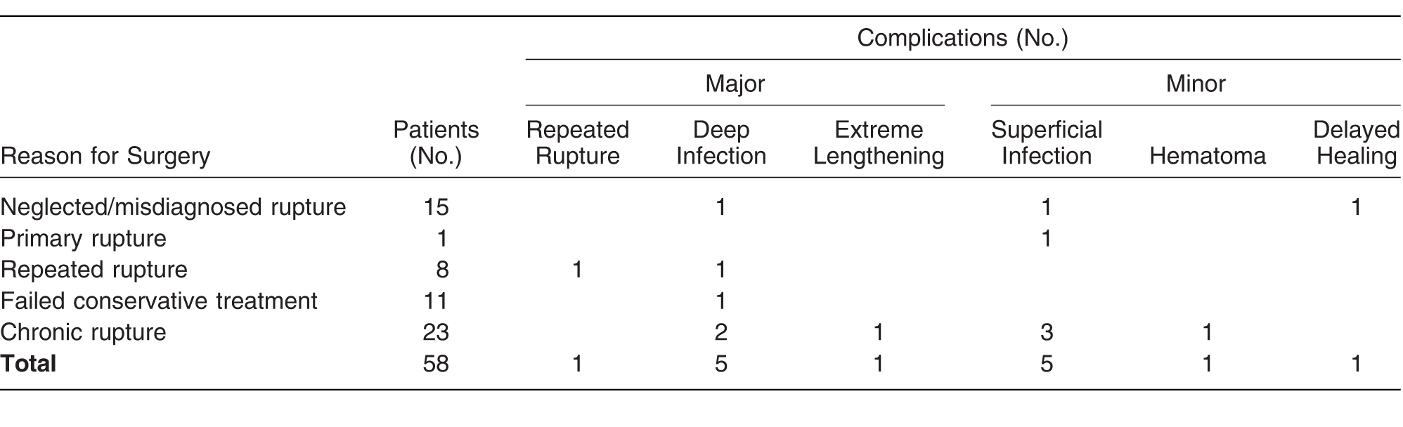
Complications were observed in 14 of the 58 patients (24%) (Table 2). Surgical site infection was the most common complication (five deep infections and five superficial infections). In the patients without complications (n = 44, 76%), follow-up was uneventful, and all were able to heel raise at the final follow-up at a mean of 5.9 months (range, 5–6 months) after injury. Patients without any complications had a mean of 4.6 clinical follow-up visits (range, 3–6). Seven patients with a complication underwent repeated operation because of skin edge necrosis and deep infection (n = 5), hematoma formation (n = 1), and a repeated rupture (n = 1).
Table 2.
Complications in 58 Patients Treated with Semitendinous Tendon Autograft Augmentation.
Major Complications
All of the patients with major complications required several revision operations (mean, 5; range, 4–6). Deep infection and skin edge necrosis was seen in five patients, of which all needed several repeated operations and negative pressure wound therapy (NPWT). The mean duration of NPWT was 25.5 days (range, 7–49 days). Two of the patients required skin transplant, and two required a microvascular flap. Both of the patients with a microvascular flap had first failed skin transplant. The infections were noted within a mean of 62 days postoperatively (range, 22–180 days). Due to the infection, surgical removal of the Achilles tendon had to be performed on two patients, and one was partially removed. One patient had a repeated rupture 5 months after the primary operation with semitendinous tendon autograft and experienced a deep infection after the second operation. One patient experienced extreme lengthening of the Achilles tendon postoperatively, which made plantarflexion absent.
Minor Complications
Postoperative superficial wound infection was seen in five patients. One of these patients had a wound hematoma in addition to the wound infection and required surgical evacuation of the hematoma. All of the patients were treated with oral antibiotic drugs and local treatment (irrigation and antiseptic agents). None of the superficial infections needed repeated operation or NPWT. The infection was noted within a mean of 37 days postoperatively (range, 6–60 days). One patient had delayed wound healing. The wound was closed after 32 days with help from local treatment and NPWT.
Patients who had systemic cortisone treatment were more likely to experience infection (7% versus 3%; P > .006). Age, sex, defect size, surgery indication, surgeon's experience, use of local corticosteroids, or other comorbidities showed no statistically significant correlation with complications.
Discussion
The complication rate associated with surgical treatment of Achilles tendon rupture with semitendinous augmentation in our institute is higher than previously reported with other methods of augmented Achilles rupture repair.[7,10,20,21] To our knowledge, the literature on semitendinous augmentation is limited, and we found only one case series[10] with 35 patients where no complications were noted.
Simple end-to-end suture is a safe and reliable treatment with low risk of complications,[22] and augmentation has not been found to provide any benefit compared with simple suture repair of Achilles tendon ruptures.[21,23] The neglected or chronically ruptured Achilles tendon presents a unique treatment challenge when a large gap is present and end-to-end sutures are challenging. In the present series, we reconstructed defects ranging from 2 to 9 cm. The advantage with using the semitendinous graft can be that the degenerated Achilles tendon can be debrided comprehensively without concern regarding the length of the defect that may be created, considering that the torn ends of the tendons are already unhealthy[24] and the healing capacity of the injured tendon is further limited due to hypovascularity at the midsection of the tendon.[25,26] Furthermore, with this technique, the Achilles tendon ends do not need to be mobilized by extensive soft-tissue dissection.
In previous studies, when using gastrocnemius flaps the complication rates have been 8% to 24%.[6,22,23,27,28] Mann et al[13] reported good or excellent results with flexor digitorum longus tendon and with a proximal fascial turndown flap. Wapner et al,[14] Den Hartog,[15] and Coull et al[17] reported good or excellent results on FHL tendon transfers for neglected Achilles tendon ruptures with minimal complications. Tawari et al[18] reported good or excellent results in 85% of patients with the use of peroneus brevis tendon but with a complication rate of 20%. Unfortunately, the complications have often been reported in a rather general way in most previous studies.
In the present series, using comprehensive criteria for documenting complications, we noted a 24% overall complication rate and a 17% infection rate. In the present study, problems related to wound healing (skin necrosis and superficial wound infection) were the most common complications. Both the superficial and deep infections were noted several weeks after surgery, and in one patient 180 days after surgery. The reason behind this remains unknown. It has been reported in earlier studies that augmentation can cause delayed wound healing due to increased tendon diameter and impaired local circulation of the skin.[1,8,23,29] We can speculate that avascular semitendinous graft might contribute to the even higher infection rate than in previously published studies with different augmentation techniques. It is possible that with local tendon transfers there is a potential benefit of local vascularity and potential additional strength.
In the present study, the only statistically significant factor for prediction of complication was the use of systemic cortisone. Impairment in wound healing is common and is potentially a serious adverse effect of systemic corticosteroid use. Corticosteroids interfere with the natural wound-healing process by inhibiting leukocyte and macrophage infiltration, decreasing collagen synthesis and wound maturation, and reducing keratinocyte growth factor expression after skin injury.[30-32]
Local cortisone injections did not have a statistically significant effect on infections. The effect of and possible complications, such as rupture and infection, related to corticosteroid injection in the treatment of Achilles tendinopathy is controversial,[33-37] but several authors state that the risk of rupture after local corticosteroid injections is real.[38-42]
The major limitations of this study are its retrospectivity and small study population. Statistical analysis was difficult due to the small number of patients, which makes beta error possible. Nonrandomization made it impossible to compare different augmentation techniques and possible outcomes and complications. We do not possess any information on long-term functional outcomes in patients with semitendinous augmentation or long-term outcomes in patients with major complications.
In light of the experience we have had with autologous semitendinous tendon graft augmentation, we cannot recommend further use of this technique. We should abandon reconstruction of Achilles tendon ruptures with autologous semitendinous tendon grafts at our institute. Instead, other augmentation techniques, such as FHL tendon transfer, should be used.
Financial Disclosure
None reported.
Conflict of Interest
None reported.
References
- MaffulliN.Augmented repair of acute Achilles tendon ruptures using gastrocnemius-soleus fascia. Int Orthop29: 134, 2005.
- BosworthDM.Repair of defects in the tendo achillis. J Bone Joint Surg Am38: 111, 1956.
- LiebermanJR.LozmanJ.CzajkaJ.: Repair of Achilles tendon ruptures with Dacron vascular graft. Clin Orthop Relat Res234: 204, 1988.
- AbrahamE.PankovichAM.Neglected rupture of the Achilles tendon: treatment by V-Y tendinous flap. J Bone Joint Surg Am57: 253, 1975.
- OzakiJ.FujikiJ.SugimotoK.: Reconstruction of neglected Achilles tendon rupture with Marlex mesh. Clin Orthop Relat Res238: 204, 1989.
- TakaoM.OchiM.NaitoK.: Repair of neglected Achilles tendon rupture using gastrocnemius fascial flaps. Arch Orthop Trauma Surg123: 471, 2003.
- MaffulliN.LeadbetterWB.Free gracilis tendon graft in neglected tears of the Achilles tendon. Clin J Sport Med15: 56, 2005.
- PaavolaM.OravaS.LeppilahtiJ.: Chronic Achilles tendon overuse injury: complications after surgical treatment: an analysis of 432 consecutive patients. Am J Sports Med28: 77, 2000.
- CettiR.ChristensenS-E.EjstedR.: Operative versus nonoperative treatment of Achilles tendon rupture. Am J Sports Med21: 791, 2016.
- Dumbre PatilSS.Dumbre PatilVS.BasaVR.et al: Semitendinous tendon autograft for reconstruction of large defects in chronic Achilles tendon ruptures. Foot Ankle Int35: 699, 2014.
- PintoreE.BarraV.PintoreR.: Peroneus brevis tendon transfer in neglected tears of the Achilles tendon. J Trauma Acute Care Surg50: 71, 2001.
- WilliamsJGP.Achilles tendon lesions in sport. Sports Med3: 114, 1986.
- MannRA.HolmesGB.SealeKS.: Chronic rupture of the Achilles tendon: a new technique of repair. J Bone Joint Surg Am73: 214, 1991.
- WapnerKL.PavlockGS.HechtPJ.: Repair of chronic Achilles tendon rupture with flexor hallucis longus tendon transfer. Foot Ankle14: 443, 2016.
- Den HartogBD.Flexor hallucis longus transfer for chronic Achilles tendonosis. Foot Ankle Int24: 233, 2003.
- WegrzynJ.LucianiJ-F.PhilippotR.: Chronic Achilles tendon rupture reconstruction using a modified flexor hallucis longus transfer. Int Orthop34: 1187, 2010.
- CoullR.FlavinR.StephensMM.Flexor hallucis longus tendon transfer: evaluation of postoperative morbidity. Foot Ankle Int24: 931, 2003.
- TawariAA.DhamangaonkarAA.GoregaonkarAB.: Augmented repair of degenerative tears of tendo achilles using peroneus brevis tendon: early results. Malays Orthop J7: 19, 2013.
- MaffulliN.LongoUG.GougouliasN.: Ipsilateral free semitendinous tendon graft transfer for reconstruction of chronic tears of the Achilles tendon. BMC Musculoskelet Disord9: 100, 2008.
- JiJH.KimWY.KimYY.: Semitendinous tendon augmentation for a large defect after Achilles tendon rupture: two case reports. Foot Ankle Int28: 1100, 2016.
- ZhangY-J.ZhangC.WangQ.: Augmented versus nonaugmented repair of acute Achilles tendon rupture: a systematic review and meta-analysis. Am J Sports Med46: 1767, 2018.
- NyyssönenT.SaarikoskiH.KaukonenJ-P.: Simple end-to-end suture versus augmented repair in acute Achilles tendon ruptures: a retrospective comparison in 98 patients. Acta Orthop Scand74: 206, 2003.
- HeikkinenJ.LanttoI.FlinkkiläT.: Augmented compared with nonaugmented surgical repair after total Achilles rupture: results of a prospective randomized trial with thirteen or more years of follow-up. J Bone Joint Surg Am98: 85, 2016.
- KannusP.JózsaL.Histopathological changes preceding spontaneous rupture of a tendon: a controlled study of 891 patients. J Bone Joint Surg Am73: 1507, 1991.
- LagergrenC.LindholmA.Vascular distribution in the Achilles tendon: an angiographic and microangiographic study. Acta Chir Scand116: 491, 1959.
- AhmedIM.LagopoulosM.McConnellP.: Blood supply of the Achilles tendon. J Orthop Res16: 591, 1998.
- ZellRA.SantoroVM.Augmented repair of acute Achilles tendon ruptures. Foot Ankle Int21: 469, 2000.
- GarabitoA.Martinez-MirandaJ.Sanchez-SoteloJ.Augmented repair of acute Achilles tendon ruptures using gastrocnemius-soleus fascia. Int Orthop29: 42, 2005.
- HaertschPA.The blood supply to the skin of the leg: a post-mortem investigation. Br J Plast Surg34: 470, 1981.
- LiuD.AhmetA.WardL.: A practical guide to the monitoring and management of the complications of systemic corticosteroid therapy. Allergy Asthma Clin Immunol9: 30, 2013.
- SchellenbergR.AdachiJDR.BowieD.: Oral corticosteroids in asthma: a review of benefits and risks. Can Respir J14 (suppl C): 1C,2007.
- PoetkerDM.RehDD.A comprehensive review of the adverse effects of systemic corticosteroids. Otolaryngol Clin North Am43: 753, 2010.
- WetkeE.JohannsenF.LangbergH.Achilles tendinopathy: a prospective study on the effect of active rehabilitation and steroid injections in a clinical setting. Scand J Med Sci Sports25: e392, 2015.
- CoombesBK.BissetL.VicenzinoB.Efficacy and safety of corticosteroid injections and other injections for management of tendinopathy: a systematic review of randomised controlled trials. Lancet376: 1751, 2010.
- FredbergU.BolvigL.Pfeiffer JensenM.: Ultrasonography as a tool for diagnosis, guidance of local steroid injection and, together with pressure algometry, monitoring of the treatment of athletes with chronic jumper's knee and Achilles tendinitis: a randomized, double-blind, placebo-controlled study. Scand J Rheumatol33: 94, 2009.
- PaavolaM.KannusP.JärvinenTA.: Treatment of the tendon disorders: is there a role for corticosteroid injection?Foot Ankle Clin7: 501, 2002.
- ShrierI.MathesonGO.KohlHW.Achilles tendonitis: are corticosteroid injections useful or harmful?Clin J Sport Med6: 245, 1996.
- NicholsAW.Complications associated with the use of corticosteroids in the treatment of athletic injuries. Clin J Sport Med15: E370, 2005.
- JonesJG.Achilles tendon rupture following steroid injection. J Bone Joint Surg Am67: 170, 1985.
- HugateR.PennypackerJ.SaundersM.: The effects of intratendinous and retrocalcaneal intrabursal injections of corticosteroid on the biomechanical properties of rabbit Achilles tendons. J Bone Joint Surg Am86: 794, 2004.
- MutoT.KokubuT.MifuneY.: Temporary inductions of matrix metalloprotease-3 (MMP-3) expression and cell apoptosis are associated with tendon degeneration or rupture after corticosteroid injection. J Orthop Res32: 1297, 2014.
- KapetanosG.The effect of the local corticosteroids on the healing and biomechanical properties of the partially injured tendon. Clin Orthop Relat Res163: 170, 1982.
© 2020 American Podiatric Medical Association