


Abstract
Hallux varus is most commonly seen iatrogenically following overaggressive lateral release, removal of the fibular sesamoid, or overaggressive removal of the medial eminence. There are several reported cases of traumatic hallux varus, although this is much less common. We present a case of traumatic hallux varus in a patient who was later found to have bilateral absence of her fibular sesamoids. We postulated that lack of her fibular sesamoid led to weakness of her lateral capsular ligaments, thereby making her more susceptible to this injury. We performed a repair using a split extensor hallucis longus tendon transfer that was transected proximally, rerouted the tendon under the deep transverse intermetatarsal ligament, and secured it to the first metatarsal with a Bio-Tenodesis (Arthrex, Inc, Naples, Florida) screw. At 22 months postoperatively, she has demonstrated maintenance of correction and has resumed use of normal shoe gear and participation in activities. Our goal was to demonstrate a repair for this condition that successfully maintained correction over time while still allowing for functionality of the first metatarsophalangeal joint.
Hallux varus is a deformity of the forefoot and is described as a triplane deformity with medially deviated hallux with varus rotation and dorsal contraction of the interphalangeal joint.[1-3] This is most commonly caused iatrogenically during hallux valgus surgery. Removing too much of the medial aspect of the metatarsal head, a traditional McBride procedure in which the fibular sesamoid is removed, and overaggressive lateral release are some of the most common causes of hallux varus. A study by Turner suggests that fibular sesamoidectomy can result in laceration of the flexor hallucis brevis attributable to the formation of hallux varus.[4] Several studies suggest rupture or release of the lateral capsular ligaments and the adductor tendon lead to hallux varus.[5-7] This is seen both iatrogenically and following trauma. In another case report by Lui and Tam,[8] they suggest that the surrounding ligamentous structures rather than absence of the hallucal sesamoids lead to hallux varus or valgus. They go on to state that it is unclear whether loss of the sesamoid (iatrogenically or congenitally) leads to weakening of the ligamentous structures.
There are a small number of reported traumatic hallux varus cases.[1,5] All cases known to date involve a significant amount of force on the toe medially to cause the injury. The varus deformity is often not noted initially but develops over the course of several months. Pain at the site and difficulty fitting into normal shoe gear are common complaints.[1-3] Our case differs in that our patient twisted her foot in a hole and was later found to have congenital absence of her fibular sesamoid. We postulate that absence of the fibular sesamoid led to weaker lateral capsular ligaments, which predisposed her to this traumatic hallux varus deformity. There are several different methods to repair hallux varus in the traumatic patient, each of which aims to recreate the lateral capsular ligaments. Ryan et al described using a suture anchor to repair the lateral capsule ligaments with noted success.[2] Another successful procedure performed by Cheung and Lui involved a split flexor hallucis longus tendon transfer sutured to the abductor hallucis following traumatic hallux varus after injuring her toe on a car door.[3] We proceeded with extensor hallucis longus transfer as described by Gradisek and Weil.[9,10] Our goal was to recreate the pull of the adductor hallucis and to recreate tension on the lateral capsular ligaments to produce a rectus toe that fit comfortably in most shoe gear.
Case Report
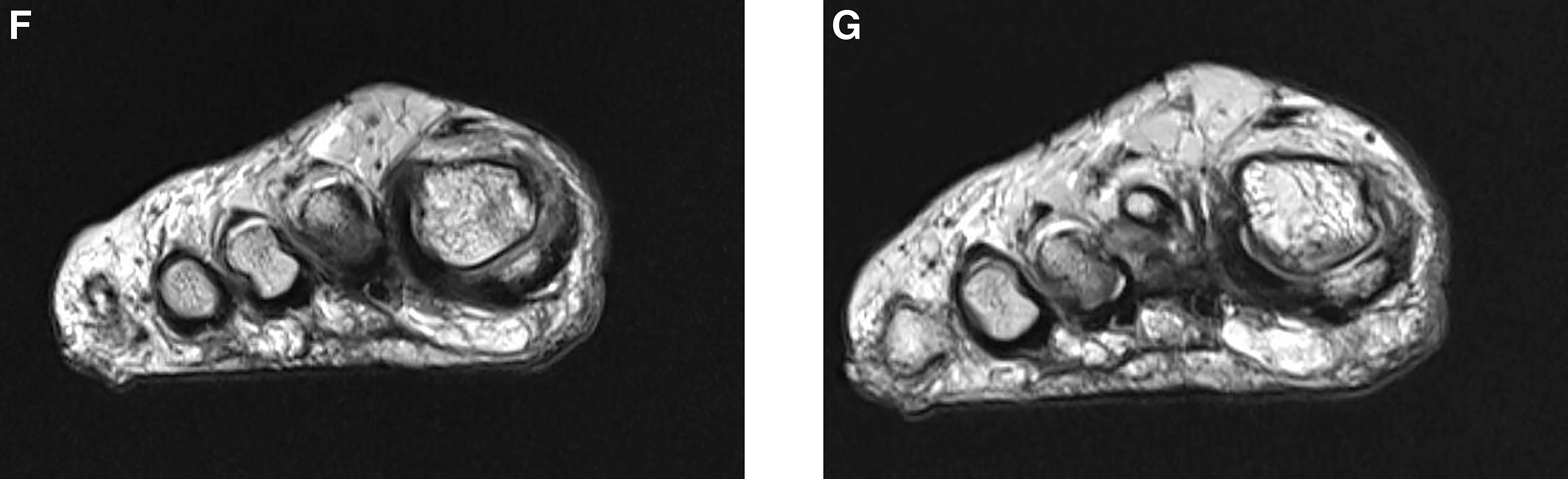
A 52-year-old woman presented to our office approximately 9 months after injuring her right foot. Her medical history was significant only for a motor vehicle accident in 1993 in which she sustained a pelvic fracture, right ankle fracture, luxated right patella, and lacerations of the right femoral artery and quadriceps. These injuries required orthopedic and reconstructive surgery. She states that she was gardening while wearing flip-flops when her foot fell into a hole. She said it felt as if her foot and toe went one way and the shoe went another direction. Pain was noted initially but her foot did not appear dramatically different at the time. Over the course of 9 months, she noted that her great toe gradually rotated medially. She began to experience increased difficulty getting into shoes and discomfort when wearing shoes. By the time she presented to our office, her great toe was in notable varus deformity, which increased in severity with weightbearing. Radiographs were taken and a varus deformity was confirmed with a 21° varus hallux abductus angle but, interestingly, absence of the fibular sesamoid was also apparent (Fig. 1A). On physical examination, she did have some discomfort with dorsiflexion, without a notable decrease in range of motion. Contralateral radiographs were also obtained and, again, the fibular sesamoid was absent. There was also a 5° varus deformity of the left foot detected by means of weightbearing radiographs that was not apparent clinically (Fig. 1B). She denied ever having surgery to either foot. Treatment options were discussed, but the patient wished to avoid surgery. A trial of taping the toe was unsuccessful. At her next follow-up, again surgical options were discussed and it was advised that a fusion of the first metatarsophalangeal joint (MPJ) would be the most reliable procedure. Soft-tissue repair procedures were also explained, and she was cautioned that they are sometimes not successful and recurrence of the deformity is possible. She had previously been in a motor vehicle accident in 1993 and endured serious injuries, leaving her more proximal lower extremity joints with limited range of motion. She was concerned that fusing the first MPJ would lead to a serious hindrance on her ability to get up from a sitting position and potentially a hindrance on her independence. She was therefore not interested in a fusion. A magnetic resonance imaging scan was later obtained and demonstrated that the adductor tendon appeared to be intact, but also demonstrated heterogeneity of the tendon, indicating injury. The lateral collateral ligament was noted to be disrupted. Medial displacement of the proximal phalanx and varus subluxation of the MPJ were also apparent (Fig. 1C). Axial views demonstrated, again, absence of the fibular sesamoid (Fig. 1D and E). After discussing the MRI results, the patient opted for soft-tissue repair.
Figure 1.
A–C, Radiographs of the injured right foot with increased varus on weightbearing and absent fibular sesamoid. D, Radiograph of the unaffected left foot with mild varus on weightbearing and notably absent fibular sesamoid. E, Magnetic resonance imaging scan of the right foot demonstrating varus position and attenuated lateral joint capsule and adductor tendon. F and G, Axial magnetic resonance imaging scans of the right foot demonstrating absence of the fibular sesamoid. (continued on next page)
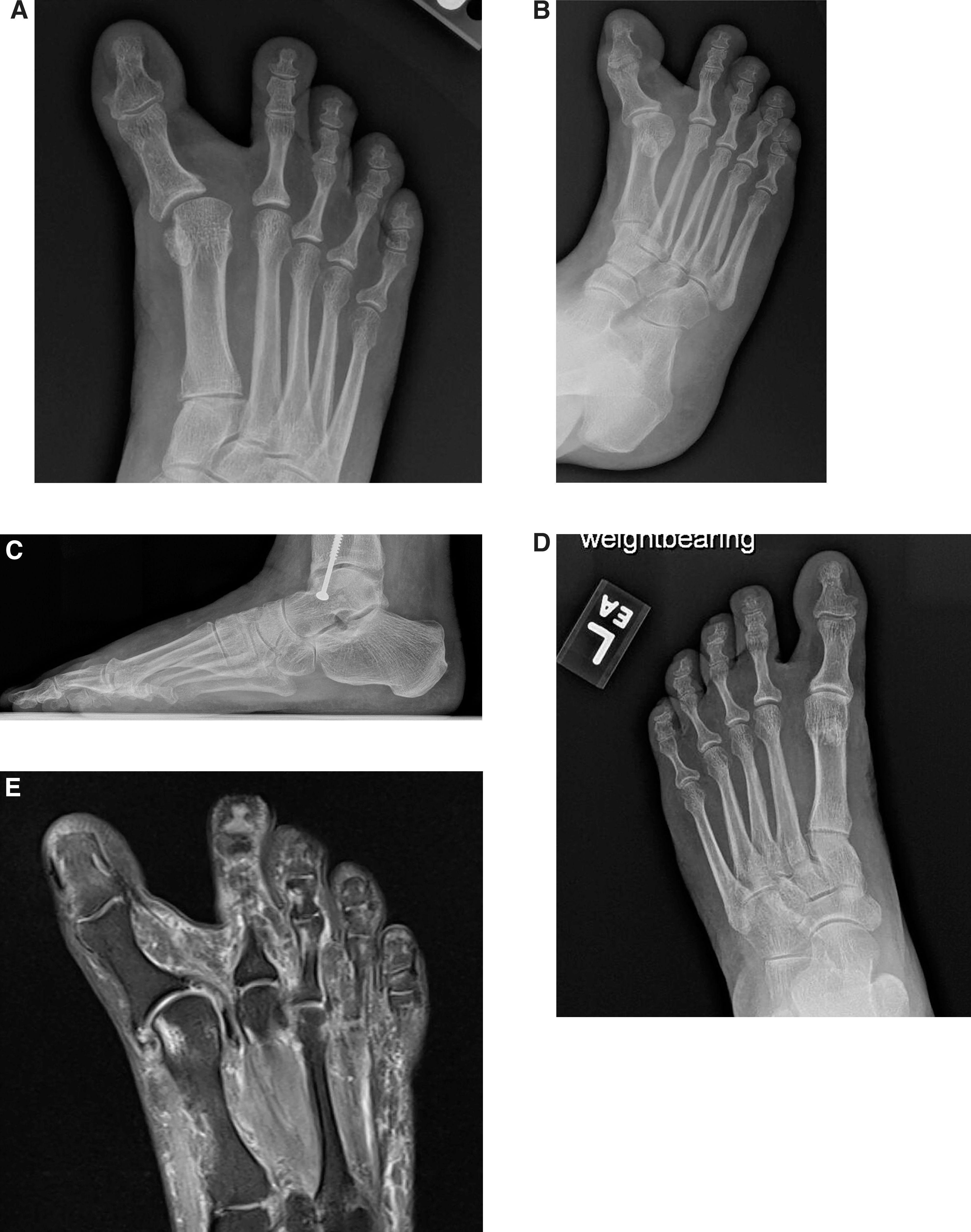
Figure 1.
Continued.
Surgical Treatment
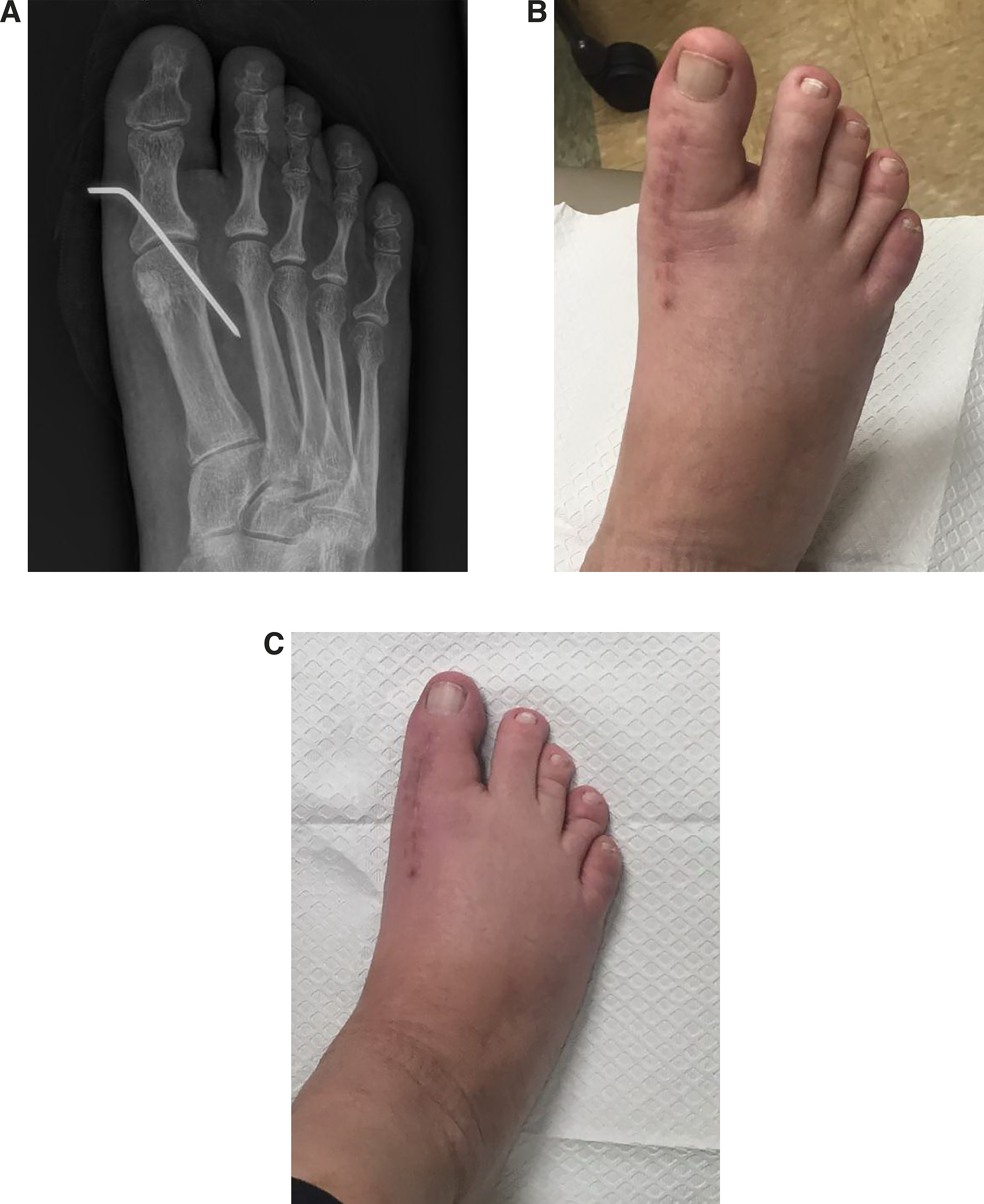
Surgical correction was approached by means of the extensor hallucis longus (EHL) as described by Gradisek and Weil where the tendon was split longitudinally and transected proximally from the lateral arm.[9,10] The tendon was then routed from distal to proximal under the deep transverse intermetatarsal ligament into a hole drilled into the first metatarsal and secured either by suturing the tendon onto itself or by an interference screw through a hole drilled into the metatarsal.[9] For our case, attention was directed to the right foot, where an approximately 8-cm incision was made dorsally over the first MTP joint. An attempt was initially made to identify the adductor tendon for primary repair, but the tissue was extremely friable and no viable tendon was usable. A medial capsulotomy was performed. This allowed the toe to drift into a more rectus position; however, varus deformity was still notable. At this time, it was decided to proceed with a split EHL tendon transfer. Attention was then directed to the EHL, where the tendon was split in half longitudinally as far proximal as possible. The lateral aspect of the split tendon was transected as far proximal as possible. The distal attachment of the EHL was left intact. The medial section of the EHL remained intact for its entire course. The tendon was rerouted under the deep transverse intermetatarsal ligament and secured into place into the first metatarsal with a Bio-Tenodesis (Arthrex, Inc, Naples, Florida) screw. A lateral capsulorrhaphy was then performed and a Kirschner wire was then placed through the proximal phalanx into the metatarsal from medial to lateral to allow the soft tissues to scar into place (Fig. 2A). She was advised to remain nonweightbearing for 2 weeks. This was followed by limited weightbearing in a fracture boot. Unfortunately, the Kirschner wire advanced into the foot because of pressure from the fracture boot and had to be removed at 3 weeks because of pain at the site. She was full weightbearing in a fracture boot at 1 month and transitioned to tennis shoes by 2 months. She was released from our care at 3 months (Fig. 2 B and C).
Figure 2.
A, One-week postoperative radiograph with Kirschner wire placed and visible bone tunnel for a Bio-Tenodesis screw. B, Two-month postoperative nonweightbearing image of the right foot. C, Two-month postoperative weightbearing image of the right foot.
Results
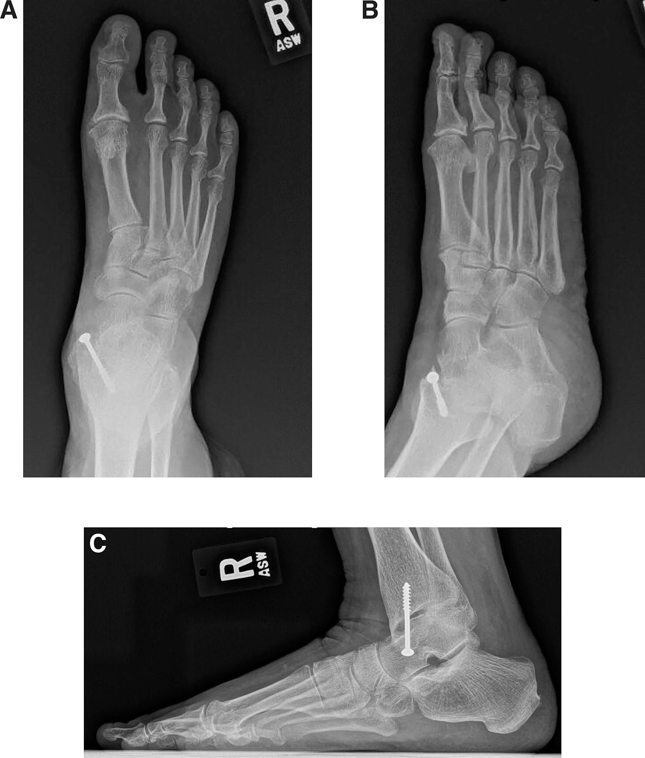
The patient was seen in the office approximately 22 months postoperatively and stated that she had experienced nearly no pain since her operation other than when the Kirschner wire advanced into her foot. She now has no pain and states that she often forgets that there was once an issue with her foot. She is now to the point that she can wear high heels all day without issue. She does relate some tightness in the joint, especially when bending to pick something up, but states this does not affect her activities. She does relate that there is loss of plantar flexion strength but that this does not impact her activities. Overall, she is very happy with her progress following the procedure. On physical examination, her correction has been maintained as evidenced by weightbearing and nonweightbearing photographs (Fig 3 A and B). Postoperatively, she can achieve approximately 30° of dorsiflexion compared to 45° in the left foot (Fig. 3C). Weightbearing radiographs of the right foot were taken at 22 months postoperatively and demonstrate maintenance of correction, but with development of arthritis in the first MPJ (Fig. 4 A–C). Her hallux abductus angle measures 9° valgus compared with her preoperative hallux abductus angle of 21° of varus. This demonstrates a maintained reduction of 30°.
Figure 3.
A, Weightbearing image of the right foot 22 months postoperatively. B, Nonweightbearing image of right foot 22 months postoperatively. C, Image demonstrating unloaded hallux dorsiflexion of the right foot 22 months postoperatively.
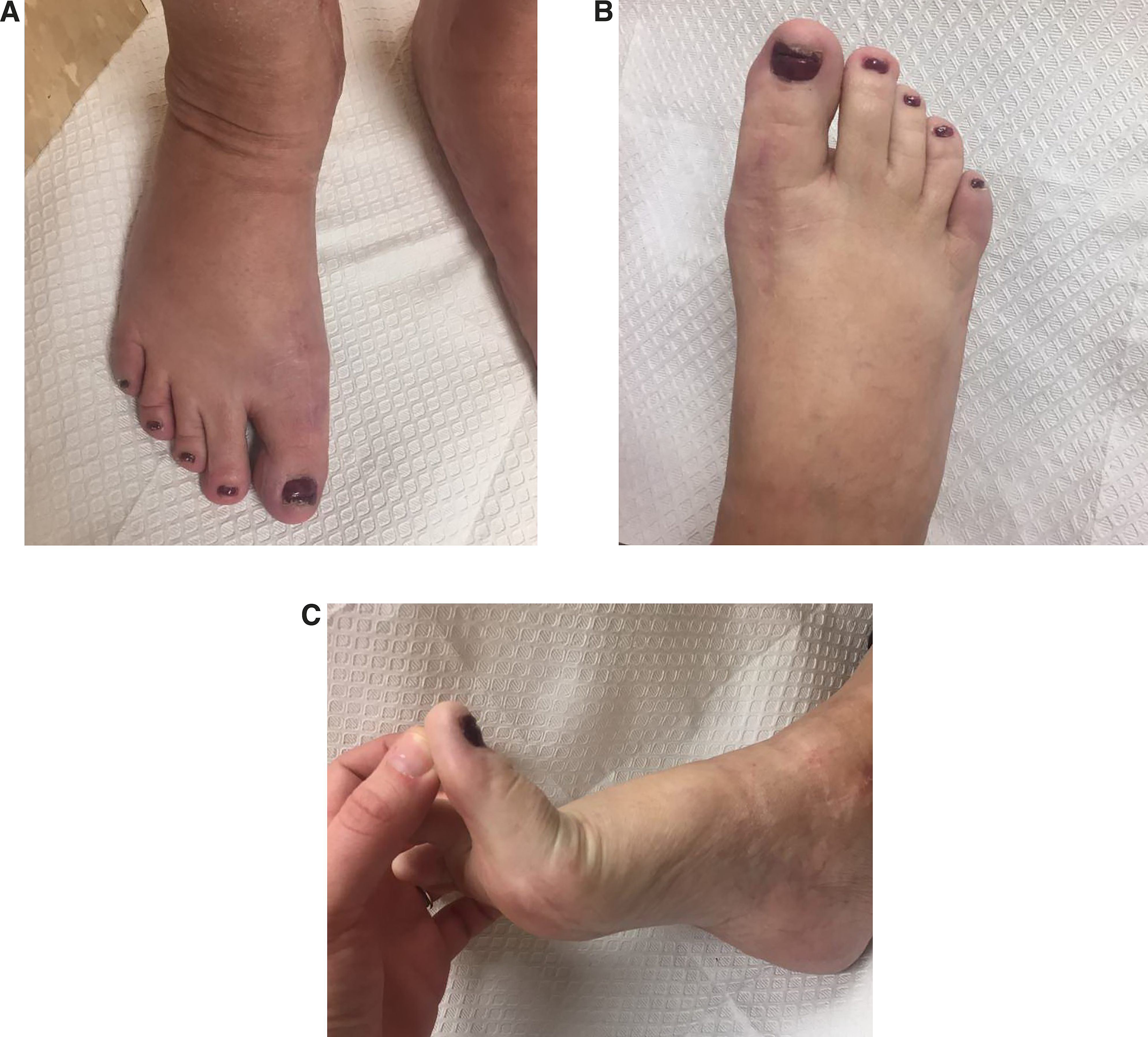
Figure 4.
A, Anteroposterior radiograph of the right foot 22 months postoperatively. B, Oblique radiograph of the right foot 22 months postoperatively. C, Lateral radiograph of the right foot 22 months postoperatively.
Discussion
In this case, it is postulated that the function of the adductor hallucis and lateral capsular ligaments were destroyed during the minor accident. This likely would have allowed the abductor hallucis tendon and medial capsule to have greater pull on the proximal phalanx. We believe that lack of the fibular sesamoid contributed to the cause of the injury in much the same way that excision of the sesamoid can lead to hallux varus. In this case, the sesamoid complex was inherently weak laterally because of the absence of the fibular sesamoid. The traumatic event in our case would have likely been a relatively low-velocity trauma, whereas many of the case studies identified for this article are much more high-velocity–type injuries. This supports our theory that lack of the fibular sesamoid was largely a contributing factor to the severity of her injury in that the lateral capsular ligaments are weakened by the lack of fibular sesamoid. This is supported by her slight varus on the left foot without injury. Jahss also described a case of a patient who presented with hallux varus and was noted to have absent fibular sesamoids.[7] This patient did not undergo trauma and was asymptomatic.
After a thorough literature review, there are many studies on iatrogenic hallux varus, but there are few studies on traumatic hallux varus. Each of these studies involves a similar mechanism of injury, and we believe our case is unique in that congenital absence of the sesamoid in conjunction with a traumatic event led to symptomatic hallux varus with a less forceful traumatic event. There have been several different methods of soft-tissue repair proposed, all of which involved some form of soft-tissue augmentation and tendinous procedures. We performed a repair using an EHL split tendon transfer as described by Gradsiek and Weil.[9,10] Our procedure differed in that we added a medial capsulotomy and lateral capsulorraphy and maintained our correction with a temporary Kirschner wire across the first MPJ. Gradsiek and Weil described a postoperative course of nonweightbearing or protected weightbearing for 6 weeks and physical therapy at 1 week. We placed our patient nonweightbearing for 2 weeks with protected weightbearing the following 4 weeks. At 2 months, she transitioned to an athletic shoe. We did not deem physical therapy necessary. Twenty-two–month follow-up demonstrated that our repair was stable, and she remains pain-free in regular shoe gear. This repair adequately recreates the lateral capsular ligaments and the pull of the adductors and balances the pull of the abductor hallucis tendon.
Conclusions
Although iatrogenic hallux varus is well known and the subject of ample publications, traumatic hallux varus is rarely reported. To our knowledge, this is the first publication of traumatic hallux varus in a patient with congenital absence of the bilateral fibular sesamoids. In most cases of traumatic hallux varus, there was a relatively forceful action leading to the eventual development of varus. In our case, the traumatic event was relatively minor. This leads us to conclude that lack of the fibular sesamoid predisposed her to this type of injury through a weakened lateral capsule. This theory is supported by an anatomical and clinical study of hallux varus by Joseph et al, who concluded that disruption of the lateral stabilizers can lead to hallux varus.[6] Our chosen procedure allowed reconstruction of the adductor tendon while still leaving the insertion of the extensor hallucis longus and its function intact. We believe this approach provided a stable reconstruction while still allowing the range of motion she needed to retain her mobility. A weakness of the procedure is that the temporary Kirschner wire became too close to the skin to remain in place without discomfort. This wire could have been cut longer or gauze could have been placed between the skin and wire to prevent discomfort. This did not seem to detract much from the case, as her correction has maintained over 22 months. A weakness of this study is that it is a case review. A formal study with multiple patient cases, either prospective or retrospective, would be more powerful.
Financial Disclosure: None reported.
Conflict of Interest: None reported.
References
- Labovitz JM, Kaczander BI: Traumatic hallux varus repair utilizing a soft-tissue anchor: a case report. J Foot Ankle Surg39: 120, 2000.
- Ryan PM, Johnston A, Gun BK: Post-traumatic dynamic hallux varus instability. J Clin Orthop Trauma5: 94, 2014.
- Cheung CN, Lui TH: Traumatic hallux varus treated by minimally invasive extensor hallucis brevis tenodesis: case report. Case Rep Orthop2015: 179642, 2016.
- Turner RS: Dynamic post-surgical hallux varus after lateral sesamoidectomy: treatment and prevention. Orthopedics9: 963, 1986.
- Barp EA, Temple EW, Hall JL, et al: Treatment of hallux varus after traumatic adductor hallucis tendon rupture. J Foot Ankle Surg57: 418, 2018.
- Joseph B, Chacko V, Abraham T, et al: Pathomechanics of congenital and acquired hallux varus: a clinical and anatomic study. Foot Ankle8: 137, 1987.
- Jahss MH: Spontaneous hallux varus: relation to poliomyelitis and congenital absence of the fibular sesamoid. Foot Ankle4: 224, 1983.
- Lui TH, Tam KF: Hallux valgus deformity associated with bilateral absence of the tibial and fibular hallucal sesamoids. J Foot Ankle Surg52: 254, 2013.
- Gradisek BP, Weil LJr: Tendon transfers and salvaging options for hallux varus deformities. Clin Podiatr Med Surg33: 85, 2016.
- Lau JT, Myerson MS: Modified split extensor hallucis longus tendon transfer for correction of hallux varus. Foot Ankle Int23: 1138, 2002.
© 2019 American Podiatric Medical Association