


Abstract
Plantar plate repairs are challenging procedures because of the small anatomy of the plantar plate. This can make them daunting, time-consuming procedures to perform. Advances in technology, such as interference screws and small suture passers, have created improved technique possibilities to decrease difficulty, correct multiple planes of deformity, create stronger constructs, and improve patient results. The plantar plate repair technique presented in this article includes a dorsal approach with a metatarsal osteotomy, a knotless repair that provides a strong construct to allow patients to protectively bear weight immediately, and can reduce operative time by presenting tips to quickly navigate the procedure. The presented technique allows for detailed correction of all three planes of deformity, maximizing patient results.
Lesser metatarsophalangeal joint (MPJ) pathology is very common in the foot and ankle. The plantar plate in particular has been extensively studied over the past decade. Originally described in 1986, a ‘‘crossover toe’’ represented an end-stage digital deformity of the lesser MPJ.[1] Recognition of this pathology in earlier stages has been described to help the surgeon identify and treat the patient before complete MPJ subluxation. The lesser MPJs are surrounded by multifarious soft-tissue structures that involve both dynamic and static forces. The dynamic forces that stabilize the lesser MPJ are the intrinsic and extrinsic muscles of the foot. However, it is mainly the plantar plate that stabilizes the MPJ in the sagittal plane.[2]
The development of lesser MPJ deformity typically occurs from either direct trauma or mechanical overuse. Etiologies of this consist of elongated second metatarsal, a hypermobile first ray, hallux valgus, hallux limitus, and pes planus, all of which result in repetitive trauma to the plantar plate.[3,4] Plantar pain present at the base of the second toe is the most common complaint for plantar plate tears.[5] Patients may feel as if they are ‘‘walking on a marble,’’ and the pain is often increased with ambulation and decreased with rest.[6] An unstable MPJ can be determined during physical examination with a modified Lachman (or ‘‘dorsal drawer’’ test) or the ‘‘paper pull-out test,’’ and transverse plane deformity can be observed clinically with medial or lateral deviation of the toe while weightbearing.[7,8] Various imaging modalities can be used for diagnosing a plantar plate, including radiography, magnetic resonance imaging, and ultrasound, although arthrography is the standard means of assessing these injuries.[6,9]
Patients usually seek out treatment once significant pain and deformity are present, which limits the success of conservative treatment options. Different nonsurgical modalities consist of low-heel shoes with a wide toe box, rocker bottom soles, metatarsal pads, graphite foot plates to stiffen the toe box, hammertoe padding, taping, Budin splints, and intra-articular corticosteroid injections.[6] Conservative treatment allows for symptomatic relief and decreases the rate of progression of the deformity, but often does not correct the underlying etiology and pathology.[10] Multiple methods and techniques have been published for repairing the plantar plate, including the plantar approach, the dorsal approach, direct repair, metatarsal osteotomies with or without suture stabilization, and others.[8,10,11,12,13,14,15,16,17] Because of the small anatomy involved, plantar plate repairs can be very challenging and time consuming, and difficult to address in both the sagittal and the transverse planes. We present a technique that is knotless in repair with a metatarsal osteotomy, provides a strong construct that allows patients to protectively bear weight immediately, provides multiplanar correction, and can reduce operative time by presenting tips so that the procedure can be quickly navigated.
Surgical Technique
The procedure is typically performed with intravenous anesthesia, a local field block, and an ankle tourniquet set at 250 mm Hg. A curvilinear incision is performed across the MPJ to decrease scar contracture and unwanted dorsiflexion. Dissection continues through the subcutaneous tissue and, once the MPJ capsule is identified and the extensor digitorum longus tendon is retracted, a linear dorsal capsulotomy is performed. The capsular attachments along the base of the proximal phalanx and metatarsal head are reflected medially and laterally. A McGlamry elevator is then used plantar to the metatarsal head and advanced proximally, with care being taken to avoid skiving cartilage or further injuring the plantar plate.
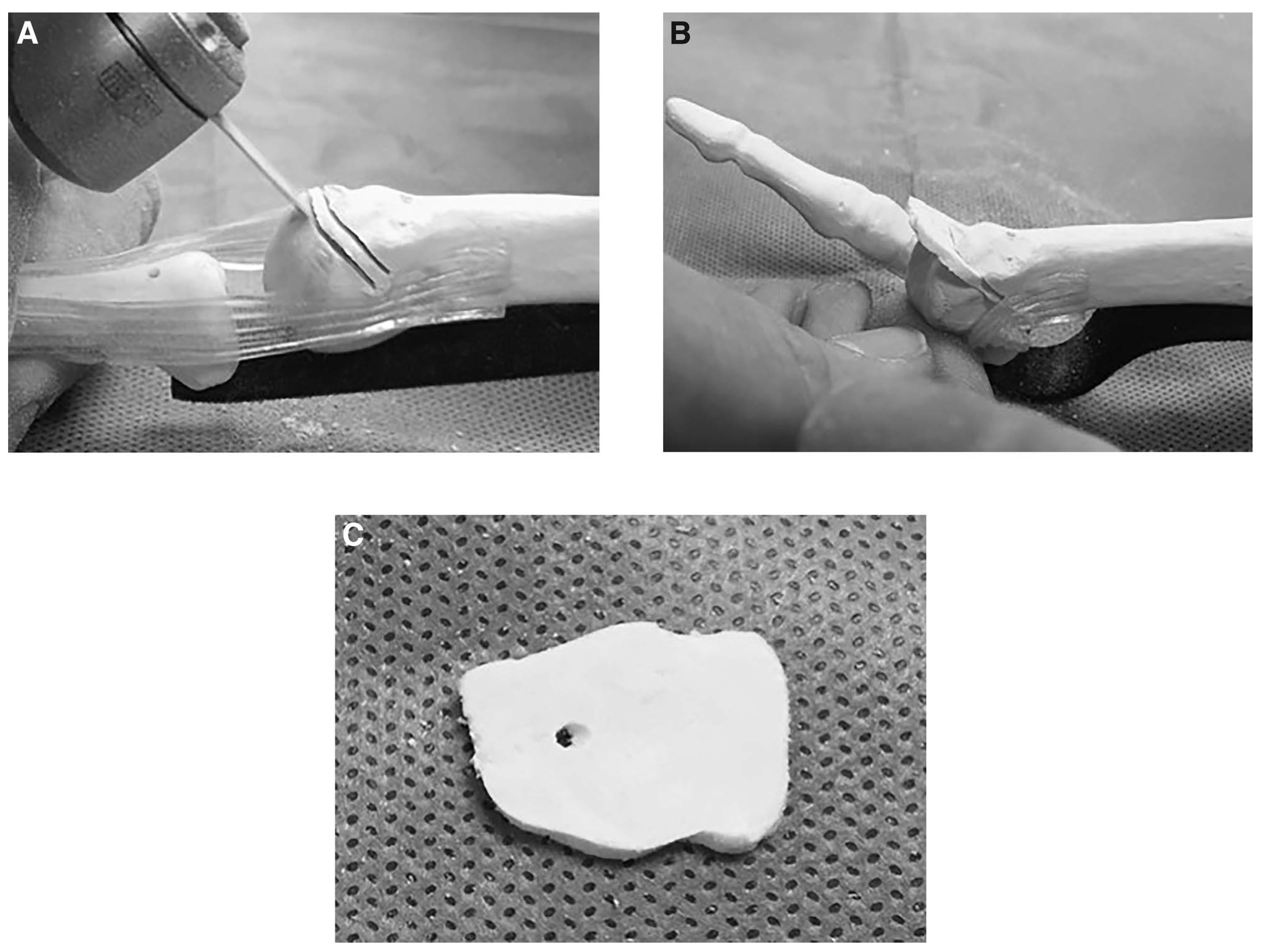
A decompression metatarsal osteotomy is often required, and we prefer to take a wafer osteotomy (or wedge if dorsiflexion is desired) (Fig. 1). The technique involves preoperative planning, where the surgeon measures the desired amount of shortening to restore the metatarsal parabola. After resection, the metatarsal head is pushed as far proximal as possible. A 1.6-mm Kirschner wire is used for temporary fixation and the proximal pin is used for joint distraction. When making a wafer or wedge osteotomy, make the proximal cut first, going halfway through before starting and completing the distal cut. Then, if making parallel cuts, complete the proximal cut (Fig. 1A and 1B). An additional tip is to place the proximal distraction wire vertically where future screw fixation of the metatarsal osteotomy will be.
Figure 1.
A and B, Metatarsal wafer osteotomy with parallel cuts. C, Rectangular wafer removed. All figures were used with polyurethane sawbones, a neoprene plantar plate, and silicone collateral ligaments.
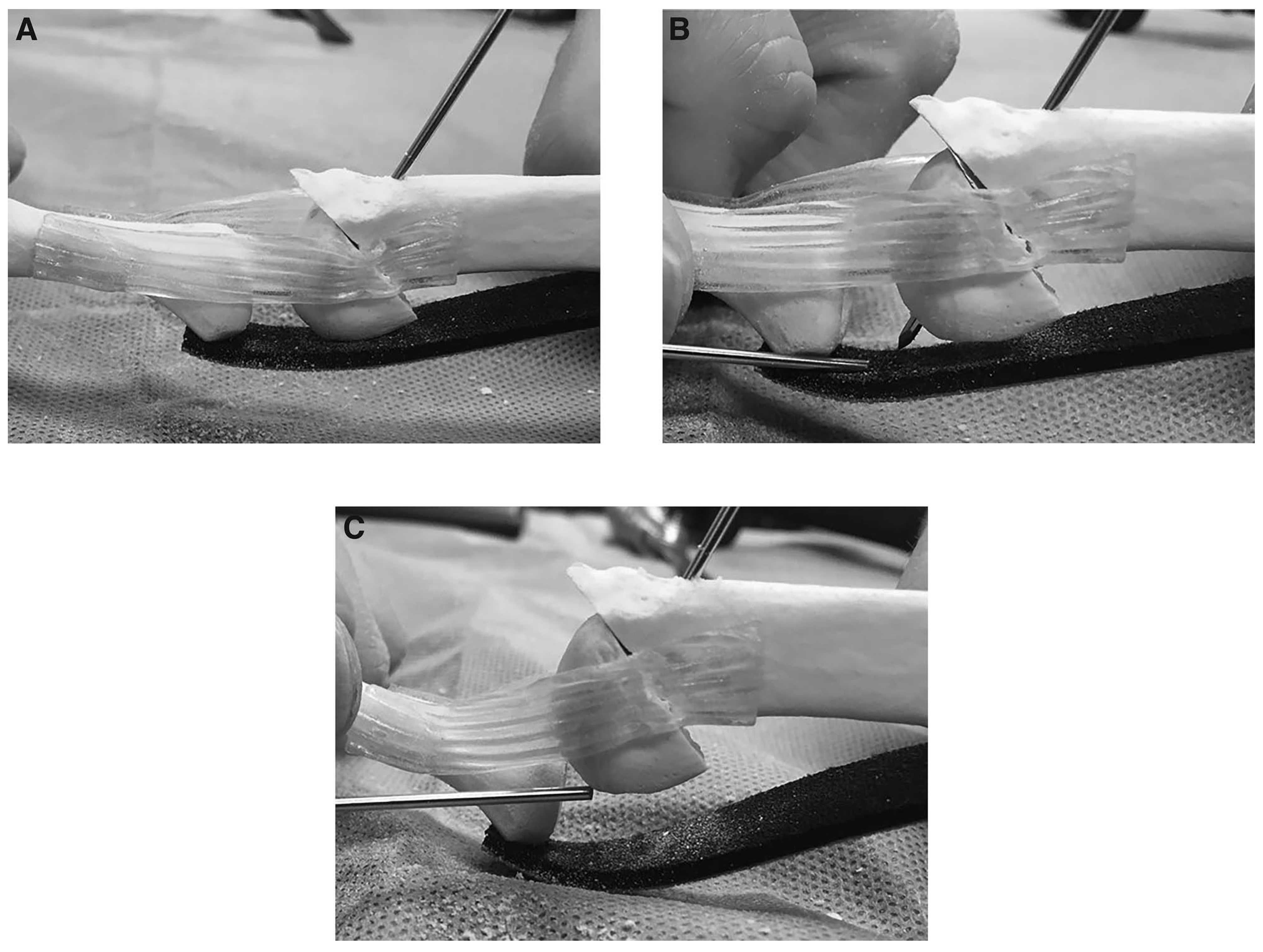
An additional 1.6-mm Kirschner wire is then placed in the midshaft of the proximal phalanx, and a joint distractor is used to visualize the plantar plate. Do not advance the distraction wires past the plantar cortex. If it is advanced through the cortex of the metatarsal, it snags the plantar plate. This makes advancing the plantar plate distally difficult, and can also damage the plantar plate, with further tearing (Fig. 2). If the tear is incomplete and the patient has isolated pain that failed conservative management, without drifting of the digit, a primary repair is all that is warranted. When the toe is subluxed or dislocated, we complete the tear at the base of the proximal phalanx. The medial and lateral borders of the plantar plate are then dissected free, along with the plantar aspect being lifted dorsally from the flexor digitorum longus tendon. Next, a 2- to 4-mm section is taken off the distal aspect of the plantar plate, up to or past the tear if appropriate. Taking this section of plantar plate greatly helps sagittal plane correction, and the authors typically remove the same amount as is taken to shorten the metatarsal. This section can also be wedged, to aid in transverse plane correction.
Figure 2.
A, Placement of 1.6-mm Kirschner wire. B, Incorrect placement of the Kirschner wire will cause further damage to the plantar plate and prevent distal advancement of the ligament. C, Correct Kirschner wire placement.
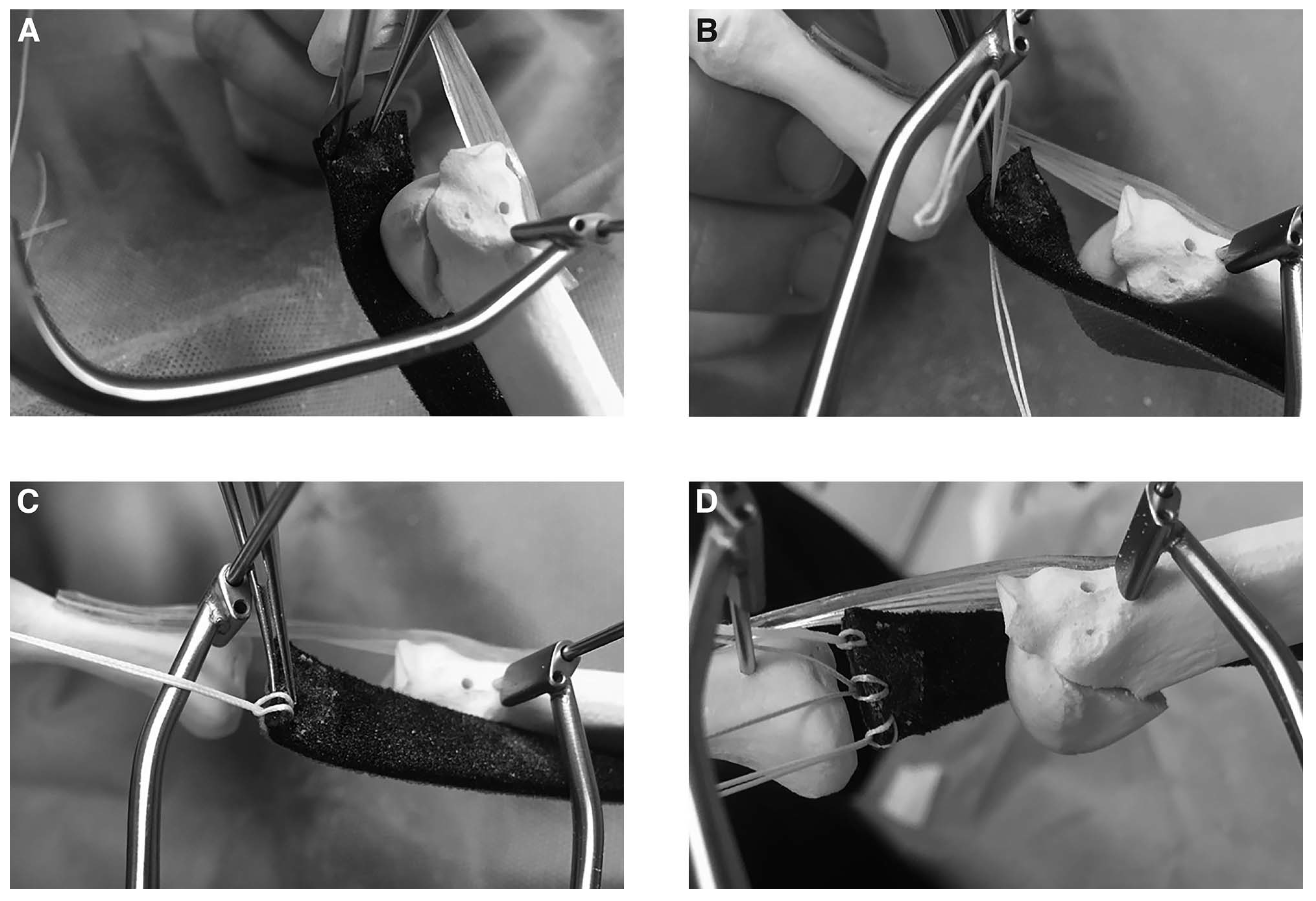
Using the Arthrex Viper suture passer (Arthrex, Inc, Naples, Florida), three separate passes with 2-0 FiberWire (Arthrex) are performed in the mobilized plantar plate (Fig. 3). The medial and lateral passes are approximately 1 to 2 mm proximal to the severed end of the plantar plate, whereas the central pass can be slightly more proximal if desired. Each loop of FiberWire is passed through the plantar plate, then locked into itself, creating three separate attachments. It is best to have curved mosquito forceps to further pull out the loop of suture before locking it to itself.
Figure 3.
A, B, and C, Arthrex Viper passing 2-0 suture through plantar plate medially, and being locked to itself. D, All three suture passes after completion.
The joint distractor is then removed, the distal wire is kept and used as a joystick, and the proximal wire is kept in the metatarsal head for temporary fixation. Any remaining plantar plate is cleared off the base of the proximal phalanx, leaving an approximately 10-mm area. A hand rasp is then used to roughen up the plantar cortex to optimize healing of the advanced plantar plate. The distal guidewire should be used to help visualize the plantar aspect of the proximal phalanx, as one can maximally plantarflex the toe to visualize the exit of Kirschner wire pilot holes.
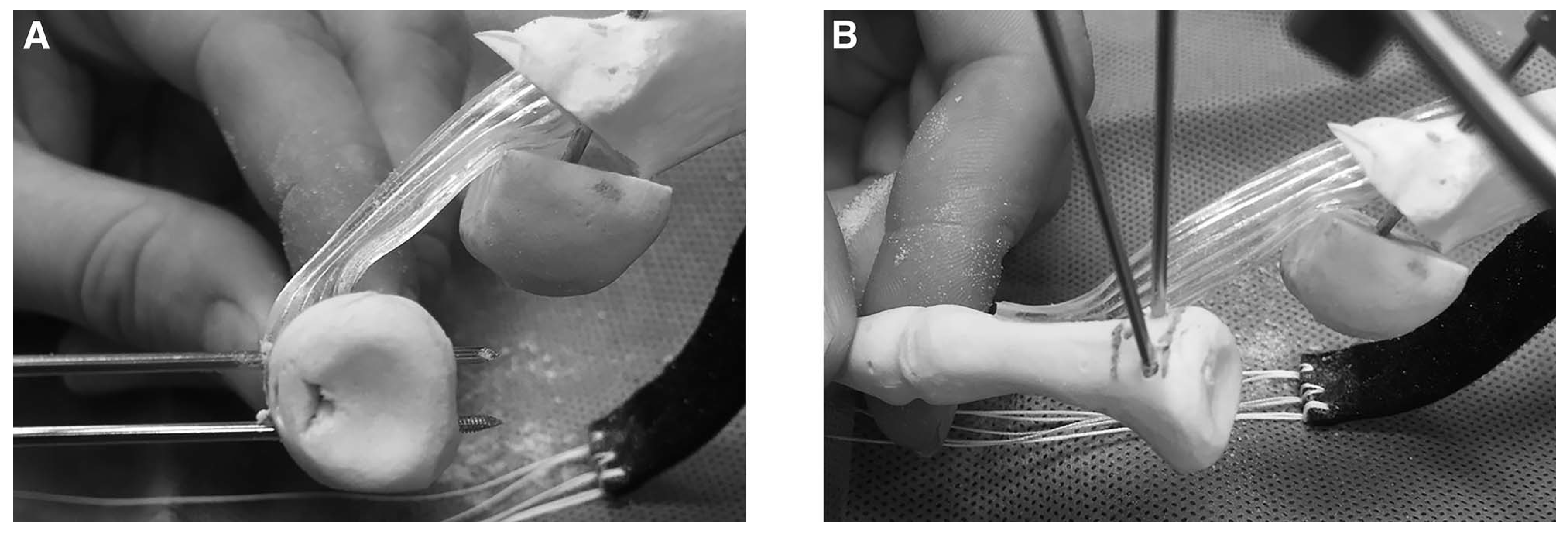
Next, dorsal-to-plantar suture holes should be drilled using a 1.6-mm Kirschner wire. It is our preference to continue using the ‘‘joystick’’ Kirschner wire to further help stabilize the phalanx. The deformity usually has a dorsal sagittal plane deformity and medial transverse plane deformity. To aid in transverse plane correction, the lateral suture hole is made more distal than the medial suture hole (Fig. 4). To further aid in sagittal plane correction, suture holes are made more distal in the proximal phalanx. The exit point of the Kirschner wire is checked plantarly to ensure ideal placement for multiplanar correction. Care should be taken that the holes in the proximal phalanx are drilled approximately 1 cm distal to the phalanx base, to prevent stress riser. The suture holes are then overdrilled using a 2.5-mm drill bit for the 2.7-mm PushLock (Arthrex) absorbable suture anchors. Suture passers are then inserted and three suture strands are passed through each respective suture hole, two from each medial or lateral side, and one strand from the central stitch is brought medially and laterally. A tip with this technique is that it is best not to pull the entire length of the FiberWire through the suture passer loop, but rather only a couple of inches. This way, there is little to no pull on the plantar plate until fixation.
Figure 4.
A, Suture hole placement in proximal phalanx. B, The wire furthest away represents the lateral wire. To aid in transverse plane correction, the lateral wire can be aimed more plantar distally than the medial wire. It is most important to focus where the wire exits plantarly, as this is where the plantar plate will be advanced to.
After the suture is passed through the proximal phalanx dorsally, all temporary Kirschner wire fixation is removed. The metatarsal head is brought out the appropriate length, and fixation of the surgeon’s choice perpendicular to the osteotomy is performed. Intraoperative fluoroscopy is used to confirm correction of metatarsal parabola. If hammertoe correction is desired, it is best to perform at this point of the procedure, before approximating and fixating the plantar plate.
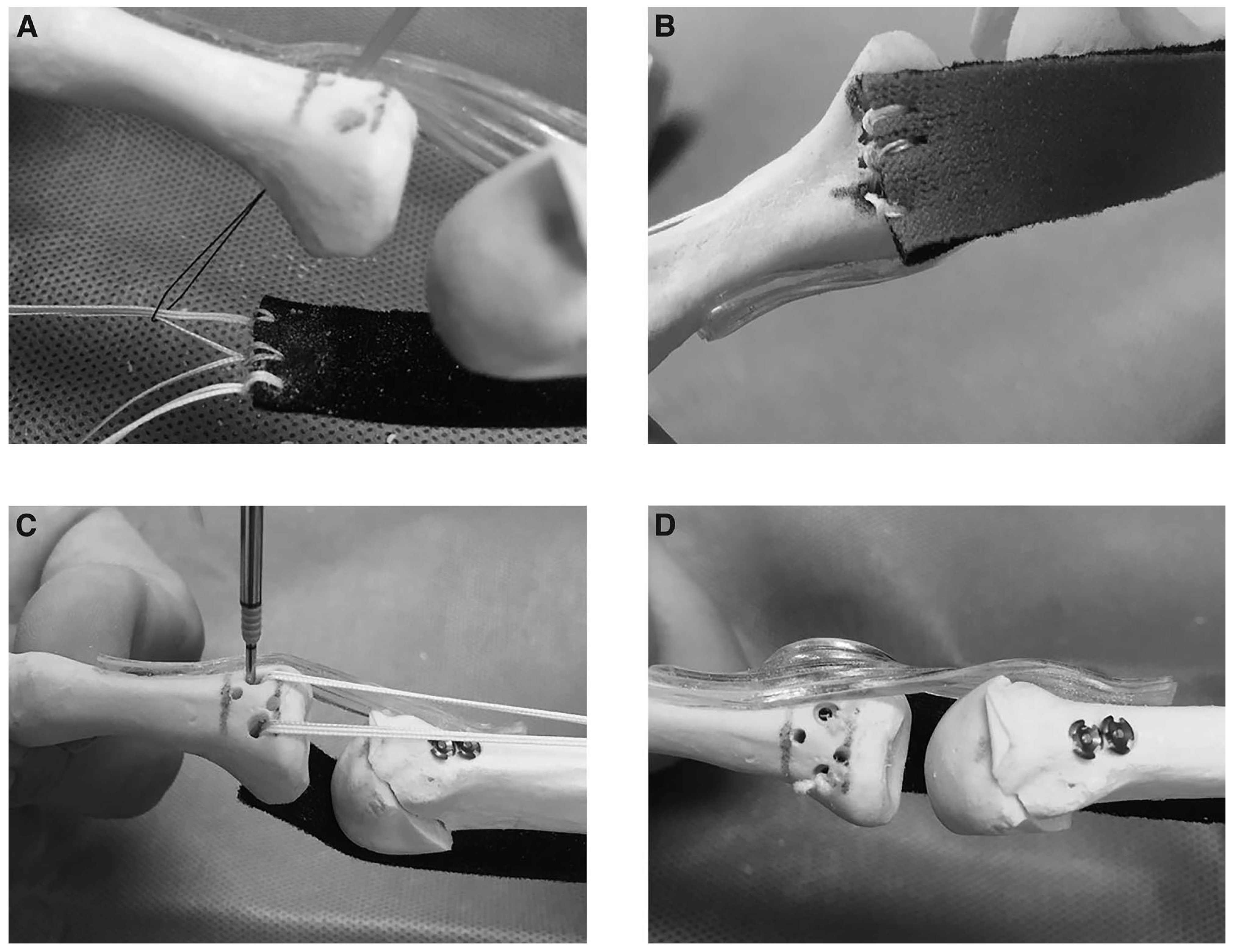
To ensure that the plantar plate is approximated close to the plantar cortex, the toe is brought into an overcorrected position, typically plantarflexed and laterally deviated. The suture is then pulled dorsally and proximally toward the metatarsal. The sagittal and frontal plane correction can be dialed in at this time, pulling first the lateral stitches and then the medial. With slight overcorrection being maintained, the 2.7-mm PushLocks are placed in the suture holes of the proximal phalanx, acting as interference screws, locking the suture into place and allowing for multiplanar correction (Fig. 5). The islet hole from the anchor is removed before insertion. The tails of the suture are then cut flush with a fresh blade, allowing a knotless fixation. The wound is irrigated copiously with sterile normal saline and closed with sutures of the surgeon’s choice. The affected toe is bandaged in an overcorrected position, and the patient can bear weight in a CAM Walker (Zinco Industries, Inc, Pasadena, California) as tolerated.
Figure 5.
A, A suture passer is used to pull the suture from the plantar to the dorsal aspect of the proximal phalanx. B and C, After metatarsal fixation, the suture should be pulled proximally to secure mul-tiplanar correction of the plantar plate. D, The 2.7-mm PushLocks should be inserted under ap-propriate tension; then, the suture should be cut flush for knotless repair.
Discussion
In recent years, surgical treatment for plantar plate pathology and MPJ instability has gained attention, as there have been multiple methods and techniques published for repairing the plantar plate.[8,10,11,12,13,14,15,16,17] Plantar approaches and direct repair of the plantar plate have been described,[16,17,18,19] but the patient must remain nonweightbearing for plantar incisions while the biomechanical causes, such as elongated second metatarsal, are not addressed. The metatarsal osteotomy has been shown to have an advantage because of these biomechanical reasons.[17,20] Nery and colleagues[17] had a similar approach to their plantar plate repair and showed excellent outcomes with postoperative American Orthopaedic Foot and Ankle Society scores of 95 and 96 in grade 1 and 2 tears.[17] However, possible complications with the osteotomy can arise, including nonunion, painful hardware, joint stiffness, and floating toe syndrome.[17,20]
The dorsal decompression wafer osteotomy that is used in this technique is a unique modification to the traditional Weil metatarsal osteotomy. It allows for additional shortening during the osteotomy and being able to angle the cut to make a wedge medially or laterally for transverse plane correction. Alternatively, wedge cuts can be made dorsally to dorsiflex the metatarsal head for sagittal plane correction. Care must be taken to not overshorten the metatarsal, as this can increase the potential for a floating toe.[21] No literature on this type of osteotomy was found during our literature review of central metatarsal osteotomies, but Gauthier and Elbaz[22] did describe a dorsal closing wedge osteotomy for Freiberg’s disease, and Pereira and colleagues[23] performed a case series on patients receiving this osteotomy with long-term follow-up.[22,23] Mean follow-up time was 23.4 years in their study, and results showed 80% excellent results and 20% good results in their clinical outcome measures.[23] We believe the osteotomy described in our technique allows for more control on different planes of correction, and further outcome studies are needed to prove its success.
Interference screws have been described in techniques on lesser MPJ pathology and provide a knotless repair that has advantages.[11,14] Feldman[14] described the use of interference screws for flexor tendon transfers in 2005, and Sung[11] described a dorsal approach plantar plate ligament repair using FiberTape with an interference screw, but did not include native plantar plate or a metatarsal osteotomy, and used only one strand of FiberTape, limiting the amount of multiplanar correction.[11,14] Interference screw fixation of ligaments and tendons in the forefoot has been described as providing a strong and stable construct.[11,14] A biomechanical study testing the strength of this construct versus simply tying the suture dorsally would aid in this discussion.
Plantar plate and predislocation repairs are sagittal plane dominant, and mostly treated as such.[2] Also, patients can commonly develop a transverse plane component, as the medial capsular structures are tighter, and the toe drifts medially. The transverse plane can be addressed in multiple ways with the proposed technique, including the following: 1) the angle of the wafer osteotomy in the metatarsal, and transverse location of the metatarsal head on fixation; 2) a wedge resection of the remaining plantar plate can be performed; 3) placement location of the three sutures in the plantar plate; 4) location and angle of the suture tunnel placement in the base of the proximal phalanx; and 5) tensioning of the suture before interference anchor placement. Frontal plane correction can also be achieved based on suture tensioning being more medial or lateral before anchor insertion. Having multiple options to correct all three planes of deformity can aid during plantar plate repair for each individualized case and maximize patient outcomes.
We have had great success with this technique, with more than 40 patients undergoing this procedure during a 3-year period (2015–2017). Subjective and functional outcomes are being closely followed on these patients and are showing great results. For a few patients, complaints of continued toe stiffness and lack of digital purchase have occurred. A cohort or comparative study would be beneficial to more thoroughly evaluate the technique and compare it to other techniques that have been presented in the literature.
Conclusions
The plantar plate technique described includes a dorsal approach, metatarsal osteotomy, and a knotless repair of the native plantar plate ligament. Advantages of the knotless repair include the following: 1) there is no loosening of the knot over time; 2) tensioning of the suture can be maintained and optimized until fixated with the interference anchor; and 3) there is a decreased risk of a prominent knot and scar tissue dorsally. The metatarsal osteotomy further corrects biomechanical causes of the plantar plate tear and prevents recurrence. This technique also allows for multiplanar correction, as various steps can be adjusted to correct a given amount of sagittal, transverse, and frontal plane deformity. Furthermore, this technique with the presented tips can improve operative time and provide a strong construct that allows the patient to bear weight immediately in a surgical shoe or boot. This plantar plate repair technique can be very useful for surgeons and can be used to treat patients with multiple planes of deformity at the lesser MPJ.
Financial Disclosure
None reported.
Conflict of Interest
Dr. McCartan consults for Arthrex, Inc.
References
- COUGHLIN, M.J. Crossover second toe deformity. Foot Ankle 1987, 8, 29. [Google Scholar] [CrossRef] [Scilit]
- CHALAYON, O.; CHERTMAN, C.; GUSS, A.D. ET AL: Role of plantar plate and surgical reconstruction techniques on static stability of lesser metatarsophalangeal joints; a biomechanical study. Foot Ankle Int 2013, 34, 1436. [Google Scholar] [CrossRef] [Scilit]
- KLEIN, E.E.; WEIL, L., JR.; WEIL, L.S., SR. ET AL: The underlying osseous deformity in plantar plate tears: a radiographic analysis. Foot Ankle Spec 2013, 6, 108. [Google Scholar] [CrossRef] [Scilit]
- DOTY, J.F.; COUGHLIN, M.J. Metatarsophalangeal joint instability of the lesser toes and plantar plate deficiency. J Am Acad Orthop Surg 2014, 22, 235. [Google Scholar] [CrossRef] [Scilit]
- COUGHLIN MJ: When to suspect crossover second toe deformity. J Musculoskel Med 1987, 4, 39.
- DOTY, J.F.; COUGHLIN, M.J.; WEIL, L., JR. ET AL: Etiology and management of lesser toe metatarsophalangeal joint instability. Foot Ankle Clin N Am 2014, 19, 385. [Google Scholar] [CrossRef] [Scilit]
- THOMPSON, F.M.; HAMILTON, W.G. Problems of the second metatarsophalangeal joint. Orthopedics 1987, 10, 83. [Google Scholar] [CrossRef] [Scilit] [PubMed]
- BOUCHE, R.T.; HEIT, E.J. Combined plantar plate and hammertoe repair with flexor digitorum longus tendon transfer for chronic, severe, sagittal plane instability of the lesser metatarsophalangeal joints: preliminary observations. J Foot Ankle Surg 2008, 47, 125. [Google Scholar] [CrossRef] [Scilit] [PubMed]
- KARPMAN, R.R.; MACCOLLUM. MS III: Arthrography of the metatarsophalangeal joint. Foot Ankle 1988, 9, 125. [Google Scholar] [CrossRef] [Scilit]
- DONEGAN, R.J.; CAMINEAR, D. Anatomic repair of plantar plate with flexor tendon sheath reinforcement. Foot Ankle Spec 2016, 9, 438. [Google Scholar] [CrossRef] [Scilit] [PubMed]
- SUNG W: Technique using interference fixation repair for plantar plate ligament disruption of lesser metatarsophalangeal joints. J Foot Ankle Surg 2015, 54, 508. [CrossRef] [Scilit]
- BLITZ, N.M.; FORD, L.A.; CHRISTENSEN, J.C. Plantar plate repair of the second metatarsophalangeal joint: technique and tips. J Foot Ankle Surg 2004, 43, 266. [Google Scholar] [CrossRef] [Scilit] [PubMed]
- CLEMENT, R.C.; ESKILDSEN, S.M.; TENNANT, J.N. Technical tip and cost analysis for lesser toe plantar plate repair with a curved suture needle. Foot Ankle Int 2015, 36, 330. [Google Scholar] [CrossRef] [Scilit]
- FELDMAN KA: The principles of interference screw fixation: application of foot and ankle surgery. J Foot Ankle Surg 2005, 44, 455. [CrossRef] [Scilit] [PubMed]
- GREGG, J.; SILBERSTEIN, M.; CLARK, C. ET AL: Plantar plate repair and Weil osteotomy for metatarsophalangeal joint instability. Foot Ankle Surg 2007, 13, 116. [Google Scholar] [CrossRef] [Scilit]
- MCALISTER, J.E.; HYER, C.F. The direct plantar plate repair technique. Foot Ankle Spec 2013, 8, 446. [Google Scholar] [CrossRef] [Scilit] [PubMed]
- NERY, C.; COUGHLIN, M.J.; BAUMFELD, D.; et al. Prospective evaluation of protocol for surgical treatment of lesser MTP joint plantar plate tears. Foot Ankle Int 2014, 35, 876. [Google Scholar] [CrossRef] [Scilit]
- BROSKY, T.A.; MCGLAMRY, M.C.; POWELL, D.R. Revisiting predislocation syndrome with direct repair of the plantar plate,’’ in Podiatr Inst Update. Chapter 10, p 45. Available online: www.podiatryinstitute.com/pdfs/ (accessed on 27 February 2019).
- JOHNSTON, R.B., III; SMITH, J.; DANIELS, T. The plantar plate of the lesser toes: an anatomical study in human cadavers. Foot Ankle Int 1994, 15, 276. [Google Scholar] [CrossRef] [Scilit]
- WEIL, L., JR.; SUNG, W.; WEIL, L.S., SR. ET AL: Anatomic plantar plate repair using the Weil metatarsal osteotomy approach. Foot Ankle Spec 2011, 4, 145. [Google Scholar] [CrossRef] [Scilit]
- SORENSEN, M.D.; WEIL, L., JR. Lesser metatarsal osteotomy. Clin Podiatr Med Surg 2015, 32, 275. [Google Scholar] [CrossRef] [Scilit]
- GAUTHIER, G.; ELBAZ, R. Freiberg’s infraction: a subchondral bone fatigue fracture. A new surgical treatment. Clin Orthop Relat Res 1979, 142, 93. [Google Scholar] [CrossRef] [Scilit]
- PEREIRA, B.S.; FRADA, T.; FREITAS, D. ET AL: Long-term follow-up of dorsal wedge osteotomy for pediatric Freiberg disease. Foot Ankle Int 2016, 37, 90. [Google Scholar] [CrossRef] [Scilit] [PubMed]
© 2019 American Podiatric Medical Association