


Abstract
Intraosseous lipomas are rare benign bone neoplasms with an incidence of less than 0.1%; origin in the calcaneus has been reported in only a few patients. First-line treatment remains conservative, but several surgical techniques have also been described. We describe a 44-year-old woman with increasing pain in her left heel for a year and a half, who noticed swelling on the lateral side of the calcaneus. The patient underwent radiography, magnetic resonance imaging, and computed tomography of her left foot, which was suspicious for an intraosseous lipoma with a threatening calcaneal fracture. We performed a surgical procedure, curettage of the tumor, spongioplastics (by autologous bone transplant and b-tricalcium phosphate), and internal stabilization with a calcaneal plate considering the goal of immediate postoperative weightbearing. Histologic examination confirmed an intraosseous lipoma of the calcaneus. The patient’s pain was relieved immediately after surgery. Internal stabilization of the calcaneus allowed the patient to immediately fully weightbear and to return to usual daily activities. Although a benign bone tumor, intraosseous lipoma can cause many complications, such as persistent pain, decreased function, or even pathologic fracture as a result of calcaneal bone weakening. Choosing an appropriate treatment is still controversial. Conservative treatment is the first option, but for patients with severe problems and threatening fracture, surgery is necessary. Internal fixation for stabilization enables immediate postoperative weightbearing and shortens recovery time.
Intraosseous lipomas are rare benign bone tumors accounting for less than 0.1% of all primary bone neoplasms [1,2,3]. They are derived from mature lipocytes and are mostly found in the metaphyses of long bones, such as the proximal femur. Only a few case studies have described its origin in the calcaneus [4,5,6]. The incidence of intraosseous lipoma of the calcaneus varies because many are asymptomatic and, therefore, undiagnosed [4]. They have been undiagnosed in the past due to their relative absence of symptoms and their radiographic similarity to a broad spectrum of other possible diagnoses [1,7,8]. Other possible differential diagnoses for foot pain are plantar fasciitis, retrocalcaneal bursitis, gout, and stress fracture, whereas the differential diagnosis based on the radiographic findings could be chondroid and myxofibrous tumors, simple and aneurysmal bone cysts, fibrous dysplasia, and bone infarcts [6,9,10].
The radiographic presentation of intraosseous lipomas depends on the stage of involution. Radiographic findings can vary from a lucent lesion with a thin sclerotic margin to a radiodense lesion with a thick sclerotic margin [1,11,12]. The pathognomonic radiographic sign is the so-called cockade sign, presenting a classic appearance of intraosseous lipoma of the calcaneus, showing a well-defined lytic lesion with a central calcification resembling a cockade [11]. Computed tomography (CT) and magnetic resonance imaging (MRI) are more definitive examinations that narrow down the differential diagnosis to fat-consisting bone lesions. However, to confirm the diagnosis, histopathologic examination of the tissue is required [1]. Tumor samples for histologic analysis are acquired with needle aspiration or curettage [12]. Classification of intraosseous lipomas is based on histopathologic and radiographic features by the Milgram classification system into three stages based on the degree of involution and necrosis and other histologic features present [4,13].
Treatment is usually conservative, consisting of clinical and radiologic follow-up of the patient. To prevent further deterioration of this condition the patient is prescribed corticosteroids or anti-inflammatory drugs, cryotherapy, and nonweightbearing devices for pathologic fractures. These measures can prevent pathologic fractures [10,14].
Operative treatment is indicated when the patient has severe pain or when the lesion is large enough to lead to a pathologic fracture [15]. Surgical therapy includes curettage, endoscopically assisted curettage, and external fixation. Conventional surgery with curettage of the lesion requires nonweight-bearing for approximately 6 weeks [10].
Although intraosseous lipomas are usually benign and have an excellent prognosis, there are several reported cases of malignant transformation to fibrous histiocytoma or liposarcoma. Malignant transformation should be suspected when rapid bone destruction is seen in stage I radiolucent lipoma [13].
Case Report
A 44-year-old woman, otherwise healthy, reported having ongoing unilateral heel pain for a year and a half, especially while wearing high heels. During that time she noticed a swelling on the lateral side of her ankle. She claimed that she did not injure herself at the time of symptom onset. Subsequently she reported having a minor injury to her heel, after which the pain increased and was present at all times, described as a visual analog scale for pain score of 10. The patient’s personal physician referred her for radiographic diagnostics, which revealed a lytic lesion in the calcaneus suspicious for a lipoma or osteoma.
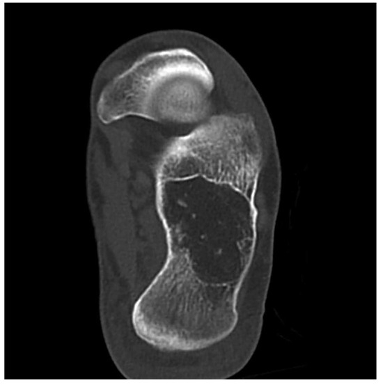
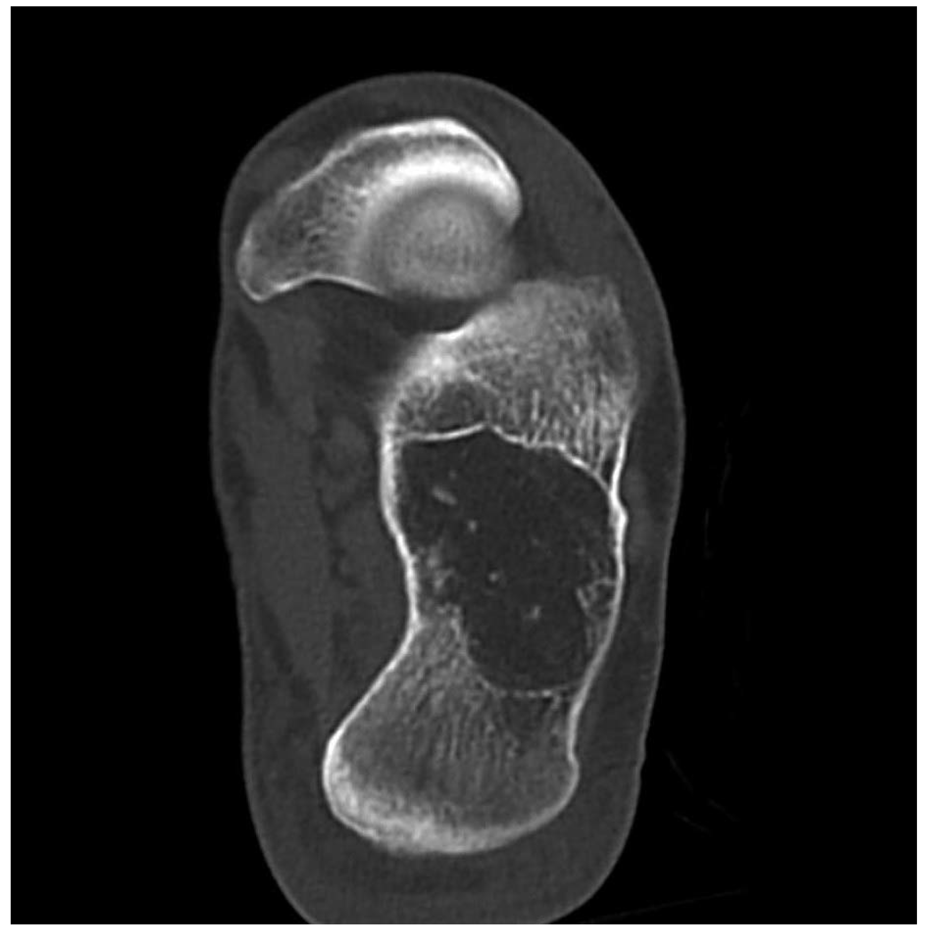
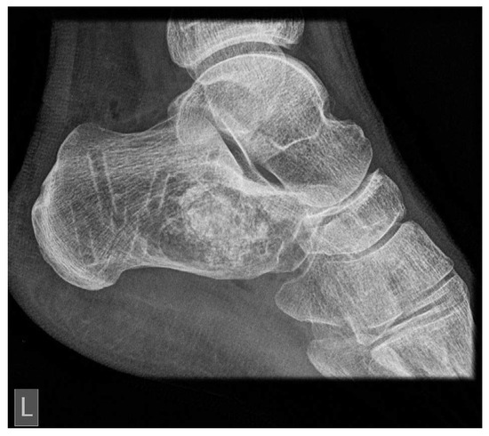
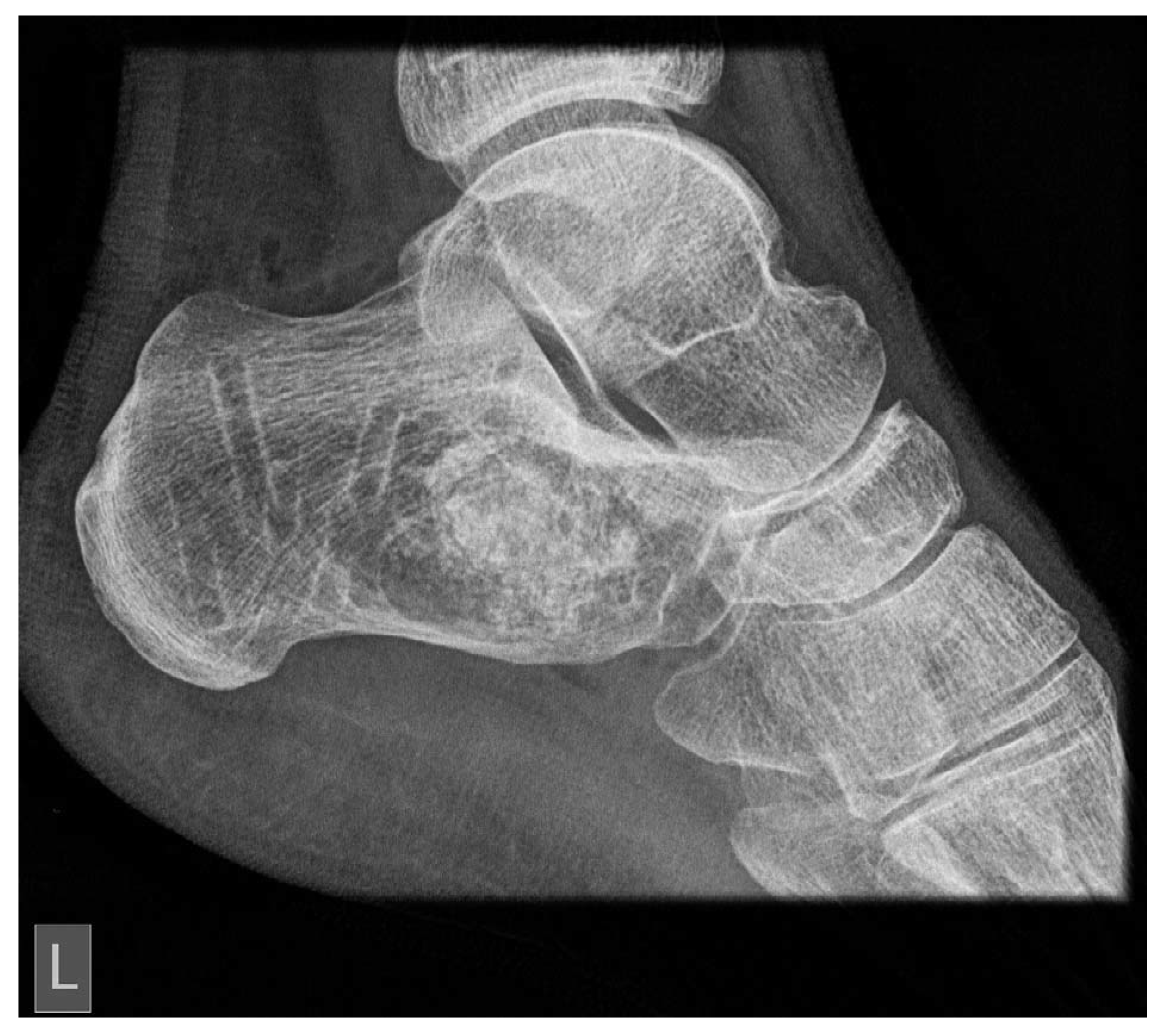
In the emergency department, findings from the overall clinical examination were normal except for the swelling under the lateral malleolus, which was painful on palpation. The patient also had typical clinical signs of plantar fasciitis, including worsening heel pain. She was referred for MRI and CT (Figure 1) of her left foot, which confirmed the diagnosis of intraosseous lipoma of the calcaneus. The volume of the intraosseous lipoma was estimated to be approximately 40% to 50% of the calcaneal volume, filling almost the complete calcaneus, from medial to lateral and inferior margin of the bone cortex. The cortex was only 1 to 2 mm wide (Figure 1).
Figure 1.
Computed tomographic image of an intraosseous lipoma in the study patient.
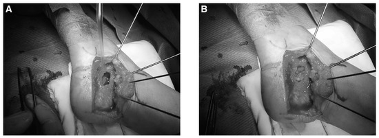
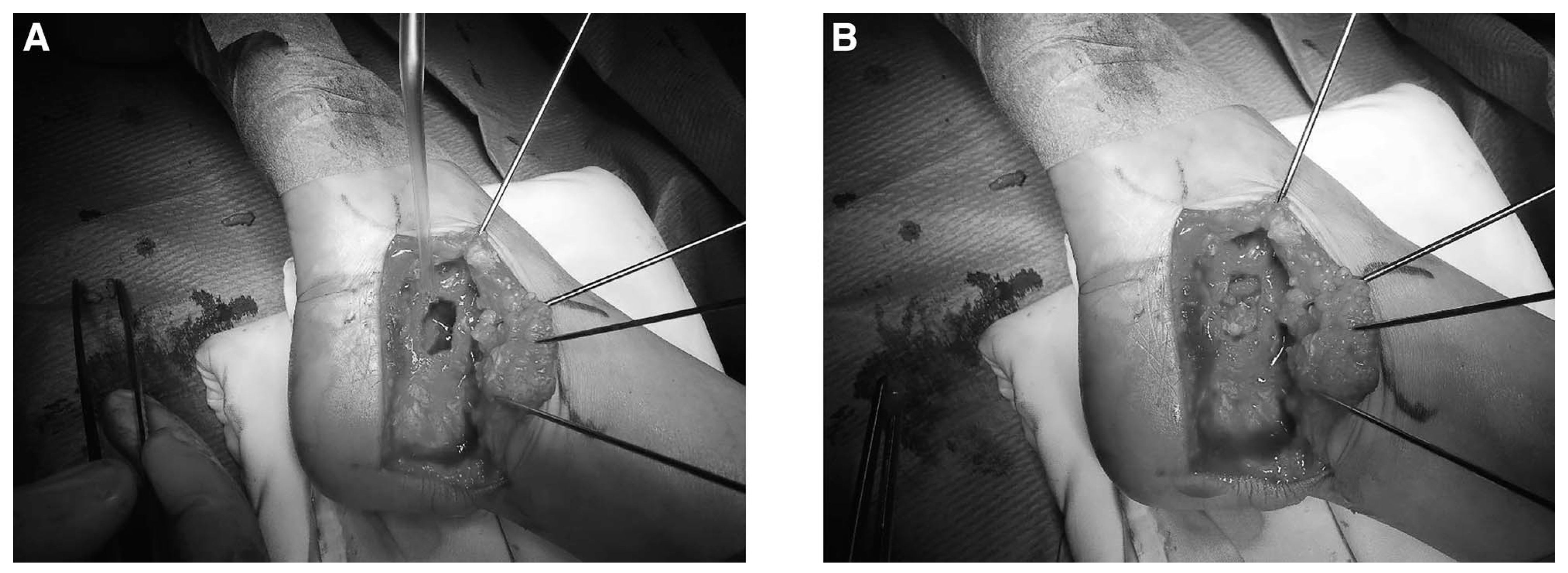
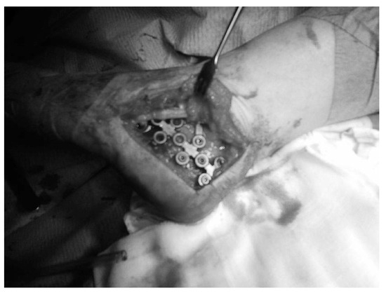
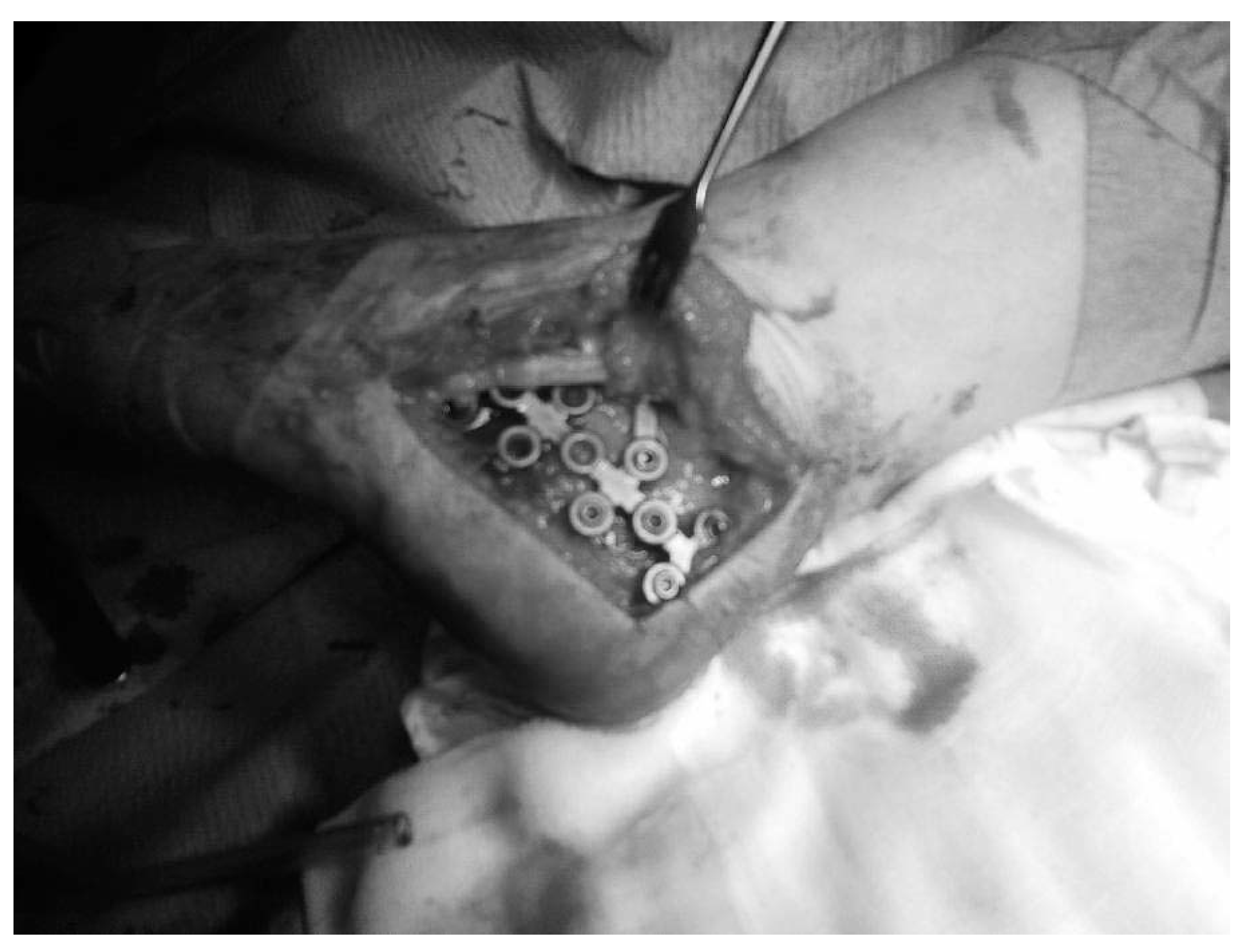
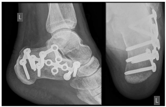
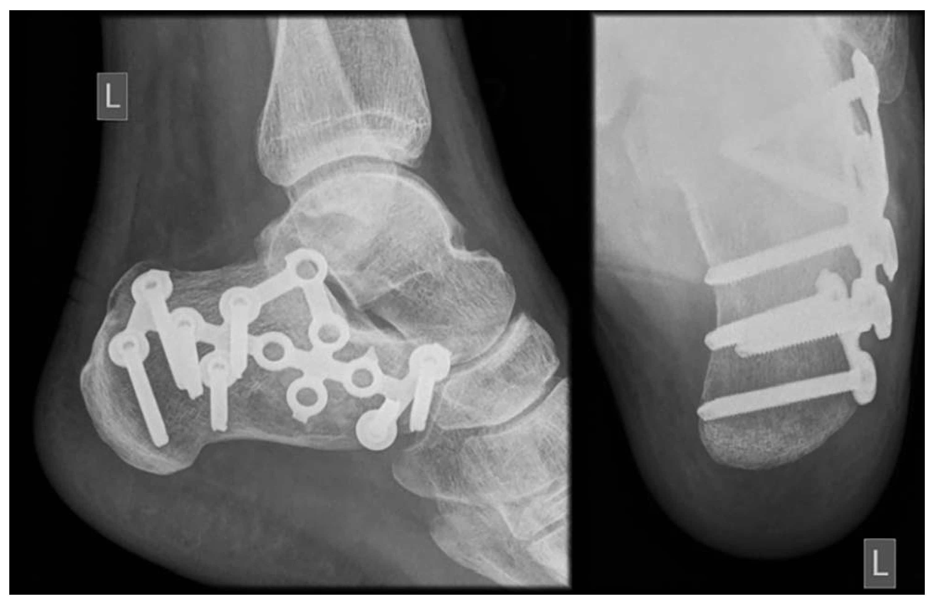
The patient was operated on under spinal anesthesia in the lateral decubitus position; we used a right-angled lateral incision to the calcaneus. Curettage of the tumor was performed through a cortical window, as well as spongioplastics of the hollow bone with autologous bone from the iliac crest (Figure 2). Because the volume of the autologous cancellous bone graft was not enough we added btricalcium phosphate. Owing to a threatening fracture of the calcaneus, internal stabilization with a calcaneal plate was necessary (Figure 3). We decided to use a lateral calcaneal locking plate (Synthes, Solothurn, Switzerland) because the patient had preserved only the bone cortex on both the medial and lateral sides, which could, therefore, lead to fracture.
Figure 2.
Intraoperative photographs of an intraosseous lipoma during curettage (A) and spongioplastics (B).
Figure 3.
Osteosynthesis of the calcaneus in the study patient using a lateral locking plate.
The histology report of tumor tissue confirmed an intraosseous lipoma. She received preoperative prophylactic doses of antibiotics and analgesics. The wound healed without complications. The patient was fully weightbearing with no aids just 2 months after surgery. Regular physical examinations were performed twice monthly, and a control radiograph of the calcaneus was performed 2 months after the operation that showed incorporation of the bone graft in the calcaneus (Figure 4). The patient was without any problems, so she was allowed to return to her daily activities.
Figure 4.
Radiograph a year after surgery showing the fully incorporated graft.
The osteosynthetic material (the calcaneal locking plate) was removed a year after the initial surgery, and radiologic control was performed, showing successfully formed new bone (Figure 5).
Figure 5.
Successfully formed new bone after osteosynthetic material removal.
Discussion
The incidence of one of the rarest benign bone tumors has increased in the past few years, mostly at the expense of widespread use of imaging diagnostics, especially CT and MRI, but also due to its typical appearance on radiography, which can be of diagnostic validity if radiologists are familiar with it [5,14]. Therefore, additional education of doctors who deal with such patients or radio- imaging should be encouraged to better recognize these radiologic features.
Because intraosseous lipoma presents with pain and sometimes swelling, it offers a wide range of differential diagnoses to a clinician. Because it is benign, misdiagnosis or failure to diagnose does not harm the patient, except when it becomes locally aggressive, consequently causing weakening of the bone and pathologic fractures [6]. In rare cases it has been described as undergoing malignant transformation, which should be concerning in patients presenting with rapid bone destruction [4,6].
However, choosing an appropriate treatment course is still controversial. Invasive treatment options, such as biopsy and surgery, are reserved for only a few patients, where symptoms such as heel pain are so severe that a patient’s life is strongly affected or there is a danger of pathologic fracture due to the large size of the intraosseous lipoma [6]. The opinion of Begatur et al [14] is that surgery is not always necessary for an intraosseous lipoma because most patients’ symptoms are relieved with conservative treatment options.
Although intraosseous lipomas are not commonly related to pathologic fractures, they can be observed in the calcaneus, as in the present patient, which describes such a course of disease. Yet, some authors report that intraosseous lipomas occur in the region called Ward’s triangle, which is a nonweight bearing area and is, therefore, unlikely to lead to fractures.[10]
We made a decision to operate based on the patient’s symptoms, the size and location of the intraosseous lipoma, and the threatening fracture. As mentioned in the study by Losito et al, [10] the present patient had a critical-size cyst, extending from the medial to the lateral cortex, filling approximately 40% of the volume of the calcaneus. That was an indication to operate because there was only calcaneus cortex left intact, exposing the patient to threatening fracture, lifelong endangerment, or even mortality [10].
In the present patient, the allograft was fully incorporated a year after the initial surgery, and the osteosynthetic material was removed because it was used only to offer more stability for the bone to heal. Also, the patient reported irritation from the osteosynthetic material, due to her thin foot, when wearing shoes and burdening the operated leg.
Futani et al [16] presented a case of bilateral calcaneal intraosseous lipomas successfully treated with endoscopic-assisted curettage, filled with B- tricalcium phosphate, allowing the patient full weightbearing the day after the surgery. The b-tricalcium phosphate dissolved 3 months after its application in the surgical procedure and was replaced by mature new bone [16].
Losito et al [10] describe treatment of calcaneal intraosseous lipoma with external fixation. Curettage was performed through the cortical window, and cancellous allograft bone was used to fill the cavity. Wound closure was followed by application of an Ilizarov external fixator for the initial postoperative period to allow early weightbearing [10].
Both cases represent good postoperative outcomes. In the case by Futani et al, [16] the lipomas were not fracture threatening; therefore, additional stabilization after curettage and filling the bone defect was not necessary. In the present patient, fixation was performed using a calcaneal plate, followed by incorporating a bone graft. Internal fixation was used considering complications found in the literature that were mentioned in connection to the Ilizarov external fixator. The only disadvantage in this patient was that she needed to undergo a second operation to remove the osteosynthetic material (the lateral locking plate). We found no published literature describing the use of internal fixation in such patients as ours.
In the case by Losito et al, [10] where postoperative weightbearing without additional stabilization could lead to calcaneal fracture, an Ilizarov external fixator was used [17]. The application of an Ilizarov external fixator is an effective way to prevent secondary fracture; however, it can be uncomfortable for the patient and also carries a high risk of pin site infection.
We decided to use a calcaneal locking plate instead of an external fixator because of the previously mentioned disadvantages. Internal fixation allowed effective stabilization until bone allograft from iliac crest mixed with b-tricalcium phosphate (which also has osteoconductive properties [18]) was fully incorporated.
Overall, the present patient’s full rehabilitation was shorter than the data found in the published literature [1,7]. As mentioned in the study by Losito et al, [10] their length of recovery was much longer than ours (9 months versus 2 months). We have not acknowledged any symptom recurrence in the present patient. The patient returned to her usual daily activities without any problems within 2 months.
There are few reports similar to the present case [6,10,15,16]. In the published literature, there are no published cases using internal fixation as a substitute for Ilizarov external fixation.
Financial Disclosure
None reported.
Conflicts of Interest
None reported.
References
- EYZAGUIRRE, E.; LIQIANG, W.; KARLA, G.M. Intraosseous lipoma: a clinical, radiologic, and pathologic study of 5 cases. Ann Diagn Pathol 2007, 11, 320. [Google Scholar] [CrossRef] [Scilit] [PubMed]
- KRISHNAN UNNI, K. Lipoma and Liposarcoma,’’ in Dahlin’s Bone Tumors: General Aspects and Data on 11.087 Cases, edited by K Krishnan Unni, p 349; Lippincott-Raven: Philadelphia, 1996. [Google Scholar]
- MIRRA, J. ‘Tumor of Fat,’’ in Bone Tumors, edited by J Mirra, p 1479; Lea & Febiger: Philadelphia, 1989. [Google Scholar]
- MILGRAM, J.W. Intraosseous lipomas: a clinicopathologic study of 66 cases. Clin Orthop Relat Res 1988, 231, 277. [Google Scholar] [CrossRef] [Scilit]
- SHIN, D.S.; KWAK, E.S.; CHOI, J.H. Intraosseous lipoma. J Korean Orthop Assoc 2003, 38, 526. [Google Scholar] [CrossRef] [Scilit]
- SORAGANVI, P.; KUKREJA, K.; RAMAKANTH, R. A rare case of aneurysmal bone cyst of the calcaneum. Foot Ankle Online J 2011, 4, 1. [Google Scholar] [CrossRef] [Scilit]
- KAPAKAYA, A.; SUBASI, M.; DABAK, N. Osseous lipoma: eleven new cases and review of the literature. Acta Orthop Belg 2006, 72, 603. [Google Scholar]
- RADL, R.; LEITHNER, A.; MACHACEK, F. Intraosseous lipoma: retrospective analysis of 29 patients. Int Othop 2004, 28, 374. [Google Scholar] [CrossRef] [Scilit] [PubMed]
- DIARD, F.; HAUGER, O.; MOINARD, M. Pseudo-cysts, lipomas, infarcts and simple cysts of the calcaneus: are there different or related lesions? JBR-BTR 2007, 90, 315. [Google Scholar] [CrossRef] [Scilit] [PubMed]
- LOSITO, J.; HERRERA, V.L.; GONZALES, R. Calcaneal intraosseous lipoma treated with external fixation: case report and review of the literature. Foot Ankle Online J 2012, 5, 1. [Google Scholar]
- BRUNI, L. The ‘‘cockade’’ image: a diagnostic sign of calcaneum intraosseous lipoma. Rays 1986, 11, 51. [Google Scholar] [PubMed]
- PROPECK, T.; BULLARD, M.A.; LIN, J. Radiologic-pathologic correlation of intraosseous lipomas. AJR Am J Roentgenol 2000, 175, 673. [Google Scholar] [CrossRef] [Scilit] [PubMed]
- MILGRAM, J.W. Malignant transformation in bone lipomas. Skeletal Radiol 1990, 19, 347. [Google Scholar] [CrossRef] [Scilit] [PubMed]
- BEGATUR, A.E.; YALCINKAYA, M.; DOGAN, A. Surgery is not always necessary in intraosseous lipoma. Orthopedics 2010, 12, 33. [Google Scholar] [CrossRef] [Scilit] [PubMed]
- ULUCAY, C.; ALTINTAS, F.; OZKAN, N.K. Surgical treatment for calcaneal intraosseous lipomas. Foot (Edinb) 2009, 19, 93. [Google Scholar] [CrossRef] [Scilit] [PubMed]
- FUTANI, H.; FUKUNAGA, S.; NISHIO, S. Successful treatment of bilateral calcaneal intraosseous lipomas using endoscopically assisted tumor resection. Antican- cer Res 2007, 27, 4311. [Google Scholar]
- MILGRAM, J.W. Intraosseous lipomas: radiologic and pathologic manifestation. Radiology 1988, 167, 155. [Google Scholar] [CrossRef] [Scilit] [PubMed]
- SCHWARZ, F.; HERTEN, M.; FERRARI, D. Guided bone regeneration at dehiscence-type defects using biphasic hydroxyapatite þ beta tricalcium phosphate (Bone Ceramict) or a collagen-coated natural bone mineral (BioOss Collagent): an immunohistochemical study in dogs. Int J Oral Maxillofac Surg 2007, 36, 1198. [Google Scholar] [CrossRef] [Scilit] [PubMed]
© 2024 American Podiatric Medical Association