


Abstract
Background: Ligamentous or joint laxity is a clinical entity characterized by increased joint mobility beyond the range of motion regarded as normal, and joint mobility is an effective indicator of the degree of laxity. We examined the influence of ligamentous laxity on the range of ankle dorsiflexion with the knee flexed and extended, comparing lax adults with a control (nonlax) group. Methods: The sample comprised 400 individuals: 200 in the control group (mean ± SD age, 32.49 ± 11.06 years) and 200 in the lax group (mean ± SD age, 29.82 ± 9.40 years). The Beighton criteria were applied to each participant to diagnose laxity or nonlaxity, and sex, age, and angle range of bilateral dorsiflexion with the knee extended and flexed were recorded. Results: The mean ± SD dorsiflexion range with the knee straight was 16.14° ± 5.29° left ankle and 21.21° ± 4.93° right ankle in the lax group and 12.94° ± 4.17° left ankle and 17.08° ± 4.40° right ankle in the control group. The respective values with the knee flexed were 15.84° ± 5.31° and 21.21° ± 4.80° in the lax group and 12.95° ± 3.95 and 17.23° ± 4.25° in the control group. Conclusions: In this sample, ankle dorsiflexion range in the lax group was 4° bigger than that in the control group in both knee positions.
Ligamentous or joint laxity is a clinical entity characterized by increased joint mobility beyond the range of motion regarded as normal.[1] Its etiology is still unknown, as is whether its isolated or benign presentation constitutes an entity in itself,[2] is simply an extreme degree of a biological variable, or is a sign of the coexistence of minor or aborted forms of other, more important, inherited entities with a discrete phenotypic expression.[3]
Genetic abnormalities have been found in collagen fibers and other proteins that form the connective tissue.[4] The alteration of these proteins makes those structures more elastic than normal but also more fragile,[5] leading to a greater risk of lesions after relatively light trauma.[6]
Joint mobility is an effective indicator of the degree of laxity,[7] but the musculoskeletal system is a complex apparatus in which other variables must be evaluated in addition to the structure of the collagen. Examples are neuromuscular tone and the configuration of the articular surfaces.[8] Furthermore, electron microscopy shows a reduction in the proportion of thick collagen fibers and concomitant increases in fine (small-diameter) collagen fibers, elastic fibers (elastin and fibrocytes), and the reticular layer.[9]
Different studies have confirmed that this clinical entity is more common in women than in men.[10-15] Its prevalence varies between 5% and 15% of the population,[16,17] a variation that is explained by the heterogeneity of the population samples in the many articles that have studied it.[18] In particular, the variation is a result of the influence of the sex, age, and race of the individuals analyzed.[12,19] For this latter parameter, for instance, the entity is more common in Asian individuals than in black Africans, and the latter are, in turn, more lax than white Europeans.[20,21]
Diagnosing laxity is purely clinical and done with a scoring system. Today, the most accurate instrument for this is the Beighton criteria.[22] These criteria are based primarily on the Beighton score (1973), which is included as an integral part of the evaluation system. Indeed, the Beighton score is still used by most authors[4,8,23-25] for their diagnosis of ligamentous laxity.
Despite not being included in this diagnostic method, range of ankle joint dorsiflexion has been proposed by several authors as a maneuver to evaluate in the diagnosis of ligamentous laxity. The first to use it were Carter and Wilkinson,[26] who added the role of the subtalar joint, specifically its increased eversion. This observation was included in their evaluation system with the item “increased ankle dorsiflexion and range of eversion of the foot,” with the two maneuvers being performed simultaneously.
Later, Kirk et al[27] used the classification of Rotés, with all of its maneuvers except that indicated previously herein, arguing that “it is a subjective maneuver” in the test denominated “feet and ankles.” Thereafter, we could find no further references to the use of hypermobility in eversion for the evaluation of this entity.
Grahame and Jenkins[28] conducted a study comparing 53 female dancers with 53 female nurses in London. They applied the Beighton maneuvers plus one more: “passive ankle dorsiflexion greater than 15° from a right angle (90° to the leg).” Silverman et al[29] mention the “usefulness of evaluating ankle dorsiflexion as a maneuver to detect ligamentous laxity.” Walker[30] included the maneuver “dorsiflexion and inversion of the ankle greater than 45°” as a clinical diagnostic test of laxity. In that study, 25.5% of the lax participants were positive in this maneuver.
Santos and Azevedo[31] used “(combined) dorsiflexion and eversion greater than 15°” as an evaluation maneuver for the presence of ligamentous laxity. Finsterbush and Pogrund[32] evaluated a sample of 100 patients (2–16 years old) to detect the presence of ligamentous laxity. They applied the Beighton criteria but replaced the maneuver of “touch the floor with the knees straight” with “increased ankle dorsiflexion.”
Ekstrand and Gilquist[33] evaluated ligamentous laxity and five movements of the lower limb, including ankle flexion with the occurrence of knee and ankle injuries in 180 soccer players (17–38 years old).
MacNeill-Shea and Mezzomo[34] also evaluated the range of ankle dorsiflexion, comparing a group of lax patients with a control group. Steele and White[35] studied 40 gymnasts aged 10 to 21 years to analyze laxity as a risk factor for injury. Their evaluation included the Carter criteria with the addition of ankle dorsiflexion with the knee both bent and straight. Francis et al[36] noted that laxity markedly affects the ankles.
Bulbena et al[17] included ankle dorsiflexion as a clinical maneuver but did not quantify the angle necessary for it to be detected as positive. They accompanied it with eversion of the foot: “excessive range of passive ankle dorsiflexion and eversion of the foot.” They thus recovered the maneuver proposed by Carter and Wilkinson. Of all of the diagnostic maneuvers evaluated, this maneuver gave the greatest frequency of positive results, distinguishing the lax group. In particular, they found positive results in 93.9% of the lax group versus 3.4% of the control group (a significant difference at P < .01). In the same vein, Bridges et al[8] examined whether, in the participants in their study, there was passive ankle dorsiflexion greater than 15°. They found 50% of the participants to be lax, with manifest hypermobility in that joint.
Subramanyam and Janaki[37] observed a high incidence of excessive ankle dorsiflexion in a group of lax children aged 6 to 15 years. Kamanli et al[25] observed 93% of ankle hypermobility in a lax group of 20 individuals (20–52 years old) that they studied in relation to a nonlax control group. Beynnon et al,[14] in an evaluation of 17 men and 17 women, found the women to have greater laxity in the ankles. O'Connor et al[38] detected a difference between men and women in the range of motion of ankle plantarflexion and described it as due to ligamentous laxity.
The objective of the present study was to examine the influence that ligamentous laxity has on the range of ankle dorsiflexion in adults, with the knee both flexed and extended, considering the possibility that this maneuver could be used as a detector of laxity.
Material and Methods
The overall study sample consisted of 400 individuals (200 in the lax group and 200 in the control group), of whom 126 were men (31.5%) and 274 were women (68.5%), aged 18 to 65 years (mean ± SD age, 31.16 ± 10.34 years). The control (nonlax) group comprised 89 men (44.5%) and 111 women (55.5%) aged 19 to 65 years (mean ± SD age, 32.49 ± 11.06 years). The lax group comprised 37 men (18.5%) and 163 women (81.5%) aged 18 to 61 years (mean ± SD age, 29.82 ± 9.40 years). The measurements were taken during 2013 and 2014.
The inclusion criteria were 1) age 18 to 65 years because epiphyseal plate closure is at 17 years of age and after 60 years of age osteoarthritic processes may limit the joints[39] and 2) white race/ethnicity because race is a factor in the prevalence of benign joint hypermobility syndrome,[40] with it being less frequent in white populations.[13,15,21]
The exclusion criteria were 1) that the individual experienced a degenerative disease (such as osteoarthritis or rheumatoid arthritis) or neuromuscular, inflammatory, or metabolic diseases such as gout; 2) surgery within the previous 6 months,[41] including the placement of an endoprosthesis[42]; 3) injury to the musculoskeletal system within the previous 6 months[25]; 4) use of lower-limb orthoses during the past 6 months, because this would influence the movement of the foot joints[43]; and 5) pregnancy, because the range of movement of the joint may be increased by greater laxity.[44]
The study protocol was in accordance with standard ethical and human research principles. Written informed consent for participation and publication was given by each patient, including for the publication of photographs. The study was approved by the Research Ethics Committee of the Faculty of Health Sciences, University of Seville (Seville, Spain).
Outcome Measurement
For the evaluation of patients, we used as the measuring instrument a two-arm plastic goniometer. This is a well-accepted and extensively used instrument for measuring the angle of articular rotation from the center of the motion.[17] Being a simple instrument of everyday use, it was important that there was no maladjustment between the arms. To this end, we selected for the study a transparent model of the instrument in which the coincidence of the arms was perfectly visible to the naked eye for all angle values.[39] Nonetheless, to avoid errors inherent in the instrument, all of the measurements were made with the same goniometer.
The Beighton laxity score was used to evaluate bilateral joint and trunk ligamentous laxity.[45] The nine items of the score were scored as “yes” (1) or “no” (0). The scores of all of the elements were summed to give an overall ligamentous laxity score ranging from 0 to 9. A participant was considered to be lax with a score of 5 or more. All of the tests were performed by the same researcher (S.B.-B.).
Procedure
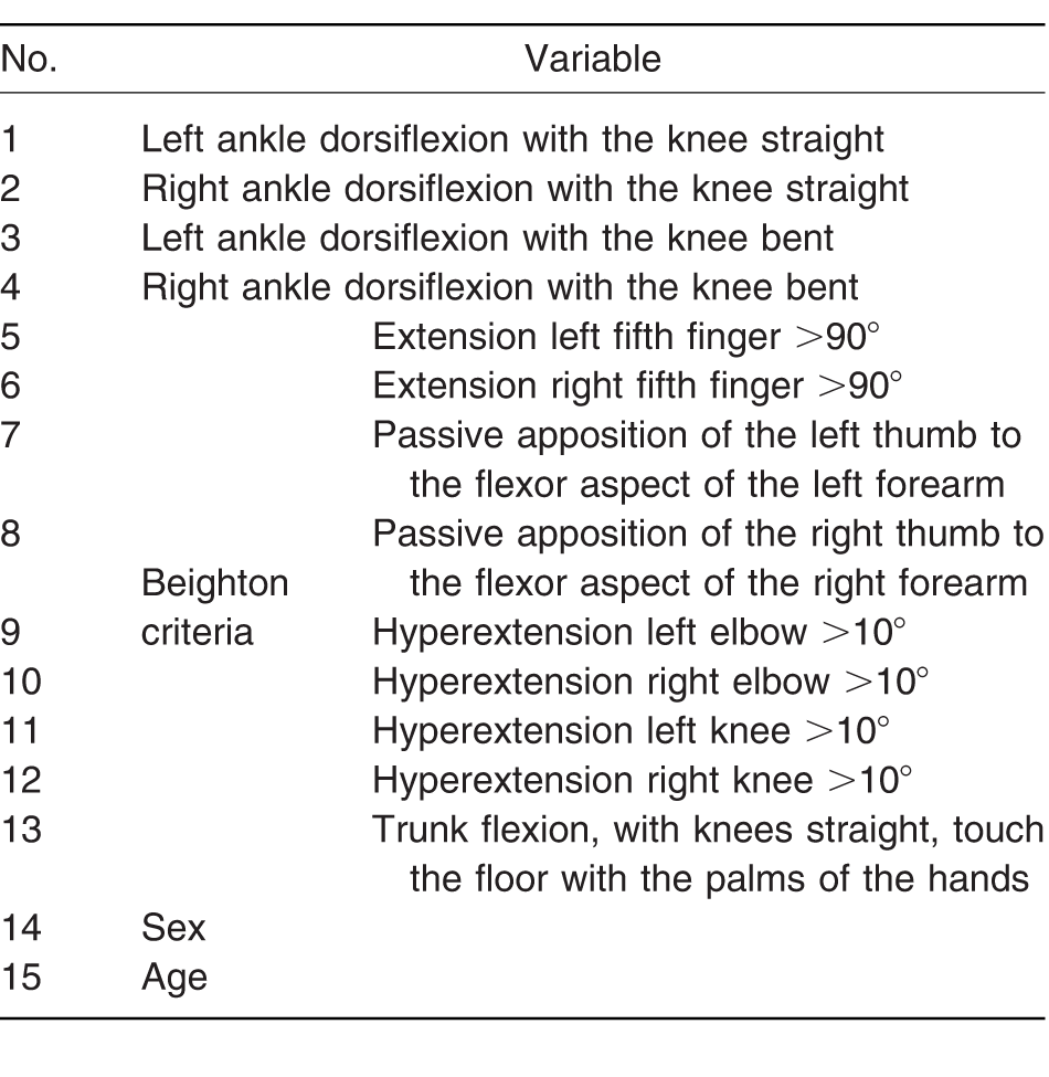
The examination protocol consisted of recording the sex, age, and 13 measurement variables (Table 1).
Table 1.
List of Variables Studied
Variable 1: Passive Dorsiflexion of the Left Ankle with the Knee Straight
The participant lies on a stretcher, supine, with the trunk flexed at approximately 30° to facilitate relaxation and comfort (Fig. 1). To quantify the angle of ankle dorsiflexion, the examiner used the goniometer as follows[34,46]: One of the arms was placed on the lateral bisection of the participant's left leg, with the individual holding it in place with the right hand for it not to move during the exploratory maneuver and, thus, avoid erroneous measurements. The other arm was placed on the lateral part of the outer edge of the foot, parallel to the plantar surface, with the center of rotation of the goniometer on the lateral aspect of the heel.[47] This arm was held in place by the examiner's right hand.
Figure 1.
Passive dorsiflexion of the left ankle with the knee straight.
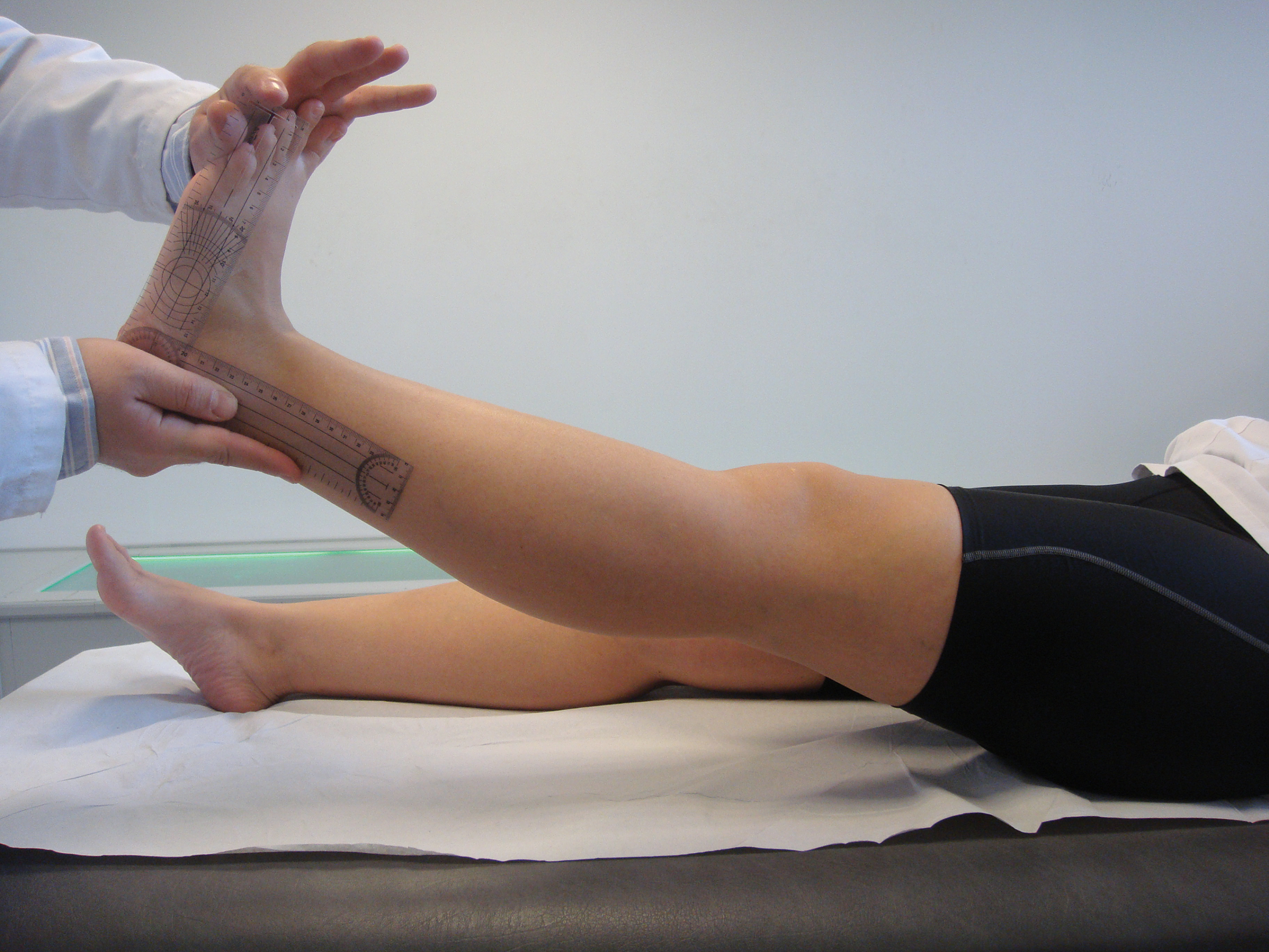
The exploratory maneuver consisted of the examiner making a pure passive dorsiflexion movement on the participant's left ankle.[48] This gesture had to avoid any pronosupination movement that the subtalar or midtarsal joints might make during the maneuver.[49] To this end, the palm of the examiner's left hand was placed in contact with the plantar and distal zone of the participant's foot while performing the ankle dorsiflexion, keeping the subtalar and midtarsal joints in an estimated neutral position during the movement.[50] The thenar zone of the examiner's right hand was responsible for holding the arm of the goniometer in place, preventing it from moving from its original position.[51] In performing the maximum passive dorsiflexion, the examiner noted the goniometer's measurement of the angle reached from the null position (considered to be the right angle formed by the foot and the leg) and recorded it on the exploration sheet.[49] Throughout this exploratory maneuver, the individual's left knee was maintained extended.[52]
Variable 2: Passive Dorsiflexion of the Right Ankle with the Knee Straight
This clinical test was performed in the same way as described for variable 1 except that the exploration was performed on the individual's right ankle. In this case, the examiner's hands were placed as follows: the left hand held the arm of the goniometer placed on the lateral aspect of the right tibia, and the right hand performed the passive dorsiflexion movement at the same time as holding the arm corresponding to the lateral part of the foot.[34]
Variable 3: Passive Dorsiflexion of the Left Ankle with the Knee Bent
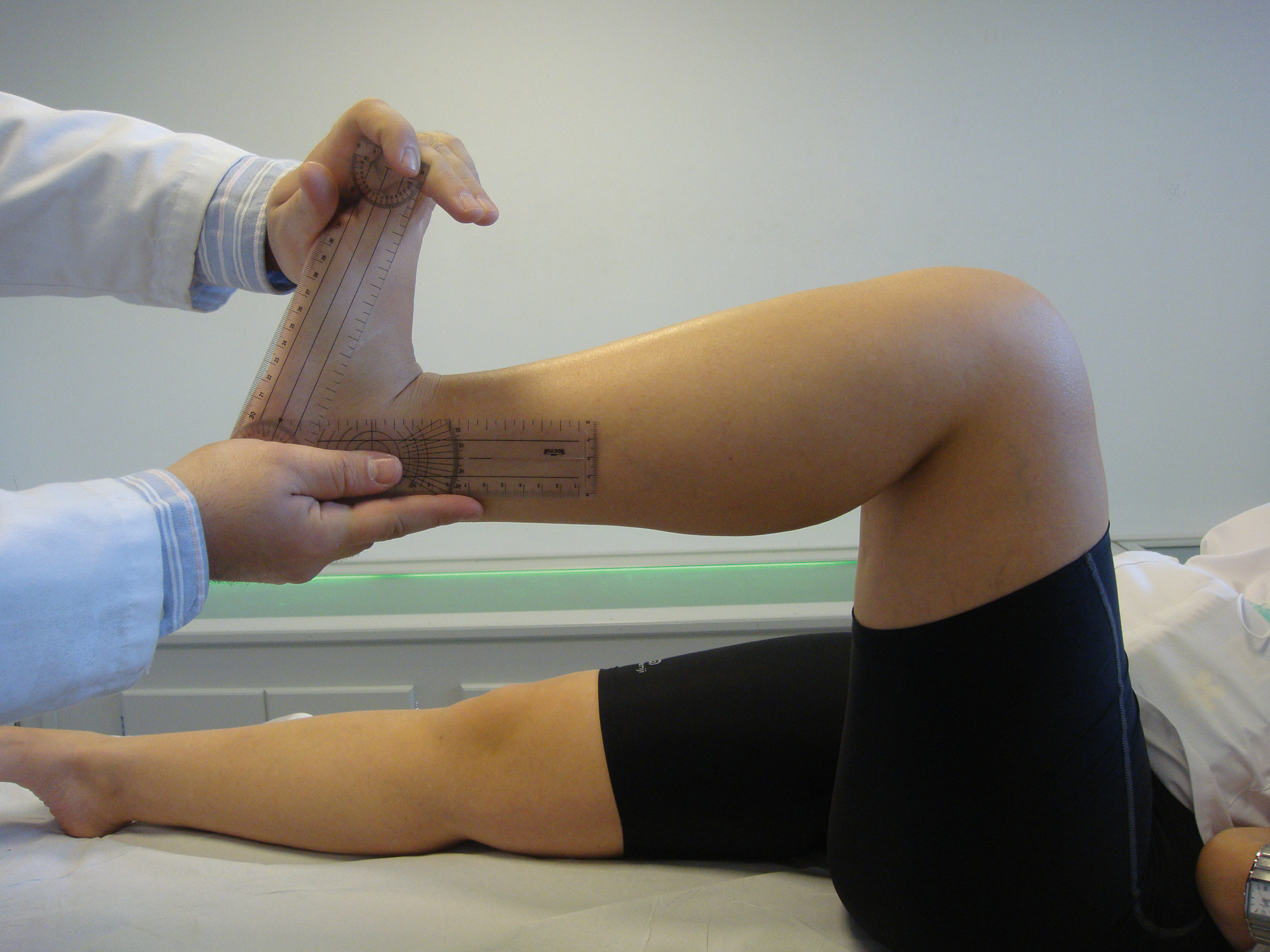
This maneuver was performed in the same way as for variable 1 except that in this case the knee was previously flexed at 90° (Fig. 2).[46,53] The 90° flexion of the knee obviates any possible role of retraction of the gastrocnemius muscles, which could limit the angle of dorsiflexion.[51,52]
Figure 2.
Passive dorsiflexion of the left ankle with the knee bent.
Variable 4: Passive Dorsiflexion of the Right Ankle with the Knee Bent
This maneuver was performed in the same way as for variable 3 except that it was on the right ankle.
Before quantifying the dorsiflexion angle, the examiner proceeded to manipulate the joint with the intention of relaxing the individual, facilitating the explanation of the maneuver to be performed and optimizing the individual's collaboration. Then the corresponding test was conducted. In this way, we first obtained a visual estimate of the maneuver, and then quantified it.[50] The dorsiflexion angle was measured thrice and the mean was recorded.[23] The three evaluations were separated by an interval of 3 sec.[53] Variables 1 to 4 were measured by a single examiner (R.G.P.). Subsequently, each participant was evaluated by another examiner (M.D.J.C.) and allocated to the control group or the lax group depending on the Beighton score[45] (variables 5–13).
Sample Size
The number of participants in the study population was determined using Epidat 4.1 software. Sample size was determined using the Freeman test, which implies the need to include, at least, ten participants for each variable studied. In the present study, 13 variables (four related to ankle flexion and nine related to the Beighton criteria) were measured, hence, 130 participants would be required. However, 200 participants per group (N = 400) were included.
Statistical Analysis
In the statistical analysis, the quantitative variables are reported as either mean ± SD or, in the case of asymmetrical distributions, medians, quartiles (P25 and P75), and ranges.
For the comparison of the quantitative variables between subgroups, the nonparametric Mann-Whitney U test was applied because the variables were not normally distributed. We determined the 95% confidence intervals for the significant differences in means or medians. Differences were considered to be statistically significant at P<.05.
Results
Table 2 presents the ankle dorsiflexion values obtained. There were significant differences (P < .001) between the two groups (lax and control), both with the knee straight and bent, and for both feet. The differences were of approximately 4° in magnitude.
Table 2.
Descriptive Statistics of the Ankle Dorsiflexion by Group, Laterality, and Knee Position
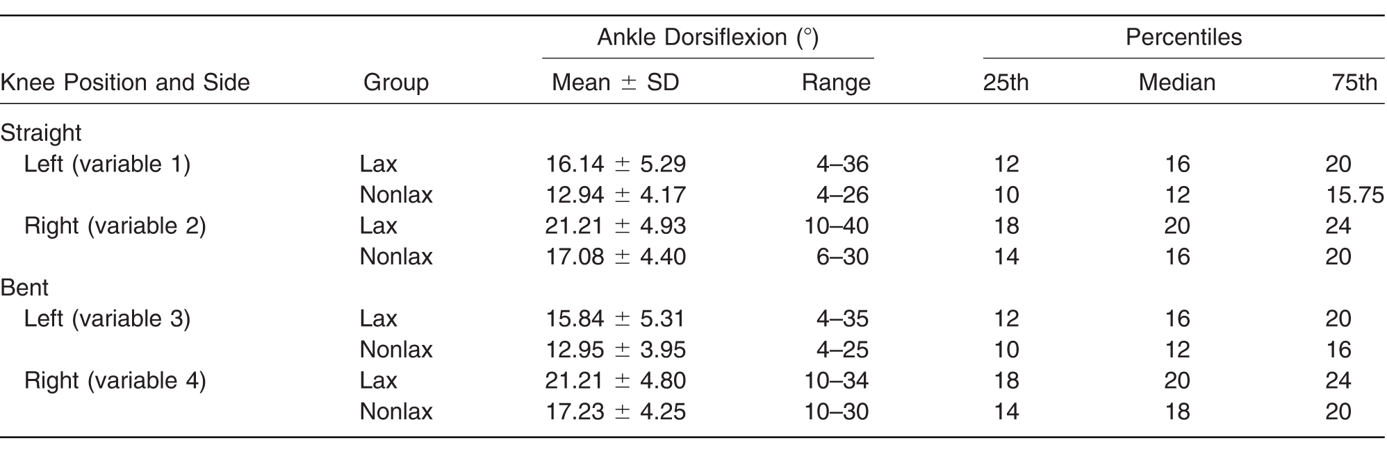
We also found significant differences (P<.05) in the values of dorsiflexion between the right ankle and the left in both groups, and in both positions of the knee. Based on laterality, differences of approximately 5° were obtained for all of the cases.
Discussion
Ankle dorsiflexion (a movement that is widely used in the protocol of clinical examination) presented greater articular ranges in the lax group, both with the knee straight and with it flexed at 90°, and for both ankles. These results are discordant with those reported by MacNeill-Shea and Mezzomo[34] in their study of 29 individuals (14–20 years old), in which no significant differences were found in dorsiflexion between the lax group and the control group.
The variability of the present results with respect to those of other studies is shown in Table 3. We would explain it as being attributable to the heterogeneity of those studies regarding the age of the sample, the method of measurement, and the position of the knee.
Table 3.
Comparison of Ankle Dorsiflexion Obtained in the Present Study Compared with Other Studies
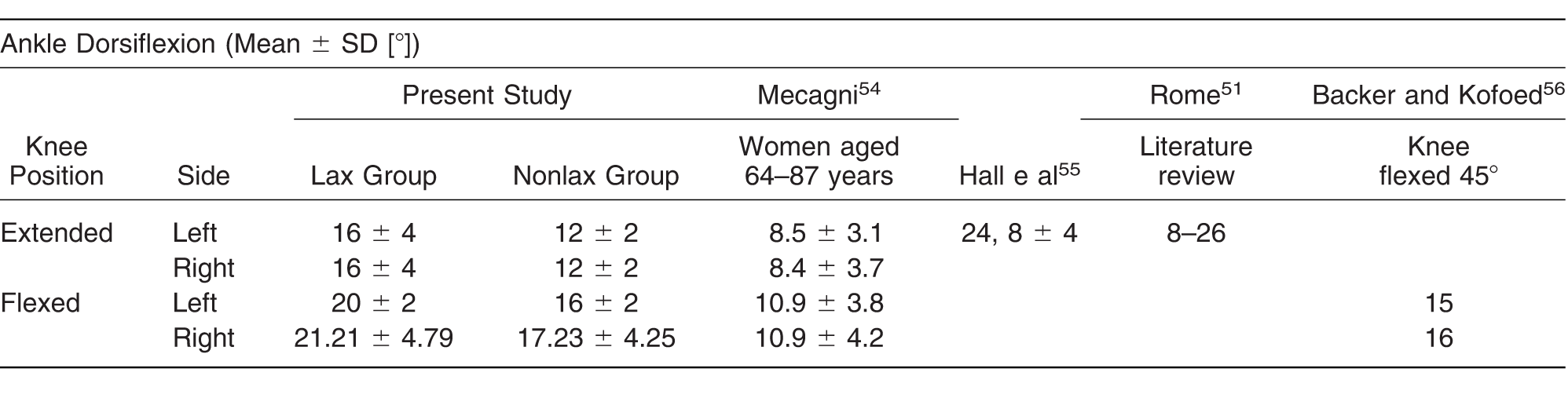
Pearsall et al[53] state that the presence of laxity in the ankle is not indicative of generalized laxity. They also suggest evaluating each joint separately rather than overall laxity. In the same sense, MacNeill-Shea and Mezzomo[34] indicate that their evaluation of dorsiflexion in lax patients did not yield any significant results.
These results contrast with studies that support the role of this joint in the diagnosis of laxity. Thus, Bulbena et al[17] report that the maneuver of “excessive ankle dorsiflexion and eversion of the foot” was positive in 93.9% of the lax group versus 3.4% of the control group (P < .001). Walker[30] indicates a 25.5% presence of the clinical diagnostic maneuver of laxity “ankle dorsiflexion and inversion greater than 45°.”
Its measurement is also referred to by other authors. Azevedo and Santos[57] explored children aged 6 to 17 years and found that girls had greater mobility than boys in the wrists, elbows, and knees but significantly less (P < .001) than boys in the ankles. But Wilkierson and Mason[58] maintain that women present greater laxity in the ankle than men.
Regarding the method used to measure ankle dorsiflexion, it is the most commonly used by the scientific community taking into account the role of the posterior muscles of the leg.[52] According to Evans and Scutter,[59] exploration applied to the extended and flexed knee presents a good intraobserver coefficient.
Many authors[46,60] set the points of location of the goniometer arms on the head of the fibula/lateral malleolus (the fixed arm) and the lateral malleolus/fifth metatarsal head (the mobile arm). We decided, however, following Jonson and Gross[61] and Mecagni,[54] to apply the fixed arm on the lateral aspect of the leg and the mobile arm on the outer edge of the foot[47] parallel to the plantar aspect so as to avoid a possible forefoot equinus affecting the result.
We noted that ligamentous laxity influences the ankle's range of dorsiflexion. It must be borne in mind that the diagnosis of this entity is purely clinical,[19,62] so that the evaluation of this articular range could be used as an element detecting a suspicion of laxity, to later be confirmed with the Beighton criteria.
Furthermore, we found differences between the mean values of dorsiflexion of the two feet. This was so in both groups and for both positions of the knee. Although it was not an objective of the study, with the identification of each participant's dominant side, the results lead us to think that laterality influences the articular range. This had already been observed by other researchers who found differences in comparing their results between the joints of the left and right sides. An example is Wood,[63] who studied 337 women (15–65 years old) to evaluate the hypermobility of the elbow. There was 30% more hyperextension in the left elbow than in the right elbow. That author concluded that the elbow's range of movement tends to be greater for the nondominant arm.
Acasuso-Díaz et al[64] used a modification of the Beighton criteria, “the nondominant Spanish modification,” applying the score, therefore, to the nondominant side. The results of Rikken-Bultman et al[10] confirmed that laxity affects most prevalently the nondominant side. Verhoeven et al[65] examined 705 African women (9–36 years old) and found a prevalence of laxity on the dominant side of 19.2% compared with 23.4% on the nondominant side.
As limitation in this study, based on the obtained results, it had been recommended to include the study of the lateral dominance in each participant to determine its influence in the laxity of each side of the body.
Conclusions
The ankle dorsiflexion range in the lax group is 4° bigger than that in the control group in both positions of the knee (straight and bent).
Financial Disclosure
None reported.
Conflict of Interest
None reported.
References
- Hudson N.Starr MR.Esdaile JM.et al: Diagnostic associations with hypermobility in rheumatology patients. Br J Rheumatol34: 1157, 1995.
- Bulbena A.Aqulló A.Pailhez G.et al: Is joint hypermobility related to anxiety in a nonclinical population also?Psychosomatics45: 432, 2004.
- De Inocencio J.Ocaña I.Benito L.Laxitud articular: prevalencia y relación con dolor musculoesquelético. An Pediatr (Barc)61: 162, 2004.
- McCormack M.Briggs J.Hakim A.et al: Joint laxity and the benign joint hypermobility syndrome in student and professional ballet dancers. J Rheumatol31: 173, 2004.
- Bravo J.Significado e importancia de estudiar a las personas con hiperlaxitud articular. Rev Chil Reumatol24: 4, 2008.
- Menéndez FM.De la laxitud a la hipermovilidad articular. Rev Cuba Reumatol7: 1, 2005.
- Bird HA.Joint hypermobility. Musculoskeletal Care5: 4, 2007.
- Bridges AL.Smith E.Reid J.Joint hypermobility in adults referred to rheumatology clinics. Ann Rheum Dis51: 793, 1992.
- Bravo J.Importancia de la hipermovilidad articular como causa frecuente de morbilidad, no sólo músculo–esquelética, sino también sistémica: criterios diagnósticos. Rev Chil Reumatol19: 33, 2003.
- Rikken-Bultman DG.Wellink L.van Dongen PW.Hypermobility in two Dutch school populations. Eur J Obstet Gynecol Reprod Biol73: 189, 1997.
- Qvindesland A.Jónsson H.Articular hypermobility in Icelandic 12-year-olds. Rheumatology (Oxford)38: 1014, 1999.
- Seow CC.Chow PK.Khong KS.A study of joint mobility in a normal population. Ann Acad Med Singapore28: 231, 1999.
- Klemp P.Williams SM.Stansfield SA.Articular mobility in Maori and European New Zealanders. Rheumatology (Oxford)41: 554, 2002.
- Beynnon BD.Bernstein IM.Belisle A.et al: The effect of estradiol and progesterone on knee and ankle joint laxity. Am J Med Sports33: 1298, 2005.
- Remvig L.Jensen DV.Ward RC.Are diagnostic criteria for general joint hypermobility and benign joint hypermobility syndrome based on reproducible and valid tests? a review of the literature. J Rheumatol34: 798, 2007.
- Yunchoy H.Síndrome de hipemovilidad articular. Rev Reumatol17: 74, 2001.
- Bulbena A.Duro JC.Porta M.et al: Clinical assessment of hypermobility of joints: assembling criteria. J Rheumatol19: 115, 1992.
- Murray KJ.Woo P.Benign joint hypermobility in childhood. Rheumatology (Oxford)40: 489, 2001.
- Van der Giessen LJ.Liekens D.Rutgers KJM.et al: Validation of Beighton score and prevalence of connective tissue signs in 773 Dutch children. J Rheumatol28: 2726, 2001.
- March L.Silman A.Joint hyperlaxity: is there a case for screening?Br J Rheumatol32: 91, 1993.
- Bird HA.Joint hypermobility in children. Rheumatology (Oxford)44: 703, 2005.
- Grahame R.Pain, distress and joint hyperlaxity. Jt Bone Spine67: 157, 2000.
- Van Andel CJ.Roescher WB.Tromp MF.et al: Quantification of wrist joint laxity. J Am Soc Surg Hand33: 667, 2008.
- Mikkelsson M.Salminen K.Kautianen H.Joint hypermobility is not a contributing factor to musculoskeletal pain in pre–adolescents. J Rheumatol23: 1963, 1996.
- Kamanli A.Sahin S.Ozgocmen S.et al: Relationship between foot angles and hypermobility scores and assessment of foot types in hypermobile individuals. Foot Ankle Int25: 101, 2004.
- Carter C.Wilkinson J.Persistent joint laxity and congenital dislocation of the hip. J Bone Joint Surg Br46: 40, 1964.
- Kirk JA.Ansell BM.Bywaters EGL.The hypermobility syndrome: musculoskeletal complaints associated with generalized joint hypermobility. Ann Rheum Dis26: 419, 1967.
- Grahame R.Jenkins JM.Joint hypermobility: asset or liability? a study of joint mobility in ballet dancers. Ann Rheum Dis31: 109, 1972.
- Silverman L.Constine WH.Grahame R.Survey of joint mobility and in vivo skin elasticity in London schoolchildren. Ann Rheum Dis34: 177, 1975.
- Walker JM.Generalized joint laxity in Iglookik Eskimos and in island lake Amerindians. Hum Biol47: 263, 1975.
- Santos MC.Azevedo ES.Generalized joint hypermobility and black admixture in school children of Bahia, Brazil. Am J Phys Anthropol55: 43, 1981.
- Finsterbush A.Pogrund H.The hypermobility syndrome: musculoskeletal complaints in 100 consecutive cases of generalized joint hypermobility. Clin Orthop Relat Res168: 124, 1982.
- Ekstrand J.Gilquist J.The frequency of muscle tightness and injuries in soccer players. Am J Sports Med10: 75, 1982.
- MacNeill-Shea S.Mezzomo JM.Relationship of ankle strength and hypermobility to squatting skills of children with Down syndrome. Phys Ther65: 1958, 1985.
- Steele V.White J.Injure prediction in female gymnasts. Br J Sports Med22: 31, 1986.
- Francis H.March L.Terenty T.et al: Benign joint hypermobility with neuropathy: documentation and mechanism of tarsal tunnel syndrome. Br J Rheumatol14: 577, 1987.
- Subramanyam V.Janaki KV.Joint hypermobility in south Indian children. Indian Pediatr33: 771, 1996.
- O'Connor K.Bragdon G.Baumhauer JF.Sexual dimorphism of the foot and ankle. Orthop Clin North Am37: 569, 2006.
- Lafuente G.Patrón rotador de la extremidad inferior: un nuevo parámetro exploratorio: relación con el hallux límitus [tesis], Departamento de Podología, Universidad de Sevilla, Seville, Spain, 2006.
- Lin CJ.Lai KA.Kuan TA.et al: Correlating factors and clinical significance of flexible flatfoot in preschool children. J Pediatr Orthop21: 378, 2001.
- Ferrari J.Parslow C.Lim E.et al: Joint hypermobility: the use of a new assessment tool to measure lower limb hypermobility. Clin Exp Rheumatol23: 413, 2005.
- Hirsch C.Hirsch M.John MT.et al: Reliability of the Beighton Hypermobility Index to determinate the general joint laxity performed by dentists. J Orofac Orthop5: 342, 2007.
- Munuera PV.Domínguez G.Palomo I.et al: Effects of rearfoot-controlling orthotic treatment on dorsiflexion of the hallux in feet with abnormal subtalar pronation: a preliminary report. JAPMA96: 283, 2006.
- Marnarch ML.Ramin KD.Ramsey PS.et al: Characterization of the relationship between joint laxity and maternal hormones in pregnancy. Obstet Gynecol101: 331, 2003.
- Beighton P.Solomon L.Soskolne C.Articular mobility in an African population. Ann Rheum Dis32: 413, 1973.
- Kilgour G.McNair P.Stott NS.Intrarater reliability of lower limb sagittal range-of-motion measures in children with spastic diplegia. Dev Med Child Neurol45: 391, 2003.
- Pandya S.Florence J.King WM.et al: Reliability of goniometric measurements in patients with Duchenne muscular dystrophy. Phys Ther65: 1339, 1985.
- Woodburn J.Video joint angle position analysis of the effects of subtalar joint position on maximum ankle joint dorsiflexion. J Br Podiatr Med46: 1, 1991.
- Scharfbillig R.Scutter SD.Measurement of foot dorsiflexion: a modified Lidcombe template. JAPMA94: 573, 2004.
- Youdas JW.Bogard CL.Suman VJ.Reliability of goniometric measurements and visual estimates of ankle joint active range of motion obtained in a clinical setting. Arch Phys Med Rehabil74: 1113, 1993.
- Rome K.Ankle joint dorsiflexion measurement studies: a review of the literature. JAPMA86: 205, 1996.
- Johanson MA.Cooksey A.Hillier C.et al: Heel lifts and the stance phase of gait in subjects with limited ankle dorsiflexion. J Athl Train41: 159, 2006.
- Pearsall AW.Kovaleski JE.Heitman RJ.et al: The relationships between instrumented measurements of ankle and knee ligamentous laxity and generalized joint laxity. J Sports Phys Fitness46: 104, 2006.
- Mecagni C.Balance and ankle range of motion in community-dwelling women aged 64 to 87: a correlational study. Phys Ther80: 1004, 2000.
- Hall MG.Ferrell WR.Sturrock RD.et al: The effect of the hypermobility syndrome on knee joint propioception. Br J Rheumatol34: 121, 1995.
- Backer M.Kofoed H.Passive ankle mobility. J Bone Joint Surg Br71: 696, 1989.
- Azevedo ES.Santos MCN.Joint mobility in children: a population study. Acta Anthropog6: 33, 1982.
- Wilkierson RD.Mason MA.Differences in men's and women's mean ankle ligamentous laxity. Iowa Orthop J20: 46, 2000.
- Evans AM.Scutter SD.Sagittal plane range of motion of the pediatric ankle joint: a reliability study. JAPMA96: 418, 2006.
- Van Gheluwe B.Kirby KA.Roosen P.et al: Reliability and accuracy of biomechanical measurements of the lower extremities. JAPMA92: 317, 2002.
- Jonson SR.Gross MT.Intraexaminer reliability, interexaminer reliability, and mean values for nine lower extremity skeletal measures in healthy naval midshipmen. J Orthop Sports Phys Ther25: 253, 1997.
- Simmonds JV.Keer RJ.Hypermobility and the hypermobility syndrome, part 2: assessment and management of hypermobility syndrome: illustrated via case studies. Man Ther13: e1, 2008.
- Wood PHN.Is hypermobility a discrete entity?Proc R Soc Med64: 690, 1971.
- Acasuso-Díaz M.Collantes-Estévez E.Sánchez P.Joint hyperlaxity and musculoligamentous lesions: study of a population of homogeneus age, sex and physical exertion. Br J Rheumatol32: 120, 1993.
- Verhoeven JJ.Tuinman M.van Dongan PW.Joint hypermobility in African non-pregnant nulliparous women. Eur J Obstet Gynecol Reprod Biol82: 69, 1999.
© 2018 American Podiatric Medical Association