


Abstract
Background: Over a 74-month period (∼6 years), 143 lower-extremity osteomyelitis locations in 125 patients were treated with a calcium sulfate/hydroxyapatite liquid bone void filler with antibiotic(s). Methods: The osteomyelitis locations were treated with a percutaneous antibiotic delivery technique delivering intraosseous antibiotic followed by either oral or intravenous antibiotics for 4 weeks. Results: There was no recurrence of osteomyelitis in 96.15% of the treatable patients. Outcomes classified by the Cierny-Mader clinical classification are discussed as well. Conclusions: A bone void filler with antibiotic(s) using the percutaneous antibiotic delivery technique is a safe, reliable, and effective means to treat lower-extremity osteomyelitis with either oral or intravenous antibiotics for 4 weeks.
If you ask ten different physicians their opinion on how to treat osteomyelitis, many different and varying answers may be received. One general consensus is that osteomyelitis management has not evolved a great amount aside from newer antibiotics. The Food and Drug Administration has given no antibiotic indication for osteomyelitis treatment. Antibiotics may be less effective in treating areas of necrotic bone with biofilm formation because the antibiotic is impaired in penetrating the infected bone.[1] Suitable drugs are not yet available for the eradication of biofilm-producing bacteria.[2] Antibiotic regimens alone do not have strong evidence and have been shown to limit success in treating lower-extremity osteomyelitis.[3,4] Studies have been performed that support lower-extremity osteomyelitis treatment with antibiotics alone, but they have reported success between 63% and 83%.[3-5] In a 50-patient study of nonsurgical treatment of osteomyelitis with antibiotics after bone biopsy, after 13 months of treatment there was a 64% remission rate.[6] However, these studies were performed on outpatients, and the extent of infection may be less severe and conservative treatment is more likely to succeed. The Infectious Diseases Society of America guidelines recommended antibiotic drug therapy for 1 to 3 weeks for any residual soft-tissue infection only, 4 to 6 weeks for residual but viable osteomyelitic bone present, or at least 3 months for nonoperative cases.[7] Also of importance is that these studies show that 13% to 28% of patients may worsen during treatment and require early surgery.
Buchholz and Klemm's surgical management of osteomyelitis has not evolved greatly from bone debridement and placement of antibiotic polymethyl methacrylate (PMMA) beads for dead space management.[8,9] When Buchholz started using gentamicin in 1971, his infection rate decreased from 5.9% to 1.1%.[10] This surgical management emphasized staged debridement, bone grafting, healing by secondary intention, long-term antibiotic administration, and rehabilitation.[11] Mader et al[12] reported a 78% success rate with bone debridement, 4 weeks of antibiotic therapy, and hyperbaric oxygen therapy. In the study by Cierny,[13] long-term outcomes of the overall success rate, classified by host type but not location, after initial treatment were 96% in A hosts and 74% in B hosts, and overall success rates in patients requiring retreatment were 99% in A hosts and 90% in B hosts. Walenkamp et al[14] reported a recurrence rate of 45.4% in 184 lower-leg/ankle cases and 2.5% in ten foot cases. In a comparison of 380 patients who received prolonged antibiotic treatment only, PMMA beads alone, or a combination of both, the success rates were 76%, 57%, and 71%, respectively.[15] There were no data regarding osteomyelitis location. Case studies and reports in the literature for antibiotic carriers other than PMMA do not offer strong data, outcomes, or long-term follow-up.[16,17] There are recent European conference reports of lower osteomyelitis reoccurrence rates and higher antibiotic bone levels in defects using a calcium sulphate/hydroxyapatite biocomposite with gentamicin.[18-21] Techniques for using a calcium sulphate/hydroxyapatite biocomposite with antibiotic as an implantable device has been previously described in treating lower-extremity osteomyelitis.[22] A limitation of PMMA and other nonabsorbable antibiotic carrier vehicles is that they do not elude enough of the their antibiotic to have a bactericidal effect on the biofilm found in osteomyelitis or to penetrate into microchannels and disvascular bone cavities.[23,24] This limitation contributes to nontraumatic lower-extremity amputation. Of those nontraumatic lower-extremity amputations, approximately 60% involve osteomyelitis.[25] Faglia et al,[26] in reporting on 350 patients with diabetic foot osteomyelitis, stated a higher rate of transtibial amputation when osteomyelitis involved the heel instead of the midfoot or forefoot in diabetic patients.
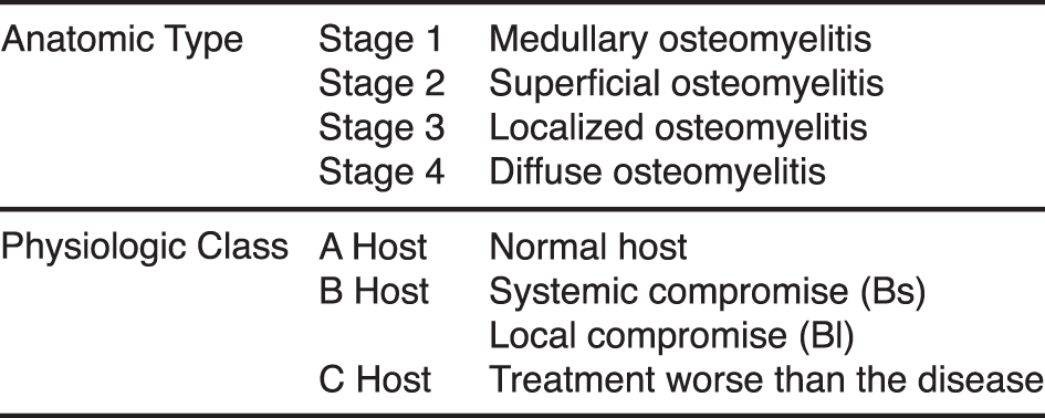
Osteomyelitis can be classified by duration (acute or chronic), pathogenesis (trauma, contiguous spread, hematogenous, surgical), site, extent, or patient type. A common classification system widely used is the Cierny-Mader clinical classification system (Fig. 1).[27] The Cierny-Mader clinical classification system is based on anatomical, clinical, and radiologic features and characterizes osteomyelitis as being in one of four anatomical stages. In stage 1 (medullary), osteomyelitis is confined to the medullary cavity of the bone. Stage 2 (superficial) osteomyelitis involves only the cortical bone and most often originates from a direct inoculation or a contiguous focus infection. Stage 3 (localized) osteomyelitis usually involves both cortical and medullary bone. In stage 3, the bone remains stable and the infectious process does not involve the entire bone diameter. Stage 4 (diffuse) osteomyelitis involves the entire thickness of the bone with loss of stability. This system further characterizes the host as either A, B, or C. A hosts are patients without systemic or local compromising factors. B hosts are affected by one or more compromising factors. C hosts are patients so severely compromised that the radical treatment necessary would have an unacceptable risk-benefit ratio.
Figure 1.
Cierny-Mader osteomyelitis classification system.
Methods
The percutaneous antibiotic delivery technique (PAD-T) was selected for patients with lower-extremity osteomyelitis involving Cierny-Mader anatomical classification types I, II, and III, with no restriction to host type. Classification type IV patients were selected on a case-by-case basis, with no restriction to host type. Patients with type IV osteomyelitis were selected for the PAD-T when there was not substantial loss of bone integrity. Patient selection in spongiosa cavity defects up to 2 to 3 cm3 was allowed. Larger defects were not treated with this technique and may have to be addressed separately through an open technique with possible external fixation. Diabetic and nondiabetic patients were treated. No restrictions for age were in place. Because the PAD-T requires a small incision, generally 3 to 5 mm, no patient restrictions were given for the presence of moderate-to-advanced peripheral arterial disease. Intravenous (IV) or oral antibiotics, per infectious disease recommendations, were continued for 4 weeks after the bone procedure.
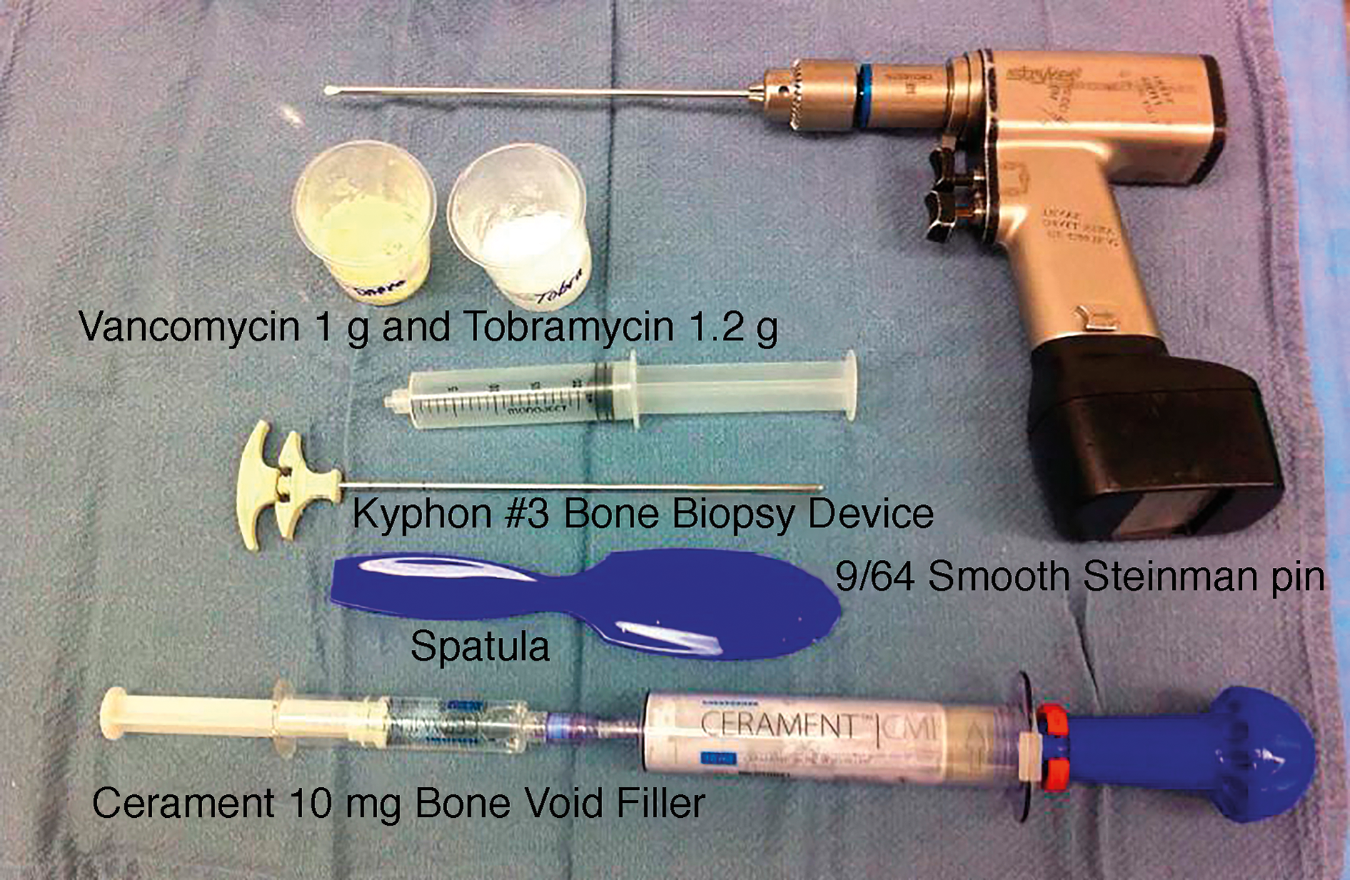
The PAD-T liquid antibiotic carrier vehicle used for this osteomyelitis treatment has been previously described.[22] Thorough familiarization of the three-dimensional anatomy of the area of osteomyelitis is paramount to successful percutaneous antibiotic delivery. The setup for this procedure is fairly simple and is illustrated in Fig. 2. A tourniquet should be used to minimize medullary bleeding. Only use antibiotics in powder form. Using antibiotics in liquid form will overdilute the bone void filler (BVF) and render the mixture difficult to handle, and there may be excessive leakage of the BVF. Unless preoperative culture results are available or drug allergies contradict their use, the antibiotics used are vancomycin and tobramycin. If a substitution for tobramycin is needed, ceftazidime, 1 g, can be used.
Figure 2.
Instrumentation tray setup.
Results
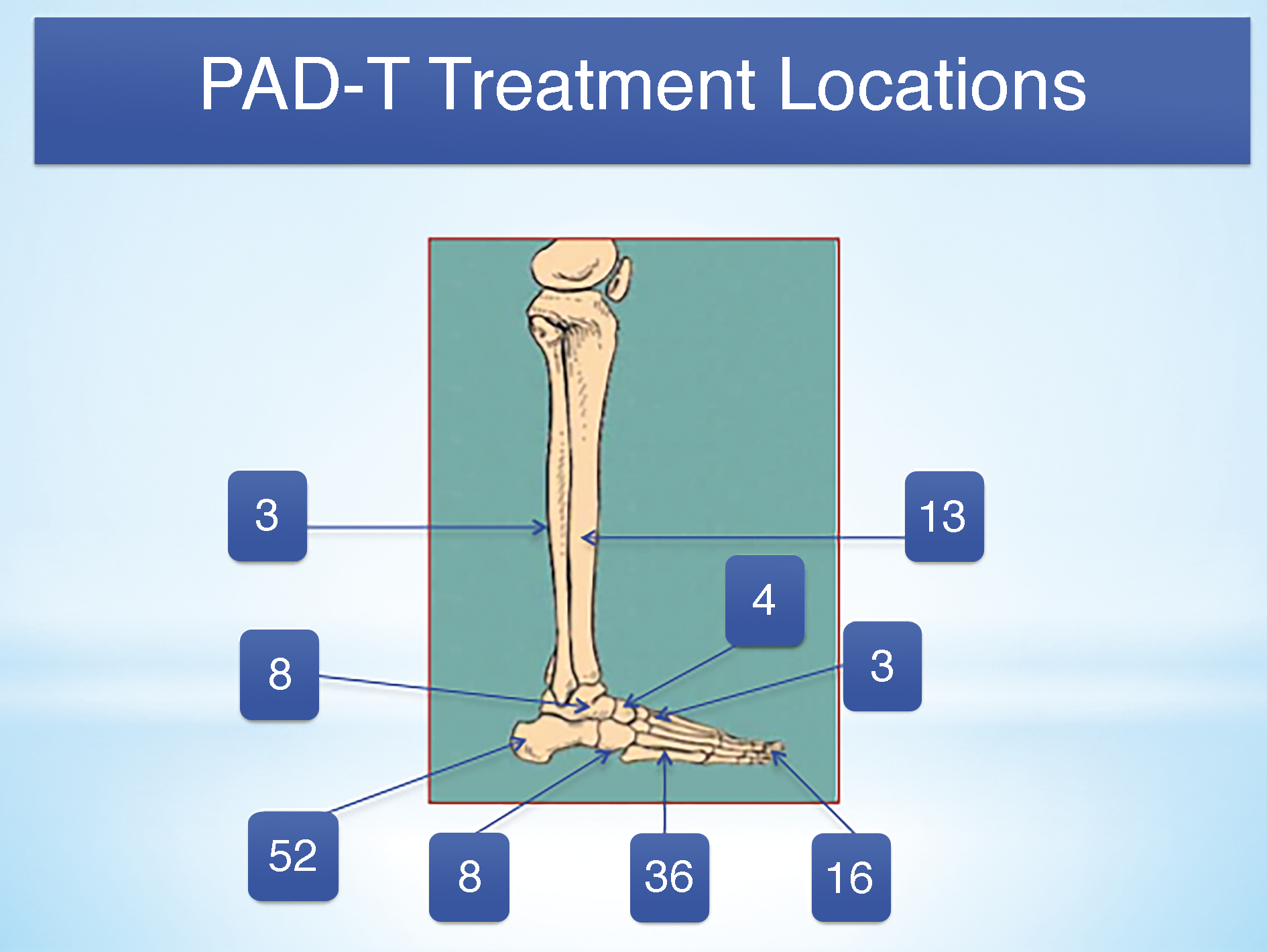
A total of 143 infected lower-extremity bones in 125 patients were treated using the PAD-T with BVF and antibiotics. Infected bone locations were the phalanx in 16 patients, metatarsal in 36, cuneiform in three, navicular in four, cuboid in eight, calcaneus in 52, talus in eight, distal tibia in 13, and distal fibula in three (Fig. 3). After the PAD-T procedure, oral or IV antibiotics were given based on bone cultures. In working with the infectious disease doctors it was felt that antibiotics were required for only 4 weeks after this bone procedure unless otherwise clinically indicated. The longest follow-up was to 74 months. Eighty-seven patients were male and 38 were female; 16 patients were using tobacco. The average patient age was 57 years (range, 12-84 years), and 104 patients were diabetic, with an average hemoglobin A1c level of 7.86. Among the 125 patients treated there were four deaths unrelated to the osteomyelitis treatment. Classifying the patient deaths by the Cierny-Mader staging system there were 3 stage III and 1 stage IV, and all four patients were B hosts. Bone locations were the cuneiform, calcaneus, talus, and distal tibia. There were an additional nine below-the-knee amputations (BKAs) due to the soft-tissue infection and necrosis overwhelming the body's ability to fight the disease process, unrelated to the osteomyelitis. Classifying the involved bone in the BKAs by the Cierny-Mader staging system there were 1 stage II, 6 stage III, and 2 stage IV; 7 were B hosts and 2 were C hosts. Bone locations were the phalanx in two patients, metatarsal in one, cuboid in one, calcaneus in three, talus in one, and distal tibia in one. The patient death and BKA incidences reflected 13 locations of osteomyelitis.
Figure 3.
Percutaneous antibiotic delivery technique (PAD-T) treatment locations.
Among the remaining 130 locations there were five failures by bone location where bone resection was necessary. Classifying the bone failures by the Cierny-Mader staging system there were three stage III and two stage IV, and all five were B hosts. Bone locations were the phalanx in one patient, metatarsal in three, and calcaneus in one. Among the remaining 125 bone locations treated with PAD-T with BVF and antibiotics, there were no recurrences, a 96.15% success rate. Classifying the successful cases by the Cierny-Mader system there were two stage I, four stage II, 112 stage III, and seven stage IV, with eight A hosts, 111 B hosts, and six C hosts. Bone locations were the phalanx in 13 patients, metatarsal in 32, cuneiform in two, navicular in four, cuboid in seven, calcaneus in 47, talus in six, distal tibia in 11, and distal fibula in three.
Discussion
The effectiveness and success of the PAD-T comes from the interactions among the BVF carrier vehicle, the selected antibiotic(s), and the infected bone. Osteomyelitis causes bone destruction, osseous vascular destruction, vascular congestion, and biofilm generation. This process erodes bone leaving a path of least resistance throughout the bone, and also leaves large and small voids of compromised bone harboring free-floating and adherent bacteria. The macrochannels and microchannels interconnect these areas of bone erosion and infection that can very easily be left in bone after traditional debridement and IV antibiotic management, serving as a future nidus for reinfection. Regarding infected bone voids larger than 3 cm3, bone grafting is required once the bone infection is resolved, otherwise a sterile cavity remains, which may be acceptable as well. Approximately 2 to 3 weeks after delivery of the BVF with antibiotics, there is no organized presence of the BVF in that cavity. This occurs because when the antibiotics are added, the antibiotic disrupts the ability of the BVF to set properly and maintain its structural integrity, and, thus, the BVF quickly reabsorbs.
Bone debridement and irrigation will remove the free-swimming bacteria, but the adherent biofilm remains.[13] Unsuccessful bone biofilm treatment is fostered by the microvascular destruction and congestion, not allowing sufficient oral or parental antibiotic concentration at the bone infection.[28,29] The PAD-T addresses this by using a liquid BVF with antibiotic(s). The liquid BVF moves nicely along the path of least resistance created by the bone infection. The movement of the liquid BVF with antibiotic(s) allows complete contact with all infected bone surfaces. With this apposition of the BVF with the infected bone, the BVF eludes enough antibiotic to address the biofilm. The amount of BVF antibiotic elusion is critical in the eradication of the biofilm. Dr. X. Yang from the Hospital for Special Surgery in New York, New York, reported over 10 days excellent calcium sulfate/phosphate vancomycin elusion rates at three separate concentrations compared with PMMA vancomycin elusion rates for both the initial burst and subsequent tail elusion.[22] The BVF calcium sulfate/phosphate vancomycin initial elusion concentration release was at 10+ mg/h versus approximately 0.25 mg/h for the PMMA vancomycin. The tail elusion concentration for the BVF calcium sulfate/phosphate with vancomycin was maintained at 0.5 mg/h for 4 weeks versus 0.05 mg/h for the PMMA with vancomycin.
As stated previously, the PAD-T using a liquid BVF provides significant antibiotic elusion. With that antibiotic elusion, the PAD-T also provides significant minimal inhibitory concentration (MIC) drug levels. The Clinical and Laboratory Standard Institute guidelines determine the MIC of bacteria by broth microdilution and macrodilution. In vitro bacterial and fungal MIC testing of drug release from the BVF calcium sulfate/phosphate discs had repeatedly uniform, extremely large, and rapid antibiotic diffusion.[22,23] In the in vitro bacterial MIC study using antibiotic BVF calcium sulfate/phosphate discs, for methicillin-resistant Staphylococcus aureus the average zone of inhibition for vancomycin, 1 g, was 30 mm, or 100% greater than the minimal inhibitory zone required (15 mm), and for tobramycin, 1.2 g, the average zone of inhibition was 25.6 mm, or 173% greater than the minimal inhibitory zone required (15 mm). For the Pseudomonas aeruginosa tested, the average zone of inhibition for tobramycin, 1.2 g, was 43 mm, or 185% greater than the minimal inhibitory zone required (15 mm).[22] Other antibiotics that have been incorporated with the BVF are rifampin, vancomycin, tobramycin, vancomycin/tobramycin combination, piperacillin-tazobactam (Zosyn; Pfizer, New York, New York), ticarcillin–clavulanic acid (Timentin; GlaxoSmithKline, Research Triangle Park, North Carolina), ceftriaxone (Fortaz; Teligent Pharma Inc, Buena, New Jersey), ancef, cefepime (Maxipime; Hospira Inc, Lake Forest, Illinois), imipenem (Primaxin; Merck Sharp & Dohme Corp, Kenilworth, New Jersey), daptomycin, minocycline, and polymyxin B.
Conclusions
The PAD-T using a liquid BVF with antibiotics is a safe and reliable method for the adjunct treatment of osteomyelitis followed by 4 weeks of oral or IV antibiotics. This method allows excellent BVF penetration into the infected bone addressing biofilm, allows higher antibiotic elusion compared with PMMA with antibiotics, and has substantially higher in vitro zones of inhibition than the stated MIC levels by the Clinical and Laboratory Standard Institute. The 96.15% success rate as reported over 7 years is a higher success rate than previously reported for surgical and nonsurgical osteomyelitis treatments.
Financial Disclosure
None reported.
Conflict of Interest
None reported.
References
- Peters EJ.Lipsky BA.Diagnosis and management of infection in the diabetic foot. Med Clin North Am97: 911, 2013.
- Walter G.Kemmerer M.Kappler C.et al: Treatment algorithms for chronic osteomyelitis. Dtsch Arztebl Int109: 257, 2012.
- Acharya S.Soliman M.Egun A.et al: Conservative management of diabetic foot osteomyelitis. Diabetes Res Clin Pract101: e18, 2013.
- Senneville E.Lombart A.Beltrand E.et al: Outcome of diabetic foot osteomyelitis treated nonsurgically: a retrospective cohort study. Diabetes Care31: 637, 2008.
- Game FL.Jeffcoate WJ.Primarily non-surgical management of osteomyelitis of the foot in diabetes. Diabetologia51: 962, 2008.
- Senneville E.Lombart A.Beltrand E.et al: Outcome of diabetic foot osteomyelitis treated nonsurgically: a retrospective cohort study. Diabetes Care31: 637, 2008.
- Lipsky BA.Berendt AR.Cornia PB.et al: Infectious Diseases Society of America clinical practice guideline for the diagnosis and treatment of diabetic foot infections. Clin Infect Dis54: 132, 2012.
- Buchholz HW.Engelbrecht H.Depot effects of various antibiotics mixed with Palacos resins [in German]. Chirurg41: 511, 1970.
- Klemm K.Antibiotic bead chains. Clin Orthop295: 63, 1993.
- Calhoun JH.Mader JT.Antibiotic beads in the management of surgical infections. Am J Surg157: 443, 1898.
- Cierny G III.Infected tibial nonunions (1981-1995): the evolution of change. Clin Orthop Relat Res360: 97, 1999.
- Mader JT.Mauro MD.Calhoun JH.Update on the diagnosis and management of osteomyelitis. Clin Podiatr Med Surg Infect Dis13: 701, 1996.
- Cierny G III.Surgical treatment of osteomyelitis. Plast Reconstr Surg127(suppl 1): 190S, 2011.
- Walenkamp GH.Kleijn LL.de Leeuw M.Osteomyelitis treated with gentamicin–PMMA beads: 100 patients followed for 1-12 years. Acta Orthop Scand69: 518, 1998.
- Blaha JD.Calhoun JH.Nelson CL.et al: Comparison of the clinical efficacy and tolerance of gentamicin PMMA beads on surgical wire versus combined and systemic therapy for osteomyelitis. Clin Orthop Related Res295: 8, 1993.
- Kanellakopoulou K.Giamarellos-Bourboulis EJ.Carrier systems for the local delivery of antibiotics in bone infections. Drugs59: 1223, 2000.
- Trombelli L.Heitz-Mayfield LJ.Needleman I.et al. A systematic review of graft materials and biologic agents for periodontal intraosseous defects. J Clin Periodontol29(suppl 3): 117, 2002.
- Pempinello C.Gennaro S.Giovanni S.et al: The efficacy of injectable biphasic calcium sulphate/hydroxyapatite bone substitute Cerament G in the treatment of severe bone infections. Poster presented at: the 34th Annual Meeting of the European Bone and Joint Infection Society, Estoril, Portugal, September 10–12, 2015.
- Whisstock C.Ninkovic S.Marin M.et al: Use of a new antibiotic bone substitute to induce healing of osteomyelitis in the diabetic foot. Poster presented at: the 34th Annual Meeting of the European Bone and Joint Infection Society, Estoril, Portugal, September 10–12, 2015.
- Dvorzhinskiy A.Perino G.Chojnowski M.et al. Cerament bone void filler with gentamicin increases bone formation and decreases detectable Infection in a rat model of debrided osteomyelitis. Poster presented at: the 34th Annual Meeting of the European Bone and Joint Infection Society, Estoril, Portugal, September 10–12, 2015.
- McNally M.Ferguson J.Kendall M.A comparative study of three bioabsorbable antibiotic carriers in chronic osteomyelitis: 313 patients with a minimum 1 year follow-up. Poster presented at: the 34th Annual Meeting of the European Bone and Joint Infection Society, Estoril, Portugal, September 10–12, 2015.
- Karr JC.An overview of the percutaneous antibiotic delivery technique for osteomyelitis treatment and a case study of calcaneal osteomyelitis. JAPMA107: 511, 2017.
- Karr JC.Lauretta J.Keriazes G.In vitro antimicrobial activity of calcium sulfate and hydroxyapatite (Cerament Bone Void Filler) discs using heat-sensitive and non-heat-sensitive antibiotics against methicillin resistant Staphylococcus aureus and Pseudomonas aeruginosa. JAPMA101: 146, 2011.
- Karr JC.Lauretta J.In vitro activity of calcium sulfate and hydroxyapatite antifungal disks loaded with amphotericin B or voriconazole for consideration for adjunctive osteomyelitis management. JAPMA105: 104, 2015.
- Centers for Disease Control and Prevention: National Diabetes Fact Sheet: General Information and National Estimates on Diabetes in the United States, 2005, US Department of Health and Human Services, Centers for Disease and Prevention, Atlanta, GA, 2005.
- Faglia E.Clerici G.Caminiti M.et al: Influence of osteomyelitis location in the foot of diabetic patients with transtibial amputation. Foot Ankle Int34: 222, 2013.
- Cierny GIII, Mader JT.Penninck JJ.A clinical staging system for adult osteomyelitis. Clin Orthop Relat Res414: 7, 2003.
- Roeder B.Van Gils CC.Maling S.Antibiotic beads in the treatment of diabetic pedal osteomyelitis. J Foot Ankle Surg39: 124, 2000.
- Gogia JS.Meehan JP.Di Cesare PE.et al: Local antibiotic therapy in osteomyelitis. Semin Plast Surg23: 100, 2009.
© 2018 American Podiatric Medical Association