


Abstract
Background: Elevated dynamic plantar pressures are a consistent finding in diabetic patients with peripheral neuropathy, with implications for plantar foot ulceration. This study aimed to investigate whether a first-ray amputation affects plantar pressures and plantar pressure distribution patterns in individuals living with diabetes and peripheral neuropathy. Methods: A nonexperimental matched-subject design was conducted. Twenty patients living with diabetes and peripheral neuropathy were recruited. Group 1 (n = 10) had a first-ray amputation and group 2 (n = 10) had an intact foot with no history of ulceration. Plantar foot pressures and pressure-time integrals were measured under the second to fourth metatarsophalangeal joints, fifth metatarsophalangeal joint, and heel using a pressure platform. Results: Peak plantar pressures under the second to fourth metatarsophalangeal joints were significantly higher in participants with a first-ray amputation (P = .008). However, differences under the fifth metatarsophalangeal joint (P = .734) and heel (P = .273) were nonsignificant. Pressure-time integrals were significantly higher under the second to fourth metatarsophalangeal joints in participants with a first-ray amputation (P = .016) and in the heel in the control group (P = .046). Conclusions: Plantar pressures and pressure-time integrals seem to be significantly higher in patients with diabetic peripheral neuropathy and a first-ray amputation compared with those with diabetic neuropathy and an intact foot. Routine plantar pressure screening, orthotic prescription, and education should be recommended in patients with a first-ray amputation.
The incidence of diabetes is increasing worldwide, and nearly 50% of patients living with diabetes will develop diabetic peripheral neuropathy, [1] a main precursor to foot ulceration, which is estimated to develop in 15% of people with type 2 diabetes. This is of concern for people with diabetes and health-care providers, with episodes of ulceration strongly associated with lower-extremity amputations, reduced quality of life, long periods of hospitalization, and substantial health-care costs. [2]
Elevated dynamic plantar pressures are a consistent finding in patients with diabetes and peripheral neuropathy, with implications for plantar foot ulceration. [3] Two measures of vertical plantar pressure are most commonly assessed: mean peak plantar pressure (PPP) represents the maximum amount of pressure during stance, and the mean pressure-time integral (PTI) represents the amount of time over which maximum pressure is applied. Research regarding plantar pressures and ulceration has been thoroughly investigated, but research regarding changes in plantar pressure patterns after a first-ray amputation is missing.
The importance of the first ray as a crucial element in gait [4] and the relative lack of research associated with the amputation of this structure highlights the need for research in this field. First-ray amputations frequently arise from an ulcer in the medial or plantar aspect of the first metatarsal head, which extends proximally to the first metatarsophalangeal joint. [5] Gait is dramatically altered after a first-ray amputation. Removal of this vital structure results in loss of the windlass mechanism, which decreases medial stability and alters transfer of weight during late stance phase. Biomechanical alterations after a first-ray amputation include loss of medial column support and formation of a planovalgus position of the foot. [6]
The incidence of reamputation after first-ray amputation is as high as 42.4%. [7] This finding prompted us to conduct a study in a population with a high prevalence rate of diabetes to determine whether a first-ray amputation affects mean PPPs and plantar pressure distribution patterns in participants living with diabetes and peripheral neuropathy. The research questions posed for this study included “Does a first-ray amputation significantly affect PPPs and PTIs in the neuropathic population with type 2 diabetes mellitus?” The null hypothesis postulated for this study specifies that first-ray amputation does not significantly affect PPPs and PTIs (P > .05) in the neuropathic population with type 2 diabetes, indicating a 5% risk of concluding that a difference exists when there is no actual difference.
Methods
Participant Selection
Ten percent of the Maltese population has diabetes compared with 2% to 5% of the populations in neighboring European countries. Expectedly, foot ulceration, ulcer recurrence, and amputations are common in this specific population. [8] This emphasizes the importance of continuous research to identify the best methods of management to ensure high-quality and effective treatment in the high-risk foot, especially in individuals with the complication of diabetic peripheral neuropathy.
Individuals were recruited from the diabetes podiatry clinic of a local general hospital. An average of 5,000 people attend the diabetes podiatry clinic annually, with approximately 500 experiencing ulceration. A convenience cohort of 20 participants was enrolled for this investigation. This study was approved by the Research Ethics Committee of the University of Malta (Msida, Malta), and all of the participants provided informed consent before data collection. All of the investigations were performed in accordance with the principles of the Declaration of Helsinki as revised in 2000. Participants eligible for this study were Maltese individuals, older than 18 years, living with diabetes according to the World Health Organization diagnostic criteria. [9]
Methodological Rigor
To test the hypothesis, a prospective, nonexperimental, matched-subject design was conducted. The clinical tools used during this research were based on validated and previously published methods after a thorough review of the literature on international guidelines and recommendations. A database was constructed to record all of the information.
Patient Characteristics
Twenty participants with type 2 diabetes who had been diagnosed by hospital consultants as having diabetic peripheral neuropathy were recruited into this study. Participants were divided into two groups. Group 1 included ten participants having a first-ray amputation and a history of neuropathic ulceration. The reason for the first-ray amputation was due to complications arising from the neuropathic ulceration. Group 2 included ten participants living with diabetic peripheral neuropathy but presenting with an intact foot and no history of ulceration. Participants in each group were matched for age, sex, body mass index (BMI; calculated as weight in kilograms divided by height in meters squared), and foot posture. Frequency distribution matching was used for age and BMI. Precision matching was used for sex and foot posture.
Foot posture was assessed and classified according to the Foot Posture Index. Joint mobility at the ankle was measured with a goniometer to exclude any participants presenting with ankle equinus.
Patients were excluded from the study if they presented with any other form of neuropathy, for example, autonomic neuropathy, ankle equinus that was determined with the goniometer (<5° of dorsiflexion), changes in foot structure such as Charcot's arthropathy and rheumatoid arthritis, or if they presented with active or a history of foot ulcerations. Participants who were unable to participate in the study owing to cognitive impairments or who showed unwillingness to participate and follow the study protocol were also excluded.
Peripheral Sensory Neuropathy
A 10-g Semmes-Weinstein monofilament was used to identify the extent of diabetic peripheral sensory neuropathy. The five-point test was used. The plantar aspect of the hallux and third digit together with the first, third, and fifth metatarsal heads were used for testing. With eyes closed, each participant related to the investigator when he or she could feel the monofilament. Participants who were unable to feel the 10-g monofilament on one of the five points tested and who presented with adequate peripheral perfusion at the time of the study, ie, who had triphasic pedal waveforms and a normal ankle brachial pressure index of 0.9 to 1.2 when assessed by the clinician, were included in this study. For the participant group with first-ray amputation, the closest site on the same foot was tested, including the second digit and second metatarsophalangeal joint. Two clinicians performed the monofilament testing independently to ensure rater reliability, and only participants who had the same score by the raters were included in the study.
Pressure Mapping Protocol
The PPPs beneath the foot were recorded using a pressure platform (HR Mat; Tekscan, Boston, Massachusetts). Initially, the participants were asked to stand on the mat to calibrate the system according to their body mass as per the manufacturer's instructions. The study protocol was discussed with the participants before data collection. A period of acclimatization was included for the participants to understand the two-step walking protocol that was adopted. This protocol has been reported to be reliable and not time-consuming to implement, [10] requiring the participants to make contact with the platform on the second step. [11] Each participant was instructed to stand on both feet exactly two steps before stepping on the pressure platform. The participants started walking by stepping first with one foot on the ground and the next step on the platform and then continued walking for the next three steps. [12] Several practice trials were performed so as to familiarize the participants with the procedure and encourage natural gait, avoiding targeting. When the participants felt confident with their walking pattern, the dynamic plantar pressures for the foot with the first-ray amputation in group 1 and the same intact foot of the matched control participant (group 2) were recorded.
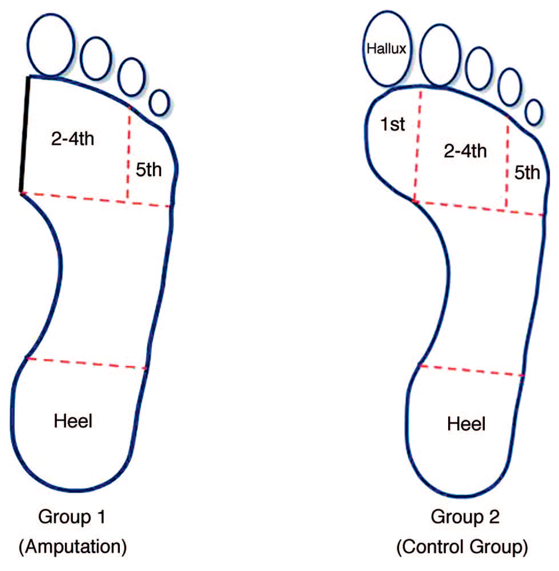
This procedure was repeated five times at the areas of interest, which included, the second to fourth metatarsophalangeal joints, fifth metatarsophalangeal joint, and heel for both groups and the first metatarsophalangeal joint and the hallux for group 2 (Fig. 1). Each trial produced five stance phases for each foot. The first and last stance phases of each trial were discarded; thus, the mean values of the three remaining stance phases for each foot were used for evaluation.
Figure 1.
Areas masked for group 1 and group 2.
Results
All the data were recorded on a spreadsheet designed in Microsoft Excel (Microsoft Corp, Redmond, Washington) to group together the information required for interpretation of the results. Statistical analyses were performed using a statistical software program (IBM SPSS Statistics for Windows, Version 20.0; IBM Corp, Armonk, New York).
Twenty participants (12 men and eight women) were included in this study. Participants were divided into two groups. Group 1 included participants living with type 2 diabetes and having a first-ray amputation, and group 2 included participants living with type 2 diabetes with an intact foot. The mean ± SD age for group 1 was 62.8 ± 9.17 years and for group 2 was 64.8 ± 6.63 years. The mean ± SD BMI for group 1 was 35.22 ± 7.81, and the mean ± SD BMI for group 2 was 31.2 ± 4.08. Participants' Foot Posture Index scores ranged from 1 to 8, with participants presenting with either a neutral or a pronated foot posture.
Ninety percent of group 1 (first-ray amputation) participants had the highest PPPs on the second to fourth metatarsophalangeal joints, and 10% displayed the highest PPPs under the heel. None of this group displayed high PPPs under the fifth metatarsophalangeal joint. Fifty percent of group 2 (intact foot) participants displayed the highest PPPs under the hallux, 30% under the first metatarsophalangeal joint, and 20% under the second to fourth metatarsophalangeal joints. No participants in this group displayed high PPPs under the fifth metatarsophalangeal joint and the heel.
Peak Plantar Pressure
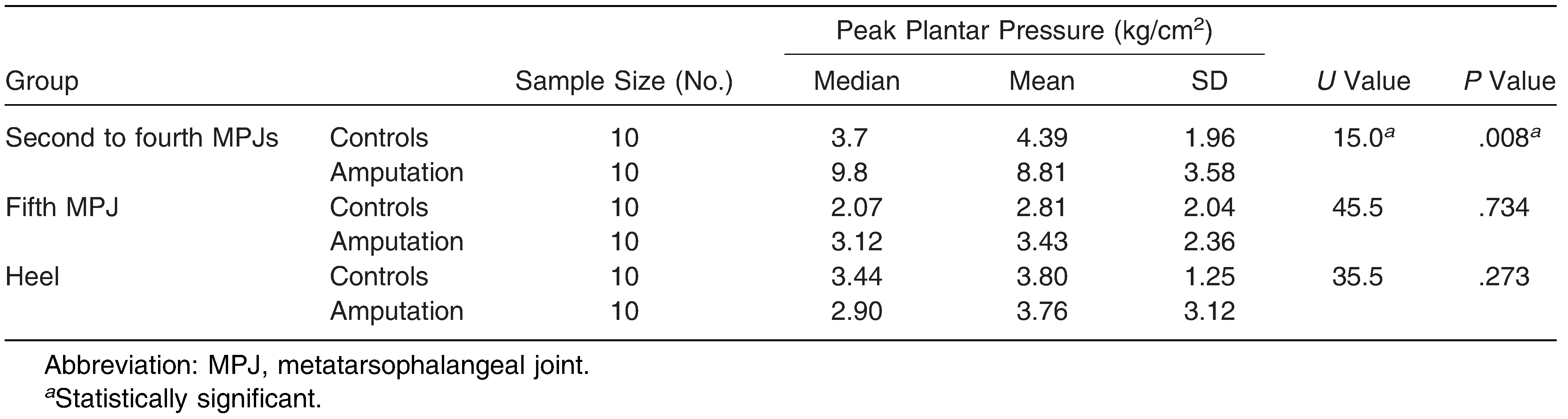
The one-sample Kolmogorov-Smirnov test was used to test normality of PPP data under the second to fourth metatarsophalangeal joints, fifth metatarsophalangeal joint, and heel. The Mann-Whitney test was used to compare PPPs between groups because the data were not normally distributed. Table 1 illustrates means and medians for each respective group and standard deviations, U values, and P values for the study group. The mean PPPs under the second to fourth metatarsophalangeal joints were significantly higher (8.81 versus 4.39 kg/cm2; total median = 5.44 kg/cm2; U = 15.0, P = .008) in participants with a first-ray amputation. However, differences under the fifth metatarsophalangeal joint (3.43 versus 2.81 kg/cm2; total median = 2.59 kg/cm2; U = 45.5; P = .734) and heel (3.76 versus 3.80 kg/cm2; total median = 3.02 kg/cm2; U = 35.5; P = .273) were nonsignificant.
Table 1.
Peak Plantar Pressures by Group (Mann-Whitney Test).
Pressure-Time Integrals
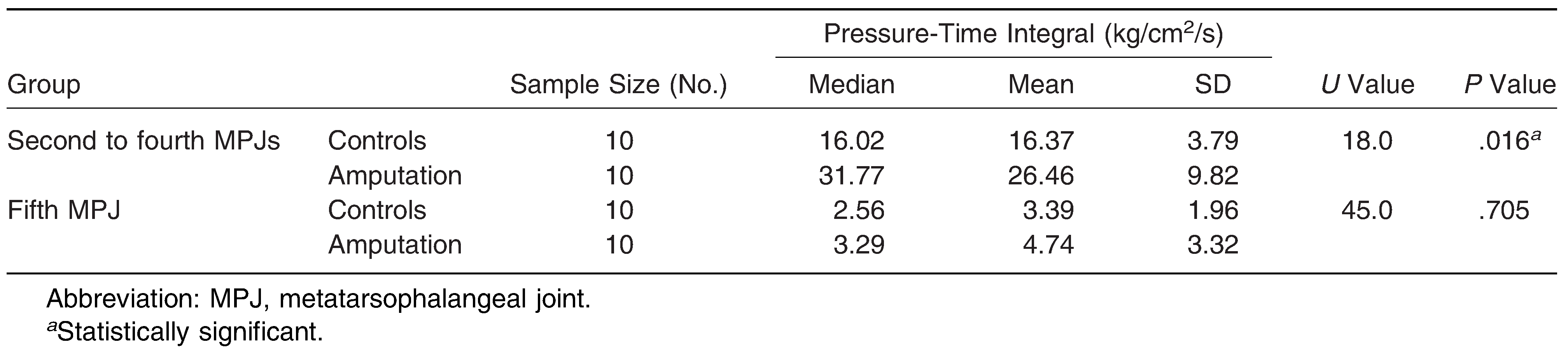
The one-sample Kolmogorov-Smirnov test was used to test normality of PTI data under the second to fourth metatarsophalangeal joints, fifth metatarsophalangeal joint, and heel. The Mann-Whitney test was used to compare PTIs under the second to fourth and fifth metatarsophalangeal joints because the data were not normally distributed (P = .024; P = .011). Mean PTIs were significantly higher under the second to fourth metatarsophalangeal joints in participants with a first-ray amputation (26.46 versus 16.37 kg/cm2/s; total median = 18.4 kg/cm2/s; U = 18.0; P = .016), but no significant difference was found under the fifth metatarsophalangeal joint (4.74 versus 3.39 kg/cm2/s; total median = 2.56 kg/cm2/s; U = 45.0; P = .705) (Table 2).
Table 2.
Pressure-Time Integrals by Group (Mann-Whitney Test).
The independent-samples t test was used to analyze mean PTIs under the heel between groups because the data had a normal distribution (P = .511). These values were significantly higher in the heel in the group without amputation (mean, 25.21 vs 18.50 kg/cm2/s; t = 2.14, P = .046) (Table 3).
Table 3.
Pressure-Time Integrals in the Heel by Group (Independent-Samples t Test).

Discussion
This study investigated plantar pressure distribution during gait between participants living with diabetic peripheral neuropathy having a first-ray amputation and a history of neuropathic ulceration (group 1) and participants also living with diabetic peripheral neuropathy but having an intact foot and no history of amputation (group 2). Comparing both groups together, mean PPPs and mean PTIs seem to be significantly higher in patients with diabetic peripheral neuropathy with a first-ray amputation compared with those with diabetic neuropathy and an intact foot. Although it was expected that an increase in PPP was to be found under the amputated foot due to a decrease in surface area, only PPPs under the second to fourth metatarsophalangeal joints were significantly increased, and while no increase was evident under the rest of the foot. Hence, it seems that plantar pressure was not evenly distributed in all areas of the foot but was concentrated under the second to fourth metatarsophalangeal joints, increasing the risk of injury to this area. Therefore, the key finding of the present study is that the PPP and PTI were transferred from the first metatarsophalangeal joint and hallux in the intact foot to the second, third, and fourth metatarsophalangeal joints in participants having a first-ray amputation. Mean PPPs under the second to fourth metatarsophalangeal joints were 4.39 kg/cm2 in the intact foot and 8.81 kg/cm2 in participants having a first-ray amputation. Pham et al [13] observed that PPPs equivalent to or greater than 6 kg/cm2 place susceptible patients at risk for foot ulceration.
In addition, PTIs under the second through fourth metatarsophalangeal joints were 16.37 kg/cm2/s in the intact foot and 26.46 kg/cm2/s in participants having a first-ray amputation. Results demonstrated that a first-ray amputation had significant effects on PPPs (P = .008) and PTIs (P = .016) under the second to fourth metatarsophalangeal joints.
Alteration in gait after an amputation is bound to lead to transfer of plantar pressures to the lesser metatarsal area, as has been reported in the literature; however, these studies do not identify the exact transfer areas and changes in pressure, making the present study the first to report these results.
Because an increase in plantar pressures increases the risk of ulceration and amputation, [14] these results suggest that after a first-ray amputation the ipsilateral limb is at an increased risk of ulceration due to elevated PPPs and PTIs under the second to fourth metatarsophalangeal joints. Izumi et al [15] stated that the ipsilateral limb is at greater risk for reamputation (50%) than the contralateral limb (29.2%) after a first-ray amputation. Furthermore, Borkosky and Roukis [16] reported that the rate of reamputation after partial first-ray amputations in participants with diabetes mellitus and peripheral sensory neuropathy is 42.4%.
Following the results of this study, we recommend routine screening of plantar pressures and immediate orthotic treatment to off load such areas in such a high-risk category of patients both before and after amputation to prevent morbidity and mortality and improve the quality of life of these patients. Proper screening and management, which includes the provision of therapeutic footwear in this specific population, is essential in the prevention of foot complications. The implementation of a structured program that includes plantar pressure screening with the use of therapeutic footwear was found to reduce the incidence of foot ulceration in patients who are at high ulcerative risk and to lower its related costs. [17]
Diabetes kills more people annually than breast cancer and AIDS combined. [18] Furthermore, the cost of treating diabetic foot ulcers is greater than that of the five most costly forms of cancer, and relative 5-year mortality after limb amputation is 68%; compared with cancer, it is second only to lung cancer (86%). [18] As the global prevalence of diabetes keeps rising due to population growth, aging of the populations, and lifestyle changes associated with urbanization, screening and management of the high-risk foot needs to keep evolving with the inclusion of the latest advances in technology, biomechanical modeling, and other innovations in clinical management to prevent amputations and reamputations. Proper screening and management in the prevention of complications is essential.
Conclusions
Research regarding plantar pressures and ulceration has been thoroughly investigated, but research regarding changes in plantar pressure patterns after a first-ray amputation is missing. Results from this study demonstrate that a first-ray amputation significantly affects PPPs and PTIs under the second to fourth metatarsophalangeal joints. The importance of increased vigilance coupled with strengthening of existing screening structures and introducing clinical guidelines regarding biomechanical assessment of the feet to reduce the incidence of diabetes foot complications and amputations is warranted.
Acknowledgments
Acknowledgment: We thank all of the participants who consented to participate in this study and Liberato Camilleri (University of Malta) for his statistical advice.
Financial Disclosure: None reported.
Conflict of Interest: None reported.
References
- Meeuwisse-Pasterkamp, S.; van der Klauw, M.; Wolffenbuttel, B. Type 2 diabetes mellitus: prevention of macrovascular complications. Exp Rev Cardiovasc Ther 2008, 6, 323. [Google Scholar]
- Boulton, A.; Vileikyte, L.; Ragnarson-Tennvall, G.; et al. The global burden of diabetic foot disease. Lancet 2005, 366, 1719. [Google Scholar]
- Fernando, ME.; Crowther, RG.; Pappas, E.; et al. Plantar pressure in diabetic peripheral neuropathy patients with active foot ulceration, previous ulceration and no history of ulceration: a meta-analysis of observational studies. PLoS One 2014, 9, e99050. [Google Scholar]
- Glasoe, W.; Yack, H.; Saltzman, C. Anatomy and biomechanics of the first ray. J Am Phys Ther Assoc 1999, 79, 854. [Google Scholar]
- Bouchard, J. Basic Principles and Techniques of Forefoot Amputations; p. The Podiatry Institute Inc: Decatur, GA, 1997; p. 276. Available at: http://www.podiatryinstitute.com/pdfs/Update_1997/1997_45.pdf. Accessed April 4.
- Paola, L.; Faglia, E.; Caminiti, M.; et al. Ulcer recurrence following first ray amputation in diabetic patients: a cohort prospective study. Diabetes Care 2003, 26, 1874. [Google Scholar]
- Borkosky, S.; Roukis, T. Incidence of repeat amputation after partial first ray amputation associated with diabetes mellitus and peripheral neuropathy: an 11-year review. J Foot Ankle Surg 2013, 52, 335. [Google Scholar]
- Galea, AM.; Springett, K.; Bungay, H.; et al. Incidence and location of diabetic foot ulcer recurrence. Diabet Foot J 2009, 12, 181. [Google Scholar]
- World Health Organization, Use of Glycated Haemoglobin in the Diagnosis of Diabetes Mellitus, World Health Organization; Geneva, Switzerland, 2011.
- Oladeji, O.; Stackhouse, C.; Gracely, E.; et al. Comparison of the two-step and midgait methods of plantar pressure measurement in children. JAPMA 2008, 98, 268. [Google Scholar]
- Hafer, JF.; Lenhoff, MW.; Song, J.; et al. Reliability of plantar pressure platforms. Gait Posture 2013, 38, 544. [Google Scholar]
- Bus, SA.; de Lange, A. A comparison of the 1-step, 2-step, and 3-step protocols for obtaining barefoot plantar pressure data in the diabetic neuropathic foot. Clin Biomech 2005, 20, 892. [Google Scholar]
- Pham, H.; Armstrong, D.; Harvey, C.; et al. Screening techniques to identify people at high risk for diabetic foot ulceration. Diabetes Care 2000, 23, 606. [Google Scholar]
- Veves, A.; Murray, H.; Young, M.; et al. The risk of foot ulceration in diabetic patients with high foot pressure: a prospective study. Diabetologia 1992, 35, 660. [Google Scholar]
- Izumi, Y.; Satterfield, K.; Lee, S.; et al. Risk of reamputation in diabetic patients stratified by limb and level of amputation: a 10-year observation. Diabetes Care 2006, 29, 566. [Google Scholar]
- Borkosky, S.; Roukis, T. Incidence of repeat amputation after partial first ray amputation associated with diabetes mellitus and peripheral neuropathy: an 11-year review. J Foot Ankle Surg 2013, 52, 335. [Google Scholar]
- Rizzo, L.; Tedeschi, A.; Fallani, E.; et al. Custom-made orthesis and shoes in a structured follow-up program reduces the incidence of neuropathic ulcers in high-risk diabetic foot patients. Int J Low Extrem Wounds 2012, 11, 59. [Google Scholar]
- American Diabetes Association, Fast facts: data and statistics about diabetes. Available at: http://professional.diabetes.org/ResourcesForProfessionals.aspx?cid=91777&loc=dorg-statistics. Accessed December 12014.
© 2018 American Podiatric Medical Association