


Abstract
Background: Tailor's bunion, or bunionette, deformity is a common condition of the lateral forefoot. To aid in assessing the deformity, radiographic angular measurements are frequently used. The objective of this study was to determine the reliability and reproducibility of these angular measurements. Methods: Thirty unique weightbearing dorsoplantar digital radiographs of pathologic feet were compiled. For these 30 radiographs and for ten repeated radiographs, six practicing clinicians measured the following angles: the fourth to fifth intermetatarsal angle, the lateral deviation angle, and the fifth metatarsophalangeal angle. Both traditional and modified versions of the fourth to fifth intermetatarsal angle were included. Results: Intraclass correlation coefficient values were calculated for each of the angles studied. Intrarater reliability was highest for the fifth metatarsophalangeal angle and lowest for the lateral deviation angle. Intrarater reliability was higher for the traditional fourth to fifth intermetatarsal angle than for the modified version. The interrater reliability calculations revealed parallel findings. Conclusions: Accurate assessment is critical when planning for surgical intervention. Awareness of the relative reliability of these radiographic angular measurements can aid in preoperative planning and may be of benefit in procedure selection.
The term tailor's bunion refers to a lateral prominence of the fifth metatarsal head. Synonymous with the term bunionette, the term was historically derived from tailors, who crossed their legs for long periods. [1] When conservative treatment for the condition fails, surgical intervention is often recommended. Selecting the appropriate surgical procedure relies heavily on radiographic assessment of the deformity. [2]
The condition is most commonly evaluated radiographically with dorsoplantar, medial oblique, and lateral views of the foot. Radiographic measurements performed on the weightbearing dorsoplantar view include the fourth to fifth intermetatarsal angle, the lateral deviation angle, and the fifth metatarsophalangeal angle. [3–5] These measurements are used to quantify the severity of the deformity, to determine the anatomical level of the deformity, and to construct a surgical treatment plan.
The fourth to fifth intermetatarsal angle is traditionally determined by the long axes of the fourth and fifth metatarsals (Fig. 1A). This angle measures the degree of separation between the two metatarsals. The fourth to fifth intermetatarsal angle has been found to average 6.2° in unaffected patients and 9.6° in symptomatic patients. [6]
Figure 1.
A, Fourth to fifth intermetatarsal angle. Axes bisect the fourth and fifth metatarsal shafts. B, Fourth to fifth intermetatarsal angle as described by Fallat and Buckholz. [6] One axis bisects the fourth metatarsal shaft and the other lies tangential to the proximal-medial cortex of the fifth metatarsal. C, Lateral deviation angle. One axis is tangent to the proximal-medial cortex of the fifth metatarsal and the other axis bisects the fifth metatarsal head and neck. D, Fifth metatarsophalangeal angle. One axis bisects the fifth metatarsal shaft and the other axis bisects the fifth toe proximal phalanx shaft.
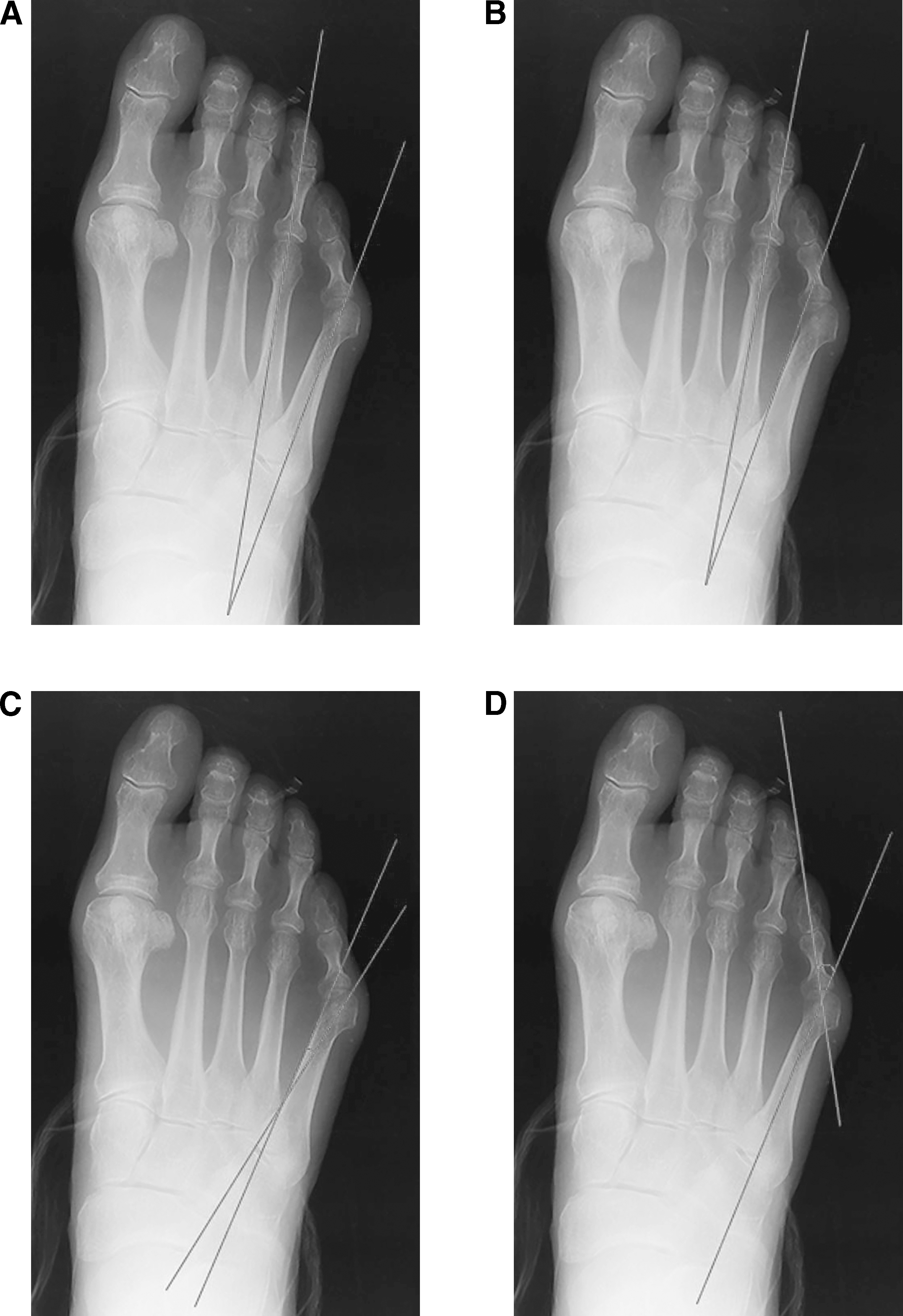
Fallat and Buckholz [6] proposed an alternate axis for the fifth metatarsal, tangential to the proximal half of the fifth metatarsal's medial cortex (Fig. 1B). They postulated that this method eliminates the effect that variations of the fifth metatarsal (bowing and a wide base) may have on the traditional fourth to fifth intermetatarsal angle measurement.
The lateral deviation angle is measured between the alternate fifth metatarsal axis (a line tangential and parallel to the medial cortex of the proximal fifth metatarsal shaft) and a line bisecting the fifth metatarsal head and neck (Fig. 1C). In the normal foot, this angle averages 6° and ranges from 0° to 7°. In the symptomatic patient, it averages 8°. [6] The lateral deviation angle is used to determine the amount of fifth metatarsal bowing. [6]
The fifth metatarsophalangeal angle is measured between the long axes of the fifth metatarsal and the proximal phalanx of the fifth digit (Fig. 1D). It typically ranges from 1° to 21° and is less than 14° in 90% of healthy patients. [7] In symptomatic patients, the angle was found to average 16°, with a range of −5° to 30°. [8] The fifth metatarsophalangeal angle is used to evaluate the deformity at the level of the fifth metatarsophalangeal joint.
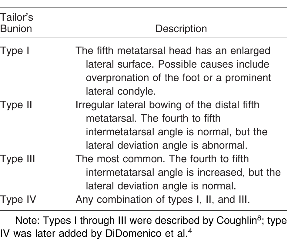
Tailor's bunion deformity has been classified by its appearance on the weightbearing dorsoplantar radiograph. [4,8] The four types are summarized in Table 1.
Table 1.
Types of Tailor's Bunion.
The purpose of this study was to determine the reliability and reproducibility of four angles used to radiographically evaluate tailor's bunion: the traditional fourth to fifth intermetatarsal angle; the modified fourth to fifth intermetatarsal angle (Fallat and Buckholz6); the lateral deviation angle; and the fifth metatarsophalangeal angle. This information may be used to improve the consistency of surgical results.
Materials and Methods
After approval from the Western University of Health Sciences institutional review board was obtained, 30 deidentified weightbearing dorsoplantar radiographs were retrospectively selected from patients diagnosed clinically as having a tailor's bunion (ie, lateral prominence of the fifth metatarsal head). All of the radiographs were dorsoplantar foot projections obtained from a single podiatric medical practice using the technique as described by Baron et al. [9] Radiographs were excluded if any of the following criteria were met: previous surgery of the foot, previous fracture of the foot, neuromuscular pathology, tumors in the lower extremities, congenital malformations of the foot, or age younger than 18 years.
Six practicing podiatric physicians were asked to participate in the study. These clinicians were provided information about the project and a detailed written and oral review of the relevant radiographic angles. The clinicians were also provided an orientation to ImageJ, a Java-based application program made readily available from the National Institutes of Health for image processing and analysis. [10] This program was provided for use in calculating the radiographic angles.
The six clinicians were then asked to review 40 digital radiographs. This included 30 unique radiographs, five of which were repeated twice. The repeated radiographs were randomly dispersed, and the clinicians were not made aware that there were repeated images. The measurements were self-recorded by the clinicians on a spreadsheet program (Microsoft Office Excel 2010; Microsoft Corp, Redmond, Washington). The clinicians were given the option of completing the radiographic assessment in one or two sessions. The clinicians made four measurements on each radiograph: the fourth to fifth intermetatarsal angle using bisections of the fourth and fifth metatarsals, the fourth to fifth intermetatarsal angle using the bisection of the fourth metatarsal and a line tangential to the medial cortex of the fifth metatarsal, the lateral deviation angle, and the fifth metatarsophalangeal angle.
Intraclass correlation coefficient (ICC) values were calculated using IBM SPSS Statistics for Windows, Version 22.0 (IBM Corp, Armonk, New York). The repeated radiographs were used to determine intrarater reliability (reproducibility), with ICCs calculated for each clinician individually. The 30 unique radiographs were used to determine interrater reliability, with ICCs also calculated for all of the clinicians combined.
Results
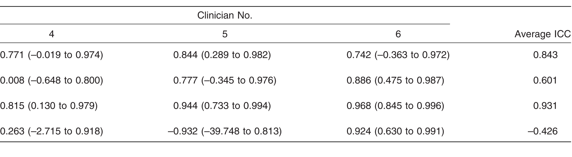
All six clinicians completed their respective radiographic assessments. Intrarater ICCs, reflecting individual reproducibility, were calculated for each of the four angular measurements (Table 2). The calculated intrarater ICC for the fifth metatarsophalangeal angle averaged 0.931, suggesting excellent reproducibility. The intrarater ICC for the fourth to fifth intermetatarsal angle using the bisection of the fifth metatarsal (traditional) was higher than the intrarater ICC using the medial tangent of the fifth metatarsal (modified) (0.843 versus 0.601). The intrarater ICC of the lateral deviation angle was notably lowest (–0.426) and indicative of poor reproducibility.
Table 2.
Mean Intraobserver Intraclass Correlation Coefficients (ICCs) and 95% Confidence Intervals for the Repeated Radiographs.
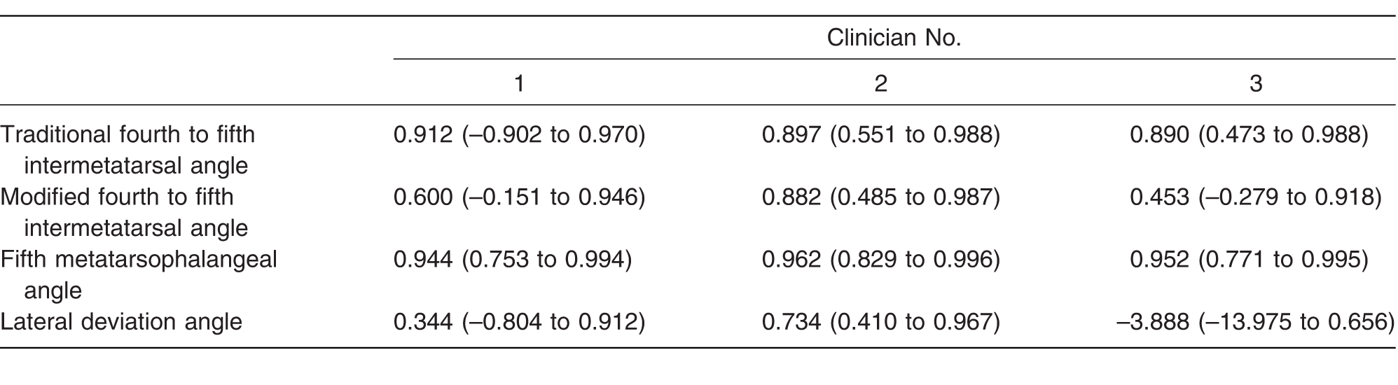
Table 2.
extended.
The interrater reliability, reflecting the overall reliability of the angular measurements, revealed similar findings (Table 3). Again, the ICC of the fifth metatarsophalangeal angle was highest (0.966; 95% confidence interval [CI], 0.941–0.981) and the ICC of the lateral deviation angle was lowest (0.091; 95% CI, –0.0571–0.357). The ICC using the bisection of the fifth metatarsal was again higher than the medial tangential method (0.909; 95% CI, 0.855–0.947 versus 0.804; 95% CI, 0.599–0.901).
Table 3.
Mean Interobserver Intraclass Correlation Coefficients (ICCs) and 95% Confidence Intervals (CIs) for Each Angle.

Discussion
Of the four angular measurements, the most reliable and reproducible angle was the fifth metatarsophalangeal angle. The measurements of this angle reflected excellent reliability between clinicians and for repeated measurements. It is possible that the precision of the fifth metatarsophalangeal angle was a result of familiarity with the similar hallux abducto valgus angle. Higher overall confidence in this angular measurement may be appropriate during preoperative planning.
The traditional method of determining the fourth to fifth intermetatarsal angle (using the bisection of the fourth and fifth metatarsals) was found to be more reliable and reproducible than using the modified method (a line tangential and parallel to the medial cortex of the proximal fifth metatarsal shaft). The medial tangential angle was initially hypothesized to be more accurate than the bisection method because it eliminates the effect of lateral deviation of the fifth metatarsal. The results of this study suggest that the traditional method of determining the fourth to fifth intermetatarsal angle provides more precision in assessing the deformity. Nonetheless, clinician familiarity should be considered in determining which method to use. Furthermore, correlation with surgical outcomes would be necessary to determine whether increased reproducibility or reliability yields a clinical benefit.
The least reliable and reproducible angle was the lateral deviation angle. Both the interobserver and intraobserver ICC values reflected poor reliability. This result may reflect poor clinician familiarity or a misunderstanding of how to accurately calculate the angle. There are undoubtedly clinicians who are very familiar with this angular measurement who find it reliable in their own practices. Nonetheless, caution should be exerted when incorporating this angular measurement into surgical decision making.
Limitations of this study include a possible lack of clinician familiarity with the ImageJ software. Although this software is generally intuitive, clinicians would presumably have greater familiarity with the radiographic software they use on a daily basis. Furthermore, clinician familiarity may be different for each of the four angles. Clinicians may have greater familiarity with the angular measurements used more routinely in surgical assessments; busy clinicians may not spend equal amounts of time calculating each of the four angles in clinical practice.
In summary, the fifth metatarsophalangeal angle was found to be the most reliable and reproducible angular measurement. Although both methods of determining the fourth to fifth intermetatarsal measurement were found to be of excellent reliability, the traditional method using the bisection of the fourth and fifth metatarsals was found to be more reliable and reproducible. The lateral deviation angle was found to be of poor reliability and reproducibility. These findings may be considered during surgical planning for the condition.
Financial Disclosure
None reported.
Conflicts of Interest
None reported.
References
- Davies H.Metatarsus quintus valgus. Br Med J1: 664, 1949.
- Roukis TS.The tailor's bunionette deformity: a field guide to surgical correction. Clin Podiatr Med Surg22: 223, 2005.
- Ajis A.Koti M.Maffulli N.Tailor's bunion: a review. J Foot Ankle Surg44: 236, 2005.
- DiDomenico L.Baze E.Gatalyak N.Revisiting the tailor's bunion and adductovarus deformity of the fifth digit. Clin Podiatr Med Surg30: 397, 2013.
- Coughlin MJ.Saltzman CL.Anderson RB.Mann's Surgery of the Foot and Ankle, 9th Ed, Elsevier, St. Louis, 2014.
- Fallat LM.Buckholz J.An analysis of the tailor's bunion by radiographic and anatomical display. JAPA70: 597, 1980.
- Steel MWIII, Johnson KA.DeWitz MA.et al: Radiographic measurements of the normal adult foot. Foot Ankle1: 151, 1980.
- Coughlin MJ.Treatment of bunionette deformity with longitudinal diaphyseal osteotomy with distal soft tissue repair. Foot Ankle11: 195, 1991.
- Baron RL.Strugielski CF.Christman RA.“Positioning Techniques & Terminology,”inFoot and Ankle Radiology, 2nd Ed, edited byRAChristman, Lippincott Williams & Wilkins, Philadelphia, 2015.
- ImageJ: Image Processing and Analysis in Java. Available at: http://imagej.nih.gov/ij/. Accessed March 27, 2018.
© 2018 American Podiatric Medical Association