


Abstract
Background: The metatarsal parabola or formula is an important principle in assessing normal foot structure and pathology. To understand what effect the positions of the distal ends of the metatarsals have on foot function, the relative length of each metatarsal must be considered. The purpose of this study was to investigate what the normal metatarsal formula is and compare these findings with previous research. Methods: Measurements of metatarsal lengths were taken from 20 feet of five male and five female cadavers. The mean lengths were used to identify each donor's metatarsal formula. Results: On average, the most common formula presented was in the order of metatarsals II>III>IV>V>I. Conclusions: These findings differ from historic measurements and raise questions regarding which metatarsal formula is considered normal. Further investigation is needed to standardize a more reliable method of measuring metatarsal lengths for evaluating foot biomechanics and planning forefoot surgeries.
The forefoot consists of five metatarsals and their respective phalanges. The general structure of the metatarsals consists of a pyramidal base, a curved long shaft, and a spherical head that together function to disperse weightbearing forces and strengthen the metatarsal arch. [1] Each metatarsal extends anteriorly at an ideal length or protrusion, parallel to one another. The second metatarsal marks the midline of the foot and is the longest metatarsal, and the first metatarsal is considered the shortest. [1] Together, the metatarsals assist in the function of ambulation and support of body weight.
Owing to their substantial proportion within the forefoot, the metatarsals are a key component in normal foot stability. Any pathologic changes in structure or positioning of the metatarsals may lead to imbalance, altered gait mechanics, and an increased chance of forefoot deformity. [2] The metatarsal parabola is measured as the distance between the tangents of two metatarsal heads as a protrusion value that quantifies the most distal points of the metatarsals. [3] It is crucial to maintain the parabola when performing osteotomies or other surgical procedures to avoid iatrogenic complications. To establish what a normal metatarsal parabola is, it is beneficial to first recognize what values are normal for metatarsal length and how these values vary by method of measurement, sex, and left and right foot.
In podiatric medicine, it is historically accepted that the metatarsal formula for length follows the pattern of II>III>V>IV>I, from longest to shortest. [4] However, minimal scientific evidence is given to support this statement. Further investigation of the relative lengths of the metatarsals shows conflicting results. Vogler and Bojsen-Møller [5] argue that a pattern of II>I>III>IV>V is expressed in most feet. Samojla [1] also references Saraffian with the most common formula as II>III>I>IV>V. In a study of 40 normal feet focusing on the difference in metatarsal lengths from the second metatarsal, Maestro et al [6] described the Lelievre formula as I<II>III>IV>V. Others, such as Domínguez et al, [7] presented only the mean metatarsal protrusions relative to the second metatarsal, omitting the complete measurements of the metatarsals themselves in their results. Most studies are inconsistent with their system of measurement. Most authors provide measurements from a straight line over Chopart's joint to a perpendicular line through the head of the metatarsals. [2] The protrusion values would then be evaluated exclusively as the relationship between the second metatarsal and the other metatarsals by way of radiographic imaging. [3]
The purpose of this study was to revisit the question of metatarsal length by taking anthropometric measurements of the metatarsals in the intrinsic foot. It was hypothesized that cadaveric metatarsal length measurements would differ from measurements presented in previous studies using radiographic imaging.
Materials and Methods
Twenty cadaveric lower-extremity specimens were used from the Anatomy Laboratory at the Barry University School of Podiatric Medicine (Miami Shores, Florida). There were five male and five female specimens. The mean age of the specimens was 73 years (range, 59–93 years). All of the specimens included an intact lower limb except for one with leg fractures and minor metatarsal fractures.
To determine metatarsal length, digits 1 to 5 were dissected respectively from the tarsometatarsal joint to the proximal phalanges. Any muscles, tendons, ligaments, and connective tissue were removed. A dissection probe was used to create a minimal space between the tarsometatarsal articulations. Distally, the sesamoids were removed and the metatarsal heads were exposed by disarticulating the proximal phalanges and releasing the metatarsal heads from their joint capsules. The metatarsals were evaluated for osseous changes or chondral damage.
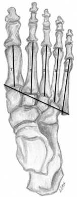
A handheld digital caliper (Neiko 01407A; Neiko Tools, Taiwan) with a measuring error of 0.02 mm was used to measure the relative lengths of the five metatarsals. Metatarsals were not disarticulated from the foot to maintain positioning. Measurements were taken from the superior and inferior aspects of each metatarsal. The length was measured from a transverse line at the most proximal midpoint at the base of the metatarsal to the most distal protruding point at the head of the metatarsal. The length was estimated as a perpendicular line from the base (Fig. 1). Three measurements were taken each from the superior and inferior aspects. The caliper was reset after each measurement to minimize error. The width of the foot was also noted and measured as the distance from the most medial position of the base of the first metatarsal to the most lateral point of the fifth metatarsal. All of the measurements were taken by the same individual.
Figure 1.
Relative metatarsal length was measured from the first to fifth metatarsals from medial to lateral from a midpoint at the proximal base of the metatarsal to the most distal protrusion of the metatarsal head. Forefoot width was also measured as the most medial aspect of the base of the first metatarsal and the most lateral protrusion of the tuberosity of the fifth metatarsal.
Statistical analysis and parameters were performed with a spreadsheet program (Microsoft Excel; Microsoft Corp, Redmond, Washington).
Results
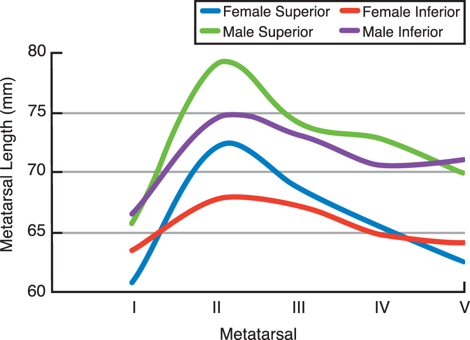
Table 1 presents the comparison between the mean ± SD metatarsal lengths measured superiorly in the left and right feet for both sexes. Table 2 shows the mean ± SD metatarsal lengths as an average of the left and right feet for both sexes separately and combined. From the mean values it is deduced that the mean lengths for the first to fifth metatarsals are 63.45, 75.64, 71.41, 69.12, and 66.27 mm, respectively, when measured superiorly. Figure 2 provides a comparison between male and female measurements. Males generally showed longer metatarsal lengths than did females. Superior measurements were also found to have greater values than inferior measurements.
Table 1.
Metatarsal Lengths Measured Superiorly in Male and Female Donors.
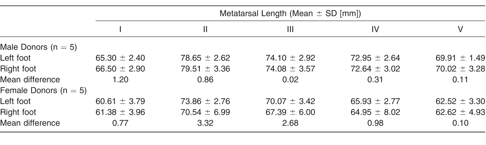
Table 2.
Metatarsal Lengths as an Average of the Left and Right Feet Measured Superiorly and Inferiorly for Male Donors, Female Donors, and Both Sexes Combined.
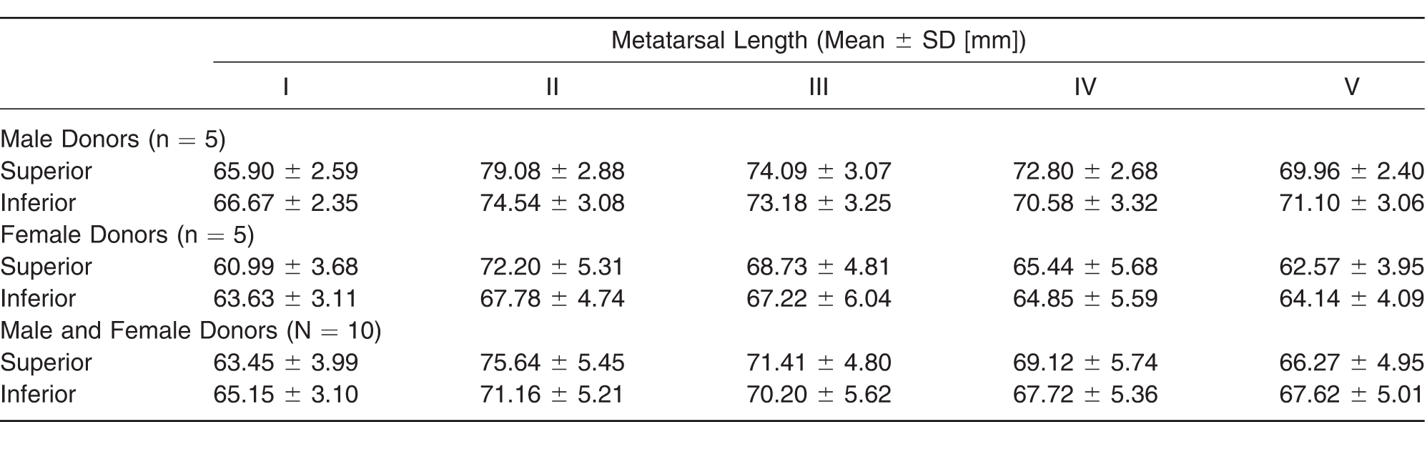
Figure 2.
A depiction of the mean metatarsal length measurements shows relative lengths to be longer in males than in females and superior measurements to be greater than inferior measurements.
From these results, it is estimated that the average formula is in the order of metatarsals II>III>IV>V>I when measured superiorly in male and females. Various metatarsal formulas are seen in both sexes when measured inferiorly. Metatarsal formulas of II>III>V>IV>I, III>II>IV>V>I, and II>I>III>V>IV are some examples.
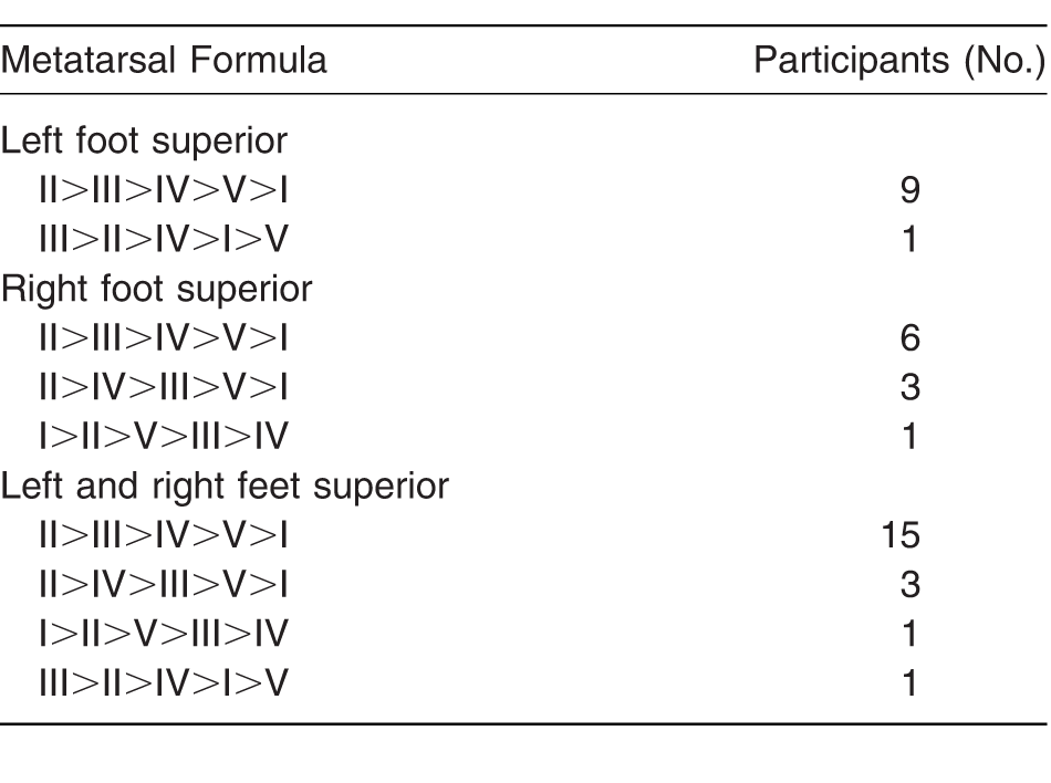
Table 3 shows the mean metatarsal formulas found in male and female donors separately. Table 4 depicts the mean metatarsal formulas when measured superiorly for both sexes when results of left and right feet are combined. Of the 20 feet, 75% shared the metatarsal formula of II>III>IV>V>I, 15% II>IV>III>V>I, 5% I>II>V>III>IV, and 5% III>II>IV>I>V.
Table 3.
Mean Metatarsal Formulas of Male and Female Donors from Superior and Inferior Measurements of the Left and Right Feet.
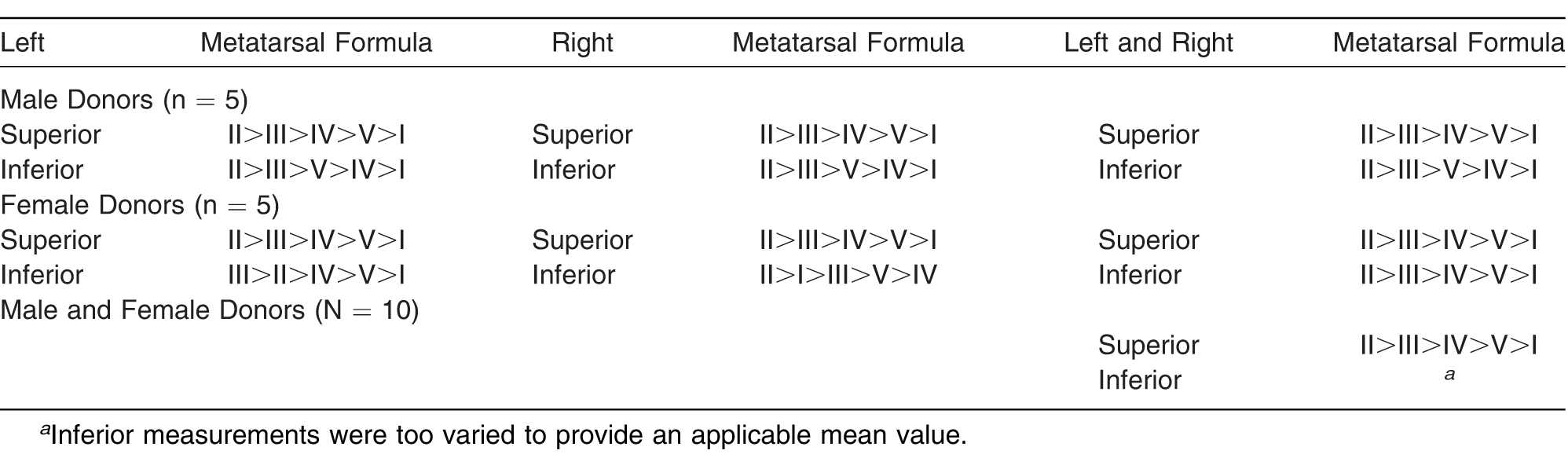
Table 4.
Mean Metatarsal Formulas for Both Sexes Combined, Presented as Left and Right Feet Separately and Combined.
Discussion
In this cadaveric study, metatarsal length measurements were obtained bilaterally on five male and five female donors. Measurements were averaged from each foot and grouped for male and female as well as superior and inferior measurements. The metatarsal lengths were then used to find the respective metatarsal formulas. In numerically comparing the metatarsal lengths in males, it is seen that the mean differences between left and right feet were minimal (≤1 mm). Superior and inferior measurements were also similar in male donors. However, in female donors, larger mean differences were noted between left and right feet and between superior and inferior aspects. This may be due to age: male donors ranged in age from 59 to 74 years, whereas female donors ranged from 71 to 93 years of age. An older range in the female donors may have led to more difficulty in measurement from more fragile metatarsals and increased likelihood of fracture.
It is also important to note that not all feet were free of pathologic abnormalities. One 67-year-old male specimen presented with hallux valgus and arthritic changes to the first metatarsophalangeal joint as seen by osteophytic changes to the metatarsal head. Another 74-year-old male specimen also had degeneration of the joint capsule. The last notable pathology was in an 81-year-old female specimen with incomplete fractures to the metatarsals. These physical changes to the metatarsal bones and joint spaces are difficult to diagnose without radiographic imaging and are subjective when being determined from clinical presentation. It is possible that the original foot structure was compromised during the transportation and preservation processes. Therefore, it is difficult to determine whether these findings are in fact endogenous and, if so, whether these differences affect overall measurements of the metatarsals.
The major findings of this study are in the metatarsal formulas calculated from the metatarsal lengths measured. From the 20 feet, the most common metatarsal formula was in the order of II>III>IV>V>I when measured superiorly. Although inferior results were too inconsistent to consider significance in the metatarsal formulas, the mean formula for males was II>III>V>IV>I and for females the same as the superior aspect, II>III>IV>V>I. These results distinguish several areas of concern. First, the method of measurement differs for superior and inferior aspects of the metatarsals. The demographics of the subject also affect metatarsal lengths, with age and sex being of utmost importance. The final measurements and conclusions of this study should be taken with caution owing to the limited sample size.
In evaluating the results of this study and others, it is noted that significant variation exists in the system of measuring the metatarsals and that little research is published on measurements in cadavers. Past studies focused on measuring from anteroposterior or oblique radiographs. This method, however, ignores the realistic structural differences found in the metatarsal bones. It is difficult to quantify a normal length value for all human feet when the metatarsals are positioned at an angle and have a shorter inferior surface. It is for these reasons that inferior measurements were difficult to obtain and will not be analyzed in detail. In the plantar view, the base of the first metatarsal overlies part of the base of the second metatarsal, making it harder to reach the base for accurate measurement. It is recommended for future reproducibility of this study to remove the first metatarsal after completing its measurement to allow easier access to the base of the second metatarsal.
A discrepancy in the method of relative metatarsal measurement is a serious concern when performing osteotomies and treating forefoot symptoms. Preoperatively, standard weightbearing radiographs are used to evaluate the metatarsal parabola or metatarsal tangent angles to plan proper treatment. [8] Chauhan et al [9] assessed three different systems of measurement as described by Coughlin, Maestro, and Hardy and Clapham and concluded that these methods were all variable and questionable. The markedly different results imply a need for more standardized methods of measurement to avoid overcorrection during surgical procedures and to provide a more precise diagnostic test that is reproducible.
Another difference to consider between radiographic measurements and cadaveric studies is the plane in which the foot is positioned and whether the foot is bearing weight. Grady and colleagues [10] compared the first and second metatarsals in the lateral and anteroposterior views and found a significant mean difference of 5.3 mm. Their study highlights the difficulty of planning procedures that shorten or lengthen the metatarsals. Another study of 81 feet with increased second metatarsal length demonstrated how the method of measurement affected clinical findings. [11] It must be remembered that it is difficult to account for metatarsal positioning in a two-dimensional image. Both the transverse and sagittal planes are required to be evaluated when assessing and managing reconstruction of the forefoot. [12]
Approximately 80% of the healthy population will experience some variation of forefoot pain during their lifetime due to biomechanical alterations. [13] Metatarsalgia is an example of forefoot pain located in the plantar region underneath the metatarsal heads. This tenderness and inflammation is usually a cause of mechanical overload and altered relationships between the metatarsals. A known characteristic factor for the development of metatarsalgia is a short first metatarsal also known as Morton's toe. However, a study comparing Morton's transverse line method and Hardy and Clapham's arc method demonstrated technical differences between the measuring methods. A short first metatarsal is, in fact, the most prevalent foot type and is consistent with this study's findings in that the first metatarsal is the shortest. [13] Measuring the metatarsal formula is not the only parameter that should be considered in surgical planning. A biomechanical assessment should also be performed to evaluate the first ray as well as overall foot function.
The Weil osteotomy is a surgical procedure indicated for the correction of relatively long or plantarflexed metatarsals, certain deformities of the metatarsophalangeal joint, and persistent metatarsalgia. [14] In a standard Weil osteotomy, the normal amount of shortening ranges from 3 to 5 mm, with a mean of 4 mm. [14,16] Delayed closure of the growth center for the metatarsals can result in a metatarsal that is longer than normal. [8] As a result, the elongated metatarsal tends to bear more weight for an extended period during the push-off phase of gait, which can lead to increased stress and pain under the metatarsal head as well as hyperkeratosis on the plantar aspect of the respective elongated metatarsal head. Of all the metatarsals, the second is the most affected due to its naturally greatest length. On the contrary, a shortened metatarsal is associated with congenital conditions or may be a result of iatrogenically induced deformity. Congenital shortening of a metatarsal is known as brachymetatarsia and leads to increased pressure at adjacent metatarsal heads. In lengthening procedures, lengthening should not exceed 50% of the original metatarsal length owing to the risk of severe complications. [15]
A more accurate description of the relative metatarsal protrusions is an asymmetrical curve. [17] Sanner [17] is in agreement with Robbins' findings that relate the distal metatarsals to one another by a semilogarithmic function. All of the feet follow the same nonlinear curve but at a different point in the curve. Therefore, the curve is standard for all feet, and weight is distributed along the curve. Any alteration in length that causes the metatarsal point to be outside of the curve will develop pathology. The main goal of forefoot surgery is to place the most distal points of the metatarsals back into the function of the curve. [17]
Overall, there is sufficient evidence demonstrating the pathologic effects of abnormal structure of the metatarsals on the biomechanics of the foot. However, more data are needed to better understand the range of measurements that compose the normal metatarsal curve. A more standardized method of measuring the metatarsals is also another factor that should be considered to achieve successful surgical results of forefoot pathology and thereby reduce iatrogenic complications.
Financial Disclosure
None reported.
Conflicts of Interest
None reported.
References
- Samojla B.“Normal Anatomy of the Forefoot,”inHallux Valgus & Forefoot Surgery, p7, Vincent J. Hetherington, Independence, MO, 2000.
- Barrôco R.Nery C.Favero G.et al: Evaluation of metatarsal relationships in the biomechanics of 332 normal feet using the method of measuring relative lengths. Rev Bras Ortop46: 431, 2011.
- Domínguez-Maldonado G.Munuera-Martinez P.Castillo-López J.et al: Normal values of metatarsal parabola arch in male and female feet. Sci World J2014: 1, 2014.
- Draves D.“Osteology and Arthrology of the Foot,”inAnatomy of the Lower Extremity, p118, Williams & Wilkins, Baltimore, 1986.
- Vogler HW.Bojsen-Møller F.Tarsal functions, movement, and stabilization mechanisms in foot, ankle, and leg performance. JAPMA90: 112, 2000.
- Maestro M.Besse J.Ragusa M.et al: Forefoot morphotype study and planning method for forefoot osteotomy. Foot Ankle Clin8: 695, 2003.
- Domínguez G.Munuera P.Lafuente G.Relative metatarsal protrusion in the adult. JAPMA96: 238, 2006.
- Thomas JL.Blitch EL.Chaney DM.et al: Diagnosis and treatment of forefoot disorders. J Foot Ankle Surg48: 239, 2009.
- Chauhan D.Bhutta MA.Barrie JL.Does it matter how we measure metatarsal length?Foot Ankle Surg17: 124, 2011.
- Grady J.Trotter K.Ruff J.et al: Radiographic investigation of the absolute and relative first metatarsal lengths in the asymptomatic foot. JAPMA105: 478, 2015.
- Bhutta MA.Chauhan D.Zubairy AI.et al: Second metatarsophalangeal joint instability and second metatarsal length association depends on the method of measurement. Foot Ankle Int31: 486, 2010.
- Barouk LS.“The Weil Lesser Metatarsal Osteotomy,”inForefoot Reconstruction, p117, Springer Science & Business Media, Paris, 2006.
- Arie EK.Moreira NS.Freire GS.et al: Study of the metatarsal formula in patient with primary metatarsalgia. Rev Bras Ortop50: 438, 2015.
- Zirm R.“The Weil Lesser Metatarsal Osteotomy,”inMcGlamry's Comprehensive Textbook of Foot and Ankle Surgery, p224, Lippincott Williams & Wilkins, Philadelphia, 2013.
- Magnan B.Bragantini A.Regis D.et al: Metatarsal lengthening by callotasis during the growth phase. J Bone Joint Surg Br77: 602, 1995.
- Nakagawa S.Kukushi J.Nakagawa T.et al: Association of metatarsalgia after hallux valgus correction with relative first metatarsal length. Foot Ankle Int37: 582, 2016.
- Sanner WH.“Foot Segmental Relationships and Bone Morphology,”inFoot and Ankle Radiology, edited byRAChristman, p283, Elsevier Science, St. Louis, 2003.
© 2018 American Podiatric Medical Association