


Ingrown toenail, or onychocryptosis, is a common inflammatory disease of the hallux. It results from the alteration of fit of the nail plate in the lateral nail fold. Ingrown toenails are usually seen in adolescents and young adults and can affect daily activities and social life. Generally, ingrown toenails are classified into three stages, including the inflammatory stage, the abscess stage, and the granulation stage. In this article, we present the rare case of a man with a diagnosis of paronychia with sporangium formation causing an ingrown toenail.
Ingrown toenail, or onychocryptosis, is a commonly encountered foot problem that often involves the hallux. It results from the alteration of fit of the nail plate in the lateral nail fold. In general, poorly fitting footwear is the most common cause. A secondary factor is incorrect cutting of the toenails. Further- more, hyperhidrosis may aggravate the condition. Patients always complain of pain, drainage with a bad odor, secondary infection, and difficulty walking. Although this problem can be seen at any point in life, the majority of patients are in their second or third decade. [1]
Case Report
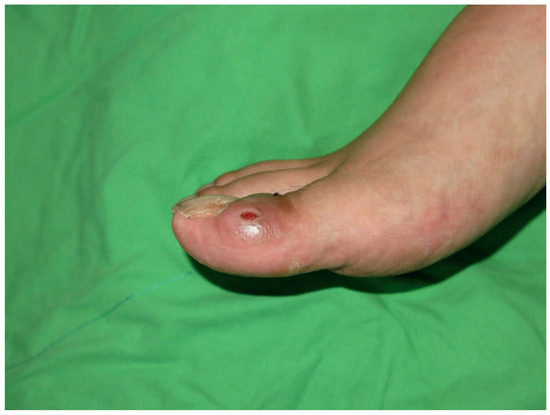
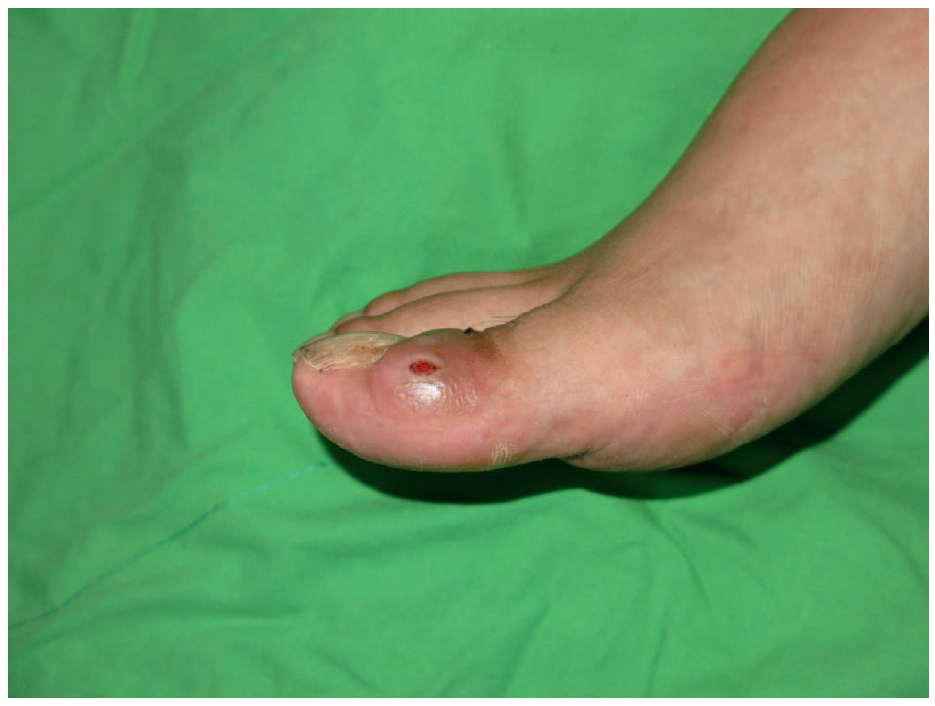
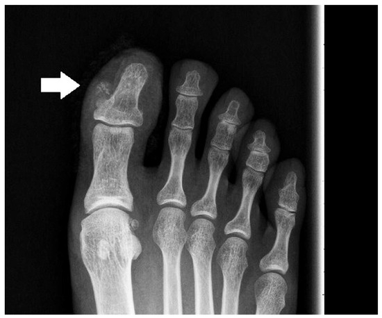
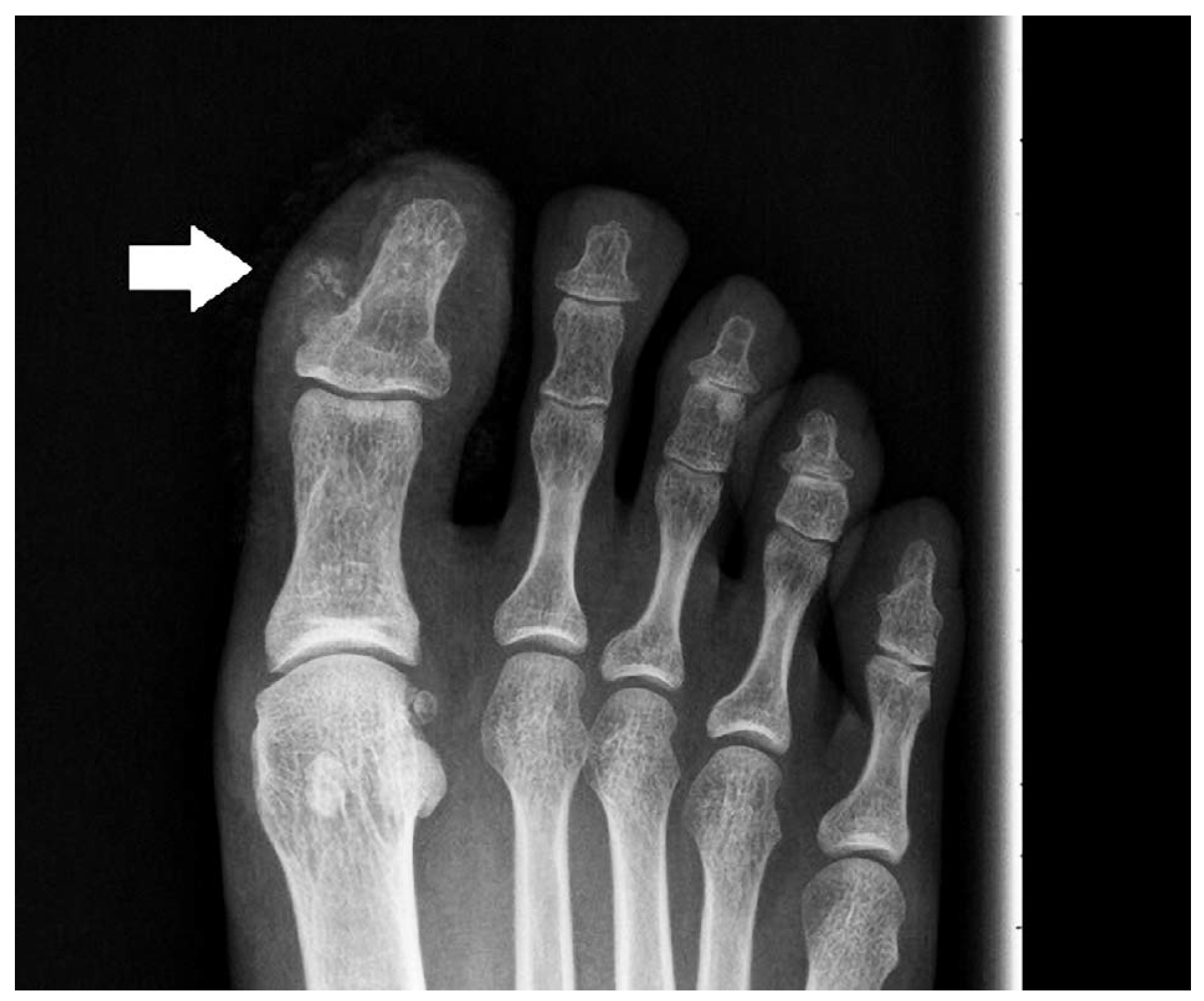
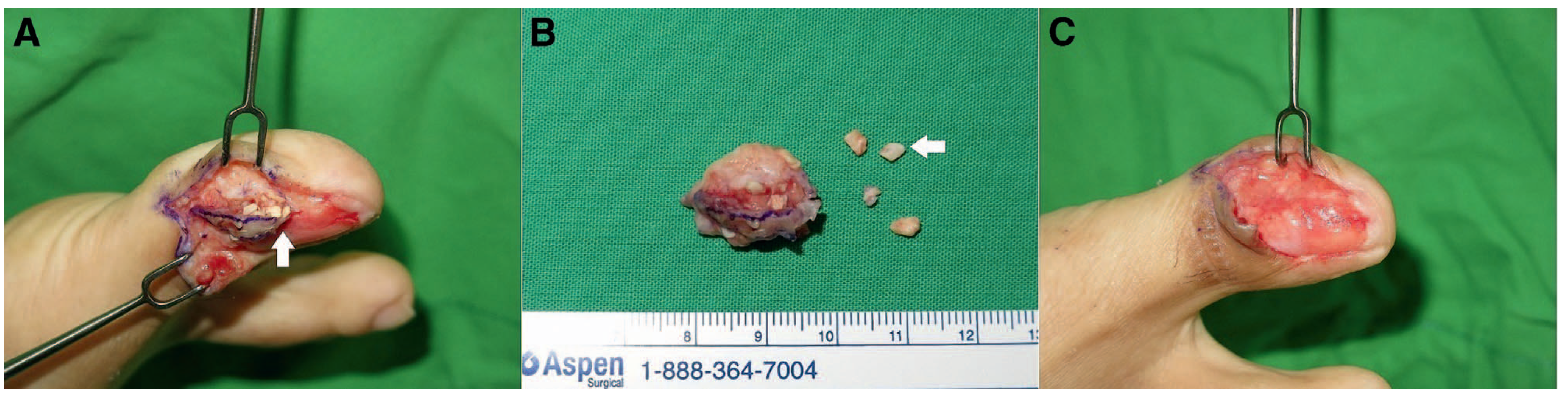
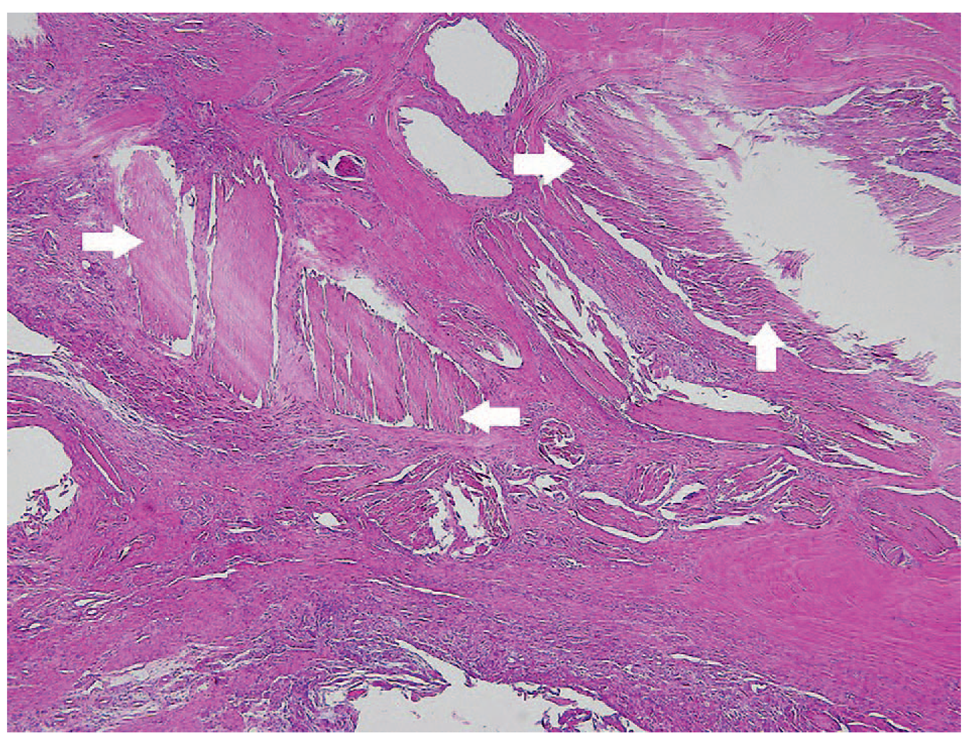
A 35-year-old man suffered from paronychia over the right hallux initially. After this, he was frequently bothered by pain arising from a protruding tumor with abscess formation over the right hallux of several months’ duration. Localized inflammation with redness and swelling was also noted over the right hallux (Figure 1). Radiography showed a nodular lesion with amorphous calcification over the medial aspect of the distal phalanx of the right hallux (Figure 2). Nail extraction and tumor excision were performed, and a tumor approximately 1 cm in diameter with broken nail pieces forming a sporangium and causing the ingrown toenail was found (Figure 3). The bone and joint were not involved. A sporangium-like form was causing the ingrown toenail and recurrent paronychia with secondary infection. Subsequently, partial matricectomy and reinsertion of a new nail plate were performed. Pathologic examination showed a cystic lesion lined with nail germinal matrix epithelium with a foreign body reaction and multiple disordered fragments of the nail plate inserted into the dermis (Figure 4).
Figure 1.
A protruding tumor with abscess formation and local inflammation is seen over the medial aspect of the distal phalanx of the right hallux.
Figure 2.
Radiograph showing a nodular lesion with amorphous calcification (arrow) over the medial aspect of the distal phalanx of the right hallux.
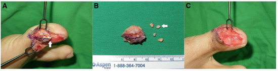
Figure 3.
(A) Before tumor excision. (B) Tumor with broken nail pieces (arrow). (C) After tumor excision and partial matricectomy. A tumor approximately 1 cm in diameter with broken nail pieces (arrow) is seen as a sporangium-like mass causing the ingrown toenail.
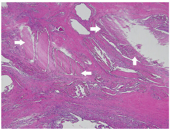
Figure 4.
Pathologic examination shows a cystic lesion lined by nail matrix epithelium with foreign body reaction and multiple disordered fragments of the nail plate inserted into the dermis (arrow) (H&E, 340).
Discussion
There are a couple of classification systems for ingrown toenails. Heifetz [2] and Mogensen [3]previously defined three stages of ingrown toenail progression. In stage I (inflammatory stage), there is erythema and slight edema and pain, particularly with pressure. In stage II (abscess stage), there is an increase in symptoms, and the wound is draining and locally infected. In stage III (granulation stage), all of the signs and symptoms are magnified and there is associated granulation tissue and nail wall hypertrophy. In 2007, Martı´nez-Nova et al [4] present- ed an expanded ingrown nail classification system as follows: stage 1, inflammatory; stage 2, abscess; stage 3, granulation and chronic nail fold hypertrophy; and stage 4, deformity of both nail folds and distal fold. However, this classification does not include the presentation of a sporangium-like tumor filled with a broken nail plate as seen in our patient. In conclusion, although ingrown toenail, or onychocryptosis, is a commonly encountered foot problem, there was no similar case presentation describing sporangium-like ingrown toenail with a broken nail plate in the literature before. In this article, we have presented the rare case of a man with a diagnosis of paronychia with sporangium formation causing an ingrown toenail.
Funding
None reported.
Conflicts of Interest
None reported.
References
- MURRAY, W.R.; BEDI, B.S. The surgical management of ingrowing toenail. Br J Surg 1975, 62, 409. [Google Scholar] [CrossRef] [Scilit] [PubMed]
- HEIFETZ, C.J. Ingrown toe-nail. Am J Surg 1937, 38, 298. [Google Scholar] [CrossRef] [Scilit]
- MOGENSEN P: Ingrowing toenail. Acta Orthop Scand 1971, 42, 94.
- Marti´nez-Nova A, Sa´ nchez-Rodri´guez R, Alonso-Pen˜ a D: A new onychocryptosis classification and treatment plan. JAPMA 2007, 97, 389. [CrossRef] [Scilit] [PubMed]
© 2018 American Podiatric Medical Association