


Abstract
Background: The aim of this work is to introduce a useful method for the clinical diagnosis of leg-length inequality: distance between the malleoli and the ground (DMG). Methods: A transversal observational study was performed on 17 patients with leg-length discrepancy. Leg-length inequality was determined with different clinical methods: with a tape measure in a supine position from the anterior superior iliac spine (ASIS) to the internal and external malleoli, as the difference between the iliac crests when standing (pelvimeter), and as asymmetry between ASISs (PALpation Meter [PALM]; A&D Medical Products Healthcare, San Jose, California). The Foot Posture Index (FPI) and the navicular drop test were also used. The DMG with Perthes rule (perpendicular to the foot when standing), the distance between the internal malleolus and the ground (DIMG), and the distance between the external malleolus and the ground were designed by the authors. Results: The DIMG is directly related to the traditional ASIS–external malleolus measurement (P = .003), the FPI (P = .010), and the navicular drop test (P < .001). There are statistically significant differences between measurement of leg-length inequality with a tape measure, in supine decubitus, from the ASIS to the internal malleolus, and from the ASIS to the external malleolus. Conclusions: This new method (the DMG) is useful for diagnosing leg-length discrepancy and is related to the ASIS–external malleolus measurement. The DIMG is significantly inversely proportional to the degree of pronation according to the FPI. Conversely, determination of leg-length discrepancy with a tape measure from the ASIS to the malleoli cannot be performed interchangeably at the level of the internal or external malleolus.
Although the study of lower-limb asymmetry is dealt with broadly in the scientific literature, there are contradictions as to the quantification, reliability, and validity of the different clinical procedures to measure them [1,2,3,4,5,6,7,8,9,10,11,12,13,14,15,16]. This also applies to the relationship of leg-length inequality with certain problems and pathologic abnormalities. The impact seems clear for most authors [10,11,12,13], but not the type and degree of leg-length discrepancy that can cause these problems [14,15]. Leg-length asymmetry is an aesthetic and functional problem; when limits of tolerance are exceeded, this leads to symptoms or is associated with certain serious pathologic abnormalities [10,11,12,13,17,18,19]: pain, disabilities, abnormalities or mechanisms of accommodating static movement and gait, etc. Among these accommodative mechanisms, the following are notable: increase in energy expenditure (because of elevation and excessive fall of the pelvis), scoliotic attitudes, and changes in foot position during static and dynamic movement. Leg-length discrepancy is an important problem that compromises the foot, vertebral column, and intermediate structures [13,18,19]. The clinical determination of this condition involves diagnostic protocols set out by some authors [2,16]. Leg-length discrepancy can have different causes: epiphyseal hypogrowth, hypergrowth because of sequelae of metaphyseal or diaphyseal trauma, infections or avascular necrosis, or paralytic, vascular, or tumor-related disorders [17,20,21]. Two major limb-length dicrepancy groups are established [22]: anatomical and functional leg-length inequality, each caused by several factors. Even today, agreement has not been reached on the most suitable clinical method for identifying the type of leg-length discrepancy experienced by an individual. It seems that the focus is on performing a broad protocol that includes different quantifiable methods by means of an exhaustive exploratory examination. In the protocols provided in the literature, there are two especially extensive clinical methods: 1) in the supine decubitus position, measuring the length of the legs with a tape measure from the anterior superior iliac spine (ASIS) to the internal [1] and external [2] malleoli and 2) in the standing position, comparing the position of the iliac crests or spines with the pelvimeter and the PALpation Meter (PALM) [4,5].
Regarding possible foot compensation, clinically it has been often, but not always, observed that in the shorter leg the foot is in varus and equinus [23] to functionally lengthen the limb; in the longer limb, hindfoot valgus is usually increased compared with the contralateral limb [16,24,25]. Both abnormalities (accommodative varus and valgus of the hindfoot) can be found combined or singularly to compensate for a leg-length inequality. Some studies have attempted to identify and measure these compensations [26,27]. The aims of the present study were to introduce a useful method for the clinical determination of leg-length asymmetry—distance between the malleoli and the ground (DMG)—and to determine whether there is a relationship between the different methods of quantifying leg-length discrepancy and foot position.
Methods
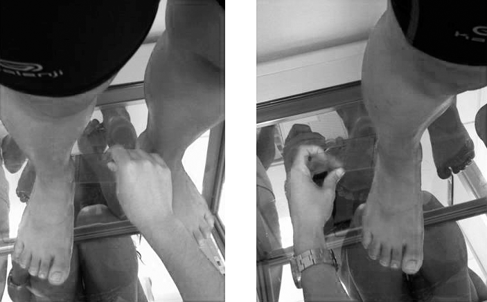
A correlational, transversal, and observational descriptive study [28] was performed on 17 patients with leg-length discrepancy selected by nonprobabilistic logic sampling. Participants were seen in the Podiatry Clinic of Seville University (Seville, Spain) between January 1, 2012, and January 1, 2013. Each participant was notified of the study aims and the methods used to measure the variables, and once understanding was reached, they gave their written consent. The study was approved by the Experimentation Ethics Committee of Seville University. The inclusion criterion was a previous diagnosis of leg-length inequality in the Department of Biomechanics and Orthopodology of the Podiatry Clinical Area of Seville University by means of the protocol set out and according to the references [2,16]. With the patient in the supine position on a stretcher with legs together and arms against the body, and previous relaxation of the pelvic muscles, the inferior borders of both malleoli were compared visually. Patients were aged 18 to 27 years (random sampling of convenience). Patients who had undergone osteo-articular surgery in the legs and feet in the previous 3 years were excluded from the study. Participants filled in a form on which demographic data and medical history were collected. The following variables were determined: 1) the length of each leg, with a tape measure from the most prominent area of the ASIS to the inferior border of the external malleolus and from the ASIS to the inferior border of the internal malleolus; 2) the difference between the iliac crests, by means of the pelvimeter; 3) asymmetry between the ASIS with the PALM (Fig. 1); 4) the Foot Posture Index (FPI), to quantify the degree of neutral, pronated, or supine position of the foot; 5) fall in navicular with the patient standing, to quantify the capacity for pronation of the subtalar joint, with the navicular drop test (NDT); and 6) the distance from both external and internal malleoli to the ground (DMG), including the distance between the internal malleolus and the ground (DIMG) and the distance between the external malleolus and the ground (DEMG). For Perthes rule (perpendicular to the foot when standing), we used a pencil mark on each border as a reference (Fig. 2).
Figure 1.
PALpation Meter: PALM.
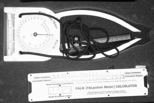
Figure 2.
The procedure for measuring the distance between the malleoli and the ground (DMG).
All of the parameters were evaluated in centimeters, except the FPI, age, and sex. Sample size was determined using CTM software (Version 1.1; Glaxo Wellcome SA, Madrid, Spain) an error (epsilon) of 2.2% was assumed for an initial estimate of 0.5 (50%) and for a confidence level of 95%. The minimum number of study legs was 30 (15 patients). Data were analyzed with a statistical software package (SPSS Statistics for Windows, Version 22.0; SPSS Inc, Chicago, Illinois). All of the variables followed a normal distribution, according to the Shapiro-Wilk test, except FPI and PALM. Numeric variables are summarized with means and typical deviations, asymmetrical variables, medians, and 25th to 75th percentiles. Nonnumeric variables are summarized with frequencies and percentages. Timely estimators and 95% confidence intervals (CIs) were obtained for the different statistics. To measure the linear relationship between pairs of numeric variables (normally distributed), Pearson or Rho Spearman correlation coefficients were calculated according to the application criteria. The correlation or independent test was applied to ascertain the statistical significance of the association both for the overall samples and the defined subgroups. To evaluate the reliability between the pelvimeter and the PALM, the intraclass correlation coefficient was calculated using the Cronbach α as a reliability statistic. To analyze the relationship between dichotomous qualitative and quantitative variables, the Student t test was performed. In the case of not complying with the requirement for equality of variance (the Levene test), the Welch-corrected Student t test was performed. In the case of the normality requirement not being compliant (the Shapiro-Wilk test), the Mann-Whitney U test was performed. If statistically significant differences were detected, CIs were determined for mean differences at 95%, which quantify these differences. Statistical significance of P = .05 was considered for all contrasts of the hypothesis.
Results
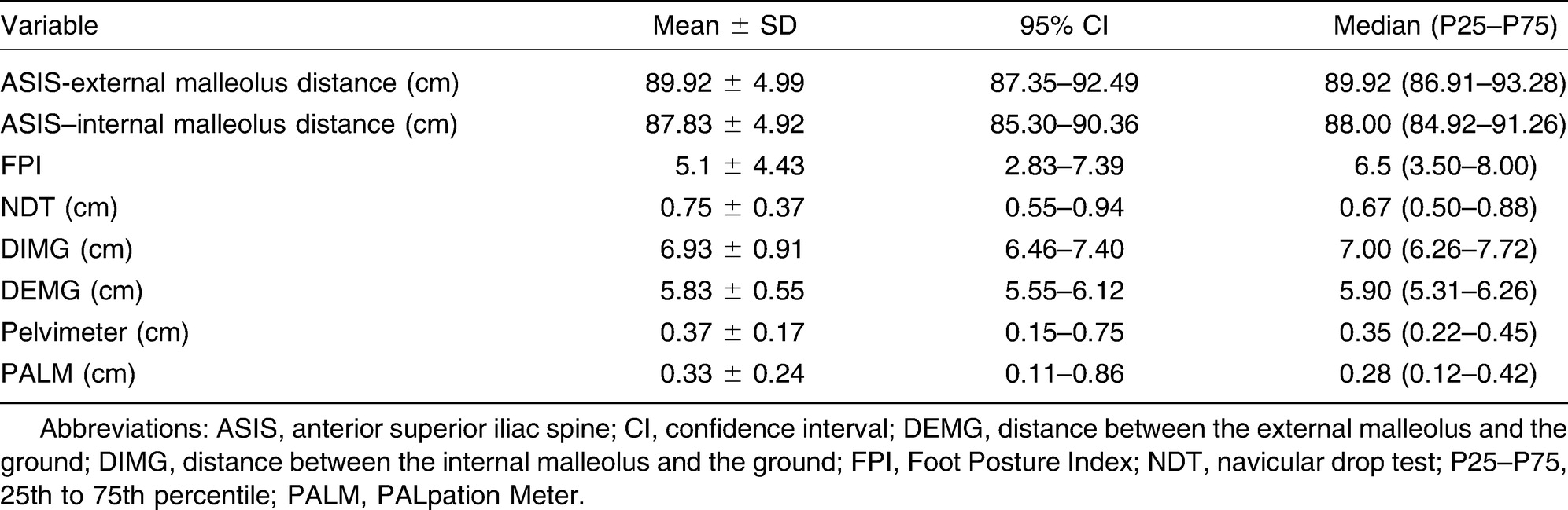
A total of 34 legs corresponding to 17 patients (11 women [64.7%] and six men [35.3%]) were analyzed; patients were aged 18 to 27 years, with a mean ± SD age of 22.47 ± 2.03 years. Regarding the descriptive analysis, the overall results, extreme values, and trends for the variables studied are shown in Table 1. The descriptive analysis includes ASIS–internal and external malleolus distance, FPI, NDT, DIMG and DEMG, pelvimeter, and PALM.
Table 1.
Descriptive Analysis of ASIS–Internal and External Malleolus Distance, FPI, NDT, DIMG and DEMG, Pelvimeter, and PALM
Leg length was considered to determine long and short legs, and we report the results obtained descriptively. The relationship of the NDT, FPI, and DMG with short and long legs was also determined. Table 2 summarizes the basic descriptive statistics that enable us to observe the extreme values in addition to robust trends. We should recall that in this study of 34 legs analyzed in the matrix, 17 were short legs and 17 were long legs.
Table 2.
Relationship Between Long Leg and Short Leg with the NDT, FPI, and DEMG and DIMG
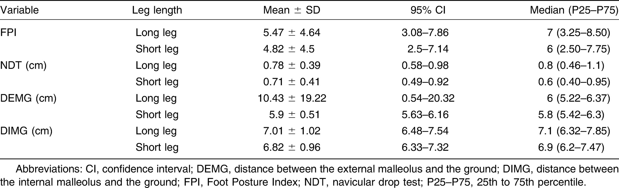
For the short leg there is a correlation between the DIMG and the FPI (r = –0.605; P = .010) such that as one increases the other decreases. The DIMG is inversely related to the NDT (r = –0.765; P < 0.01) and the DEMG (r = 0.613; P = .009). For the long leg there is no correlation between the FPI and the NDT (r = 0.157; P = .033); that is, only 2.4% of the variability in the NDT can be accounted for by the variability in the FPI. There is no correlation between the other measurements.
To determine the reliability of the pelvimeter in the quantification of leg-length discrepancy, we calculated the statistical test that provides us with the intraclass correlation coefficient using the Cronbach α reliability coefficient. The data obtained tell us that there is no concordance between the measurements made with the pelvimeter and those made with the PALM (intraclass correlation coefficient [ICC] = 0.566; 95% CI, –0.614 to 0.883; P = .102).
The results regarding the relationship between the measurement of leg-length discrepancy with a tape measure from the ASIS to the external or internal malleolus reveal that significant differences were found between both variables (P = .019); that is, there is a linear relationship between the measurement of distance from the ASIS to the external malleolus and from the same origin to the internal malleolus (r = 0.992; P < .001) such that the further the ASIS is from the external malleolus, the greater the distance from the same origin to the internal malleolus. A total of 98.4% (r2 = 0.984) of the variability in the first measurement can be accounted for by the variability in the other measurement (bivariate analysis). The data reveal that there are no statistically significant differences by sex, with some levels of significance very far removed from P = .05 in all cases.
During analysis of the correlation between ASIS–external malleolus distance and the other variables, for both short and long legs, we found that only the correlation between ASIS–external malleolus distance and the DIMG was determined; that is, as the value of the ASIS–external malleolus distance increases, the DIMG also increases.
Discussion
Some seldom-studied factors were analyzed along with leg-length inequality. A new reference parameter was also designed and quantified: DMG, DIMG, and DEMG. One factor that led us to study this was considering that the estimate of height between the malleoli and the ground could be directly related to the position adopted by the subtalar joint, in one foot or the other, according to the possible compensation. Therefore, for example, there are studies that report how supination and plantarflexion of the foot are used to compensate for the shorter leg.23 In any case, we have not found any study that shows normality ranges for both measurements or their relevance; this aspect is, thus, an aim to study in future research.
By analyzing the correlations of all of the variables in this study, we determined that the DIMG and the FPI are the factors that have led to the most correlation with the other variables. The linear correlation analyses between the different foot variables and the long leg show a relationship between the FPI and the DIMG. The significant (P = .011) inverse correlation (–0.602) leads us to consider that as foot valgus increases, the distance between the internal malleolus and the ground decreases. The same occurs between the FPI and the NDT. However, the coefficient of correlation has a low value (0.157), with a level of significance of approximately P = .033; this translates into the higher the valgus, the more capacity for subtalar pronation for the NDT according to the FPI [29] classification criteria.
For the short leg we found the following correlations: the DIMG variable is related to the other three in the following way: with the FPI we obtain a moderate inverse correlation (–0.605) with significance of approximately P = .010; that is, the DIMG decreases with more foot valgus. With the NDT, the correlation, which is also negative, is greater (–0.765) and significant (P = .000). Put otherwise, the NDT increases with increasing DIMG.
Finally, as for the correlation between the two measurements taken from the respective malleoli to the ground, we found a moderate coefficient (0.613) and a clear level of significance (P = .009). The DEMG increases with a higher DIMG, which accounts for the anthropometric characteristics of the study patients.
For the short leg, we stress that this is related to a lower degree of valgus (according to the FPI), which coincides with the study by Blustein and D'Amico [23]. This is despite the fact that the sample's characteristics do not enable us to determine whether these data are statistically significant.
In the final correlations section, in which we list the ASIS distance variable with its two reference points and that of both legs, the DIMG parameter becomes a protagonist again.
For the long leg, the coefficient of correlation for the ASIS–external malleolus distance is moderate (0.680) and significant (P = .003); that is, the greater the distance between the ASIS–external malleolus, the higher the DEMG. For the short leg, this is related to the DMG from both malleoli. This leads to an external malleolus with a moderate coefficient (0.617) and a high level of significance (P = .008).
Regarding ASIS–internal malleolus distance, the long leg leads to a high level of significance (P = .003), with a coefficient of 0.674. For the short leg, we found lower correlation coefficients (0.584 and 0.434) and positive significance (P = .014 and .082) for the DMG and each one of the malleoli, respectively.
Long left legs predominated (58.82% of the sample). Olmedo [21] reports that the left leg is normally more affected because it is usually found to be in a higher degree of adduction in the uterine bed, but the author does not clarify that this is the reason for the left leg lengthening. We have not found any studies that report sex-related differences.
Lower-limb asymmetry ranges from approximately 2 to 9 mm according to the pelvimeter and the PALM; the means are 3.7 and 3.3 mm, respectively. The longer leg usually has a higher FPI, that is, a more pronated foot—a compensation mechanism that has been identified in several studies [16,24,25]. However, Rothbart30 found that the shorter leg coincides with the foot with the higher degree of valgus. At the start of this study we wondered whether there was a statistically significant difference between the measurement of leg-length inequality taking as a reference point the ASIS with the external malleolus or taking the distal reference of the internal malleolus. The results indicate that there is a significant difference between both measurements (P = .000), that is, if different benchmarks were used, the result would be different. To this we have to add that tackling the reference points is more or less complicated, according to the anthropometric characteristics of each individual.
We found studies in which a tape measure is used to measure from the anterior superior iliac spine to the medial malleolus and lateral malleolus. According to these, the method used by the tape measure has both variants, which determines the distance between the ASIS and the external malleolus, used, among others, by Woerman and Binder-Macleod,2] and the other used by authors such as Beattie et al. [1] However, the most extensive method is determining the length of the leg with a tape measure from the anterior superior iliac spine to the lateral malleolus. No agreement has been reached on either the reliability of the measurement or which of the two is most appropriate. After reviewing works from several authors, we can verify that there is clear disagreement regarding validity and reliability between the two direct methods using the tape measure [1,2,6,7,8]
We used the pelvimeter as an additional aspect to the fieldwork. This is a commonly used instrument in our field but lacks validity. This has led us to take an interest in its validation. To our regret, this aim was not attained in this work. Moreover, we did not find any relationship whatsoever between the pelvimeter variable and the variables quantified in the foot for either of the two legs.
Given the reduced sample, we cannot extrapolate the results to the population. We have not found any studies that correlate these variables and offer similar results whereby we could make comparisons.
Conclusions
The variable most related to the remaining variables studied is the DMG. There is a direct relationship between ASIS–internal and external malleolus distance and the DMG, suggesting that this may be a useful clinical method for studying lower-limb asymmetry. In longer legs, we obtained a higher FPI for most of the sample (70.1%). Despite this finding, the results are not statistically significant: 58.82% of participants had a longer left leg, and no statistically significant differences were found according to sex. Leg-length discrepancy in healthy individuals cannot be measured interchangeably at the level of the internal or external malleolus given that there are statistically significant differences between both methods.
Financial Disclosure
None reported.
Conflicts of Interest
None reported.
References
- BEATTIE, P.; ISAACSON, K.; RIDDLE, D.L.; et al. Validity of derived measurements of leg-length differences obtained by use of a tape measure. Phys Ther 1990, 70, 150. [Google Scholar] [CrossRef] [Scilit]
- WOERMAN, A.L.; BINDER-MACLEOD, S.A. Leg length discrepancy assessment: accuracy and precision in five clinical methods of evaluation. J Orthop Sports Phys Ther 1984, 5, 230. [Google Scholar] [CrossRef] [Scilit] [PubMed]
- KNUTSON, G.A. Anatomic and functional leg-length inequality: a review and recommendation for clinical decision-making: part II. The functional or unloaded leg-length asymmetry. Chiropr Osteopat 2005, 13, 12. [Google Scholar] [CrossRef] [Scilit]
- HAGINS, M.; BROWN, M.; COOK, C.; et al. Intratester and intertester reliability of the palpation meter (PALM) in measuring pelvic position. J Manual Manip Ther 1998, 6, 130. [Google Scholar] [CrossRef] [Scilit]
- PETRONE, M.R.; GUINN, J.; REDDIN, A.; et al. The accuracy of the palpation meter (PALM) for measuring pelvic crest height difference and leg length discrepancy. J Orthop Sports Phys Ther 2003, 33, 319. [Google Scholar] [CrossRef] [Scilit] [PubMed]
- GROSS, M.T.; BURNS, C.B.; CHAPMAN, S.W.; et al. Reliability and validity of rigid lift and pelvic leveling device method in assessing functional leg length inequality. J Orthop Sports Phys Ther 1998, 27, 285. [Google Scholar] [CrossRef] [Scilit] [PubMed]
- FRIBERG, O.; NURMINEN, M.; KORHONEN, K.; et al. Accuracy and precision of clinical estimation of leg length inequality and lumbar scoliosis: comparison of clinical and radiological measurements. Int Disabil Stud 1998, 10, 49. [Google Scholar] [CrossRef] [Scilit]
- CLARKE, GR. Unequal leg length: an accurate method of detection and some clinical results. Rheumatol Phys Med 1972, 11, 385. [Google Scholar] [CrossRef] [Scilit]
- MANN, M.; GLASHEEN-WRAY, M.; NYBERG, R. Therapists agreement for palpation and observation of iliac crest heights. Phys Ther 1984, 64, 334. [Google Scholar] [CrossRef] [Scilit]
- CARLSON, M.; WILKERSON, J. Are differences in leg length predictive of lateral patello-femoral pain? Physiother Res Int 2007, 12, 29. [Google Scholar] [CrossRef] [Scilit]
- BUSQUET, L. I: Las Cadenas Musculares Tomo IV, Miembros Inferiores, Vol I; Paidotribo, Barcelona, 2007. [Google Scholar]
- RICARD, F. Tratamiento Osteopa´tico de las Algias Lumbope´lvicas, 3rd Ed ed; Panamericana: Buenos Aires, 2005. [Google Scholar]
- FRIBERG, O. Leg length asymmetry in stress fractures: a clinical and radiological study. J Sports Med Phys Fitness 1982, 22, 485. [Google Scholar]
- NAVAS, J.; UELO, JA; CAN˜ UELO, CB. : Valoracio´n posicional de la pelvis en el diagno´stico de las asimetrı´as. Salud del Pie: Rev Andal Pod 2009, 2, 52. [Google Scholar]
- SUBOTNICK, SI. Limb length discrepancies of the lower extremity (the short leg syndrome). J Orthop Sports Phys Ther 1981, 3, 11. [Google Scholar] [CrossRef] [Scilit]
- LAFUENTE, B.; LAFUENTE, G.; MUNUERA, P.V. Protocolo de exploracio´n de disimetrı´as. Podol Clı´n 2009, 10, 140. [Google Scholar]
- MORENO, JL. Podologı´a General y Biomeca´nica, p 119. Masson, Barcelona, 2003.
- GILES, L.G.; TAYLOR, J.R. Low-back pain associated with leg length inequality. Spine 1981, 6, 510. [Google Scholar] [CrossRef] [Scilit] [PubMed]
- GOFTON, J.P.; TRUEMAN, G.E. Studies in osteoarthritis of the hip: II. Osteoarthritis of the hip and leg-length disparity. Can Med Assoc J 1971, 104, 791. [Google Scholar] [PubMed]
- GARRIDO, R.; LUACES, C. Cojera en la infancia: protocolos diagno´sticos y terape´uticos en pediatrı´a. Urgencias en pediatrı´a 2002, 3, 131. [Google Scholar]
- OLMEDO, JL. Exploracio´n ortope´dica ba´sica. Pediatr Integral 2006, 10, 475. [Google Scholar]
- GURNEY, B. Leg length discrepancy. Gait Posture 2002, 15, 195. [Google Scholar] [CrossRef] [Scilit]
- BLUSTEIN, S.M.; D’AMICO, J.C. Limb length discrepancy: identification, clinical significance, and management. JAPMA 1985, 75, 200. [Google Scholar] [CrossRef] [Scilit]
- RESTOY JL, PUN˜ ET PG. Dismetrı´a en extremidades inferiores y equilibrio de la pelvis. El Peu 2009, 29, 196. [Google Scholar]
- LANGER, S. Structural leg shortage: a case report. JAPA 1976, 66, 38. [Google Scholar] [CrossRef] [Scilit]
- D’AMICO, J.C.; DINOWITZ, H.D.; POLCHANINOFF, M. Limb length discrepancy: an electrodynographic analysis. JAPMA 1985, 75, 639. [Google Scholar] [CrossRef] [Scilit]
- BLAKE, R.L.; FERGUSON, HJ. Correlation between limb length discrepancy and asymmetrical rearfoot position. JAPMA 1993, 83, 625. [Google Scholar] [CrossRef] [Scilit] [PubMed]
- ARGIMO´ NJM, JIME´NEZ J. Parte II, Me´todos de Investigacio´n Clı´nica y Epidemiolo´gica, 3rd Ed; Edide, S.L., Ed.; Elsevier: Madrid, 2004. [Google Scholar]
- REDMOND, A.C.; CRANE, Y.Z.; MENZ, HB. Normative values for the Foot Posture Index. J Foot Ankle Res 2008, 1, 6. [Google Scholar] [CrossRef] [Scilit] [PubMed]
- ROTHBART, BA. Relationship of functional leg-length discrepancy to abnormal pronation. JAPMA 2006, 96, 499. [Google Scholar] [CrossRef] [Scilit]
© 2017 American Podiatric Medical Association