


Abstract
This case report highlights a novel approach to strengthening the repair of a split peroneus brevis tendon tear with a peroneus quartus muscle autograft. We describe a 51-year-old woman with a longitudinal split tear of the peroneus brevis tendon confirmed by magnetic resonance imaging. Intraoperatively, a peroneus quartus muscle was appreciated, resected, and used as an autograft in the repair of the peroneus brevis tendon. Use of a peroneus quartus muscle as an autograft in peroneal tendon repair has not been documented in the literature, to our knowledge.
The peroneus quartus muscle is an accessory muscle that is unique to human beings [1,2]. The presence of a variant peroneus quartus muscle has been known to cause lateral ankle pain and instability, peroneal tenosynovitis, subluxation of the peroneal tendons, and longitudinal splitting of the peroneus brevis tendon [3,4]. According to Demetracopoulos et al [5] and Prakash et al [6], accurate diagnosis of peroneal tendon disorders is 60% at the first clinical evaluation. Cadaveric research has shown a 37% incidence of peroneus brevis tendon tears [2,7]. Injuries to the peroneal tendons can cause lateral ankle and hindfoot pain. Recurrent ankle sprains and hindfoot varus can cause ankle instability and dislocation of the tendons, leading to tearing of the tendons [8]. Anatomical anomalies, including a shallow retromalleolar groove, a low-lying peroneal muscle belly, a peroneus quartus muscle, a prominent peroneal tubercle, laxity in the superior retinaculum, and bone spurs on the posterior lateral edge of the fibula, are shown to be an additional source of peroneal injuries [9]. The location of the peroneus brevis tendon in the retromalleolar groove when associated with a low-lying peroneal muscle belly is a source of injury to the tendon as it courses around the fibula by mass effect [10].
The exact anatomical location of the peroneus quartus muscle is described as variable in the literature. In 1923, Hecker first termed the peroneus quartus muscle and classified its various forms by its insertion point: peroneocalcaneus externum (calcaneus), peroneocuboideus (cuboid), and peroneoperoneolongus aka peroneus accesorius (peroneus longus tendon) [1]. In a cadaveric study by Sobel et al [2] and Zammit and Singh [4], origins were noted from the peroneus brevis and peroneus longus tendons and the posterior aspect of the distal fibula. The incidence of the peroneus quartus muscle varies from 6.6%, as described by Zammit and Singh [4], up to 21.7%, by Sobel et al [2]. The use of a peroneus quartus muscle in surgical reconstruction is poorly reported in the literature. Mick and Lynch [11] reported the use of a peroneus quartus muscle in the reconstruction of the retromalleolar groove to stabilize the peroneal tendons in recurrent peroneal tendon dislocation. To our knowledge, the peroneus quartus muscle has not been documented as an autograft in the reconstruction of a split tear of the peroneus brevis tendon.
In end-to-end repair of peroneus brevis tendon tears, interposition grafts of gracilis muscle, semitendinosus muscle, and a strip of the iliotibial band have been described to bridge the defect [8]. Mook et al [12] described using frozen peroneal tendon or semitendinosus muscle allograft in the repair of 14 peroneal tendon tears.
Case Report
A 51-year-old woman presented with a painful swollen left ankle of 2.5 years' duration. The pain causes difficulty walking and trouble with footwear. The patient has a history of ankle sprains but no recent trauma. Before her presentation to our office the patient received vascular testing, treatments by physical therapists, and an evaluation by an orthopedist without resolution of her symptoms. The patient had no previous advanced imaging studies.
The patient's medical history is significant for previous deep venous thrombosis, attention deficit disorder, glaucoma, Hashimoto's thyroiditis, and autoimmune hepatitis that was in remission. On physical examination, mild edema was appreciated on the left side. The patient had a stable foot posture with a cavovarus foot type (Fig. 1). Good muscle strength was appreciated in all prime movers of the foot and ankle except the peroneal tendons. Pain was elicited on palpation of the lateral ankle ligaments and on insertion of the peroneus brevis tendon on the fifth metatarsal tuberosity. Plain radiographs of the foot and ankle revealed no acute fractures or dislocations. Magnetic resonance imaging showed sinus tarsi and ankle inflammation, a peroneus brevis tendon longitudinal tear with a characteristic C-shaped morphology, and tenosynovitis to the peroneus brevis and peroneus longus tendons. Four weeks after initial presentation, the patient underwent surgical management of a split tear of the peroneus brevis tendon, ankle joint synovitis, and subtalar joint synovitis.
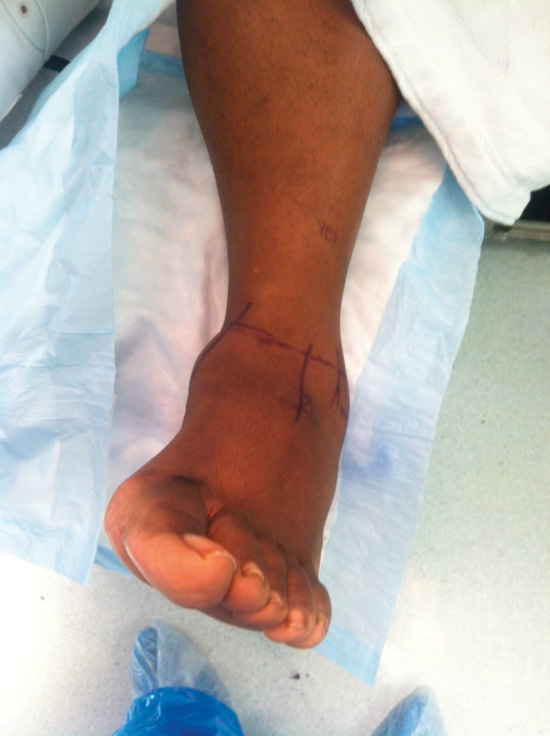
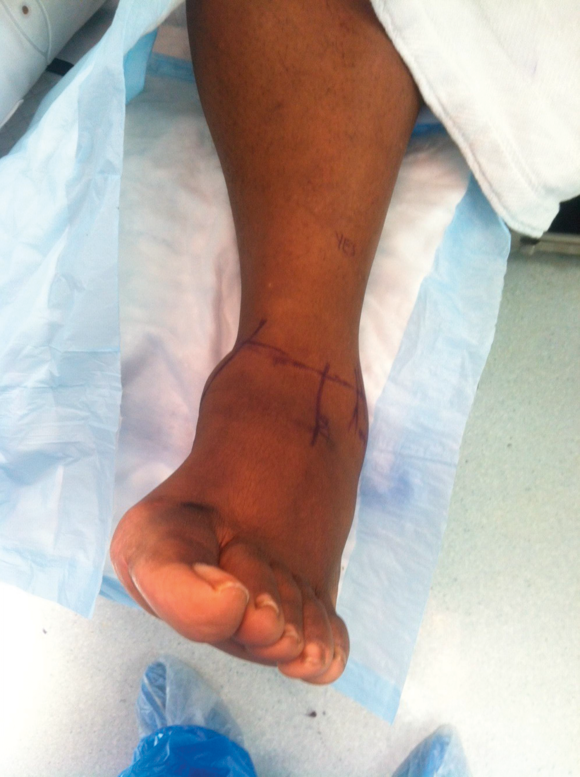
Figure 1.
Preoperative image of the cavovarus foot type.
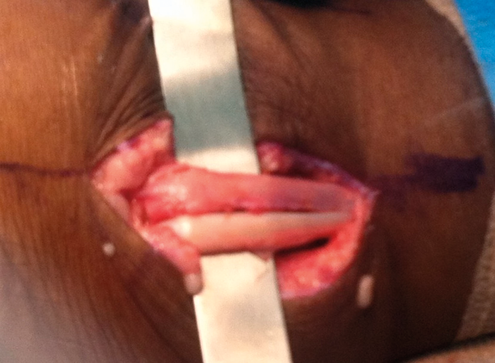
Initially, a subtalar joint and ankle joint arthroscopy was performed in the typical manner. Once dissection was completed down to the level of the peroneal tendon sheath, the sheath was opened, and the peroneal tendons were visualized. The peroneus longus muscle was noted to be tubular and healthy, whereas the peroneus brevis muscle was flattened, with a longitudinal tear from approximately 4 cm proximal to the distal edge of the lateral malleolus that extended distally 2 cm to the lateral edge of the lateral malleolus (Fig. 2). At this time, a peroneus quartus muscle and a low-lying muscle belly of a peroneus brevis muscle were noted, likely causing a mass effect on the lateral aspect of the foot (Fig. 3). The low-lying muscle belly was resected using electrocautery, and hypertrophied synovitis was removed from the tendon and sent to the pathology laboratory. Using 2-0 FiberWire (Arthrex Inc, Naples, Florida), with a running interlocking technique, the peroneus brevis tendon was retubularized along the entire aspect of the injured tendon. Intraoperatively, the decision was made to strengthen the peroneus brevis tendon repair using the peroneus quartus muscle as an onlay autograft. The peroneus quartus tendon was resected as distal as possible and was sutured to the peroneus brevis tendon using 2-0 FiberWire (Figs. 4 and 5). The peroneal tendon sheath, followed by the peroneal retinaculum, was then repaired using 2-0 Vicryl (Ethicon Inc, Somerville, New Jersey) in a taut position to prevent further dislocation of the peroneal tendon. The incision site was flushed with normal saline and closed in a layered manner. Postoperatively, the patient was placed in a posterior splint with the foot in an everted position and was nonweightbearing with crutches to the left foot. The patient was given aspirin 325 mg/d for deep venous thrombosis prophylaxis until ambulation started.
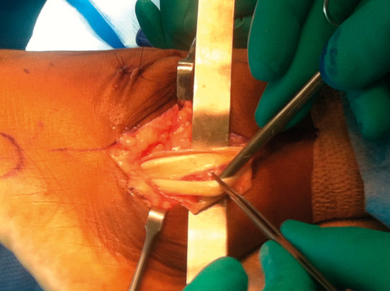
Figure 2.
Split tear in the peroneus brevis tendon.
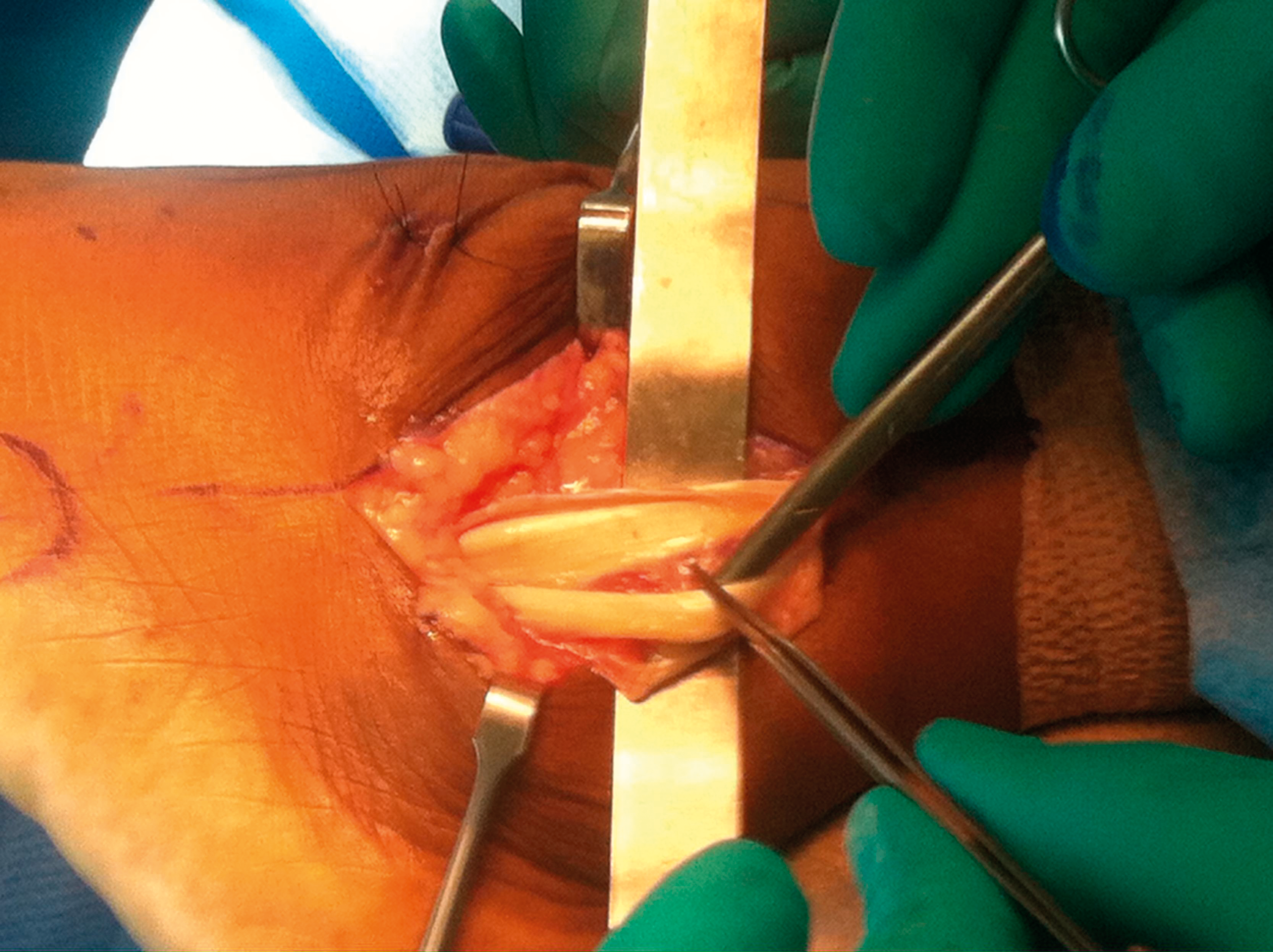
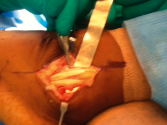
Figure 3.
Isolated peroneus quartus muscle with peroneus brevis and peroneus longus tendons.
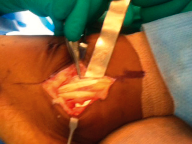
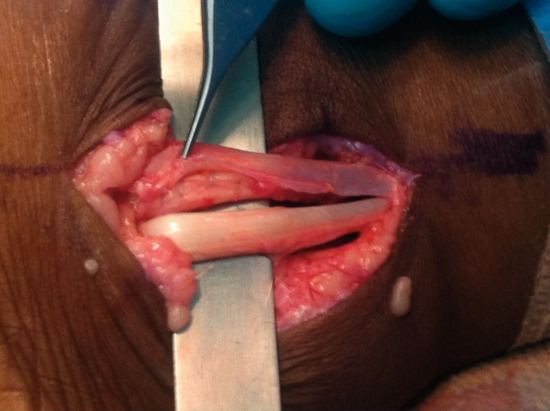
Figure 4.
Preparing the peroneus quartus muscle graft for the peroneus brevis tendon repair.
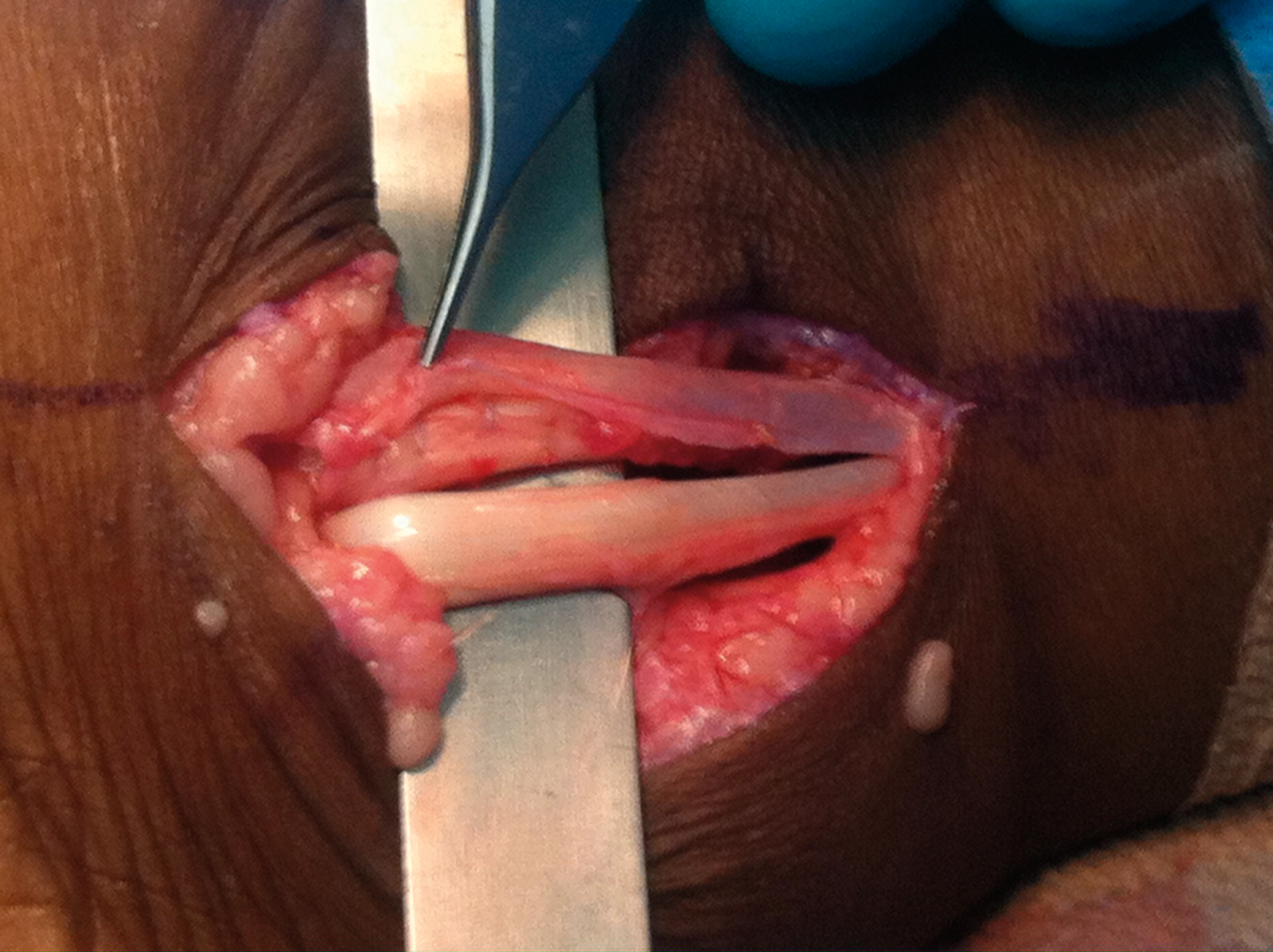
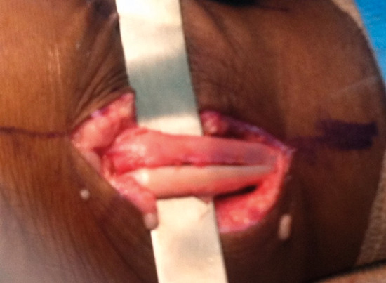
Figure 5.
Peroneus quartus muscle autograft sutured to the peroneus brevis tendon.
The postoperative course was uneventful except for slight dehiscence at the distal and proximal wound margins after the bandage had gotten wet. The wound was treated with a gauze dressing soaked in povidone-iodine (Betadine; Purdue Products, Stamford, Connecticut) and healed uneventfully, with suture removal at 3 weeks. Passive range of motion was started at 3 weeks and active range of motion at 6 weeks, when the patient was without pain to the peroneal muscles. In addition, at 6 weeks the patient was casted for orthotic devices, and physical therapy was started.
Three months postoperatively, the patient had returned to routine daily activities without restrictions with the use of her orthotic devices. At 6 months, the patient returned to all preinjury activities without pain or discomfort. Eighteen months postoperatively the patient still had no pain or activity limitations.
Discussion
This was a case report of a 51-year-old woman with long-standing bilateral ankle edema and pain to the left foot. She was diagnosed as having a split tear in her left peroneus brevis tendon, which was surgically repaired and augmented with a previously undetected autogenous peroneus quartus muscle graft. In this case, the peroneus quartus tendon was incorrectly reported on magnetic resonance imaging as a portion of the split peroneus brevis tendon.
Longitudinal split tears of the peroneus brevis tendon require surgical intervention when associated with pain and discomfort. Options for surgical intervention include primary repair of the tendon with retubularization, use of a graft for strengthening, and tenodesis to the peroneus longus tendon. Krause and Brodsky [13] graded tears to guide treatment. Grade 1 tears have damage to less than 50% of the cross-sectional area of the tendon, should be debrided, and should undergo primary repair. Grade 2 tears have damage to more than 50% of the cross-sectional area of the tendon and should have the damaged tendon excised, followed by tenodesis to the peroneus longus tendon. Along with the grade of the tendon tear, the volume of structures in the peroneal tunnel should be evaluated for their contribution to the mass effect. Overcrowding in the peroneal tunnel by a low-lying peroneus brevis muscle belly or the presence of a peroneus quartus muscle and their removal should be considered to decompress the tunnel [2]. Often an accessory peroneus quartus muscle is asymptomatic and is discovered only during magnetic resonance imaging or intraoperatively [14]. Lateral pain and swelling related to a peroneal pathologic abnormality can occur spontaneously but are often triggered by acute trauma such as an ankle sprain. Lotito et al [14] discussed several cases in which simple resection of the peroneus quartus muscle eliminated the symptoms. Kassim and Rosenfeld [15] described tendoscopic debridement for peroneal pathologic abnormalities, including resection of a peroneus quartus tendon and muscle belly. In the present case presentation, the patient had both a low-lying muscle belly and a peroneus quartus muscle likely causing a mass effect with a resultant peroneus brevis tendon split tear. The low-lying muscle belly was resected, and the peroneus quartus muscle was dissected out and used as an onlay graft in the repair of the peroneus brevis tendon tear, adding strength to the repair. We believe that in patients with the combination of a low-lying peroneus brevis muscle belly, peroneus brevis split tear, and peroneus quartus muscle, this novel surgical approach could be used to strengthen the tendon repair and eliminate the mass effect to the lateral aspect of the foot.
There are several limitations to this study. This is a case report involving only one patient. It would benefit us to see a larger number of patients with long-term follow-up to evaluate the use of a peroneus quartus muscle as an onlay autogenous graft in peroneus brevis tendon repair. The peroneus quartus muscle is an anatomical variant with a low prevalence of 6% to 21% as described in the literature. The low incidence of the peroneus quartus muscle limits its use as an autogenous graft in the repair of a peroneus brevis tendon tear. In addition, there is a limited patient population for researching where this surgical technique could be applied.
This article presents a positive surgical outcome for the novel approach to repair a peroneus brevis tendon split tear with use of the anatomical variant accessory peroneus quartus muscle as an onlay graft. Additional documentation of a peroneus quartus muscle used as an autograft and larger studies are needed to further support this novel surgical technique.
Financial Disclosure:
None reported.
Conflicts of Interest:
None reported.
References
- Cheung YY, Rosenberg ZS, Ramsinghani R, et al: Peroneus quartus muscle: MR imaging features: Radiology202: 745, 1997.
- Sobel M, Lvy M, Bohne WH: Congenital variations of the peroneus quartus muscle: an anatomic study. Foot Ankle11: 81, 1990.
- Clarkson M, Fox JN, Atsas S, et al: Clinical implications of novel variants of the fibularis (peroneus) quartus muscle inserting onto the cuboid bone: peroneocuboideus and peroneocalcaneocuboideus. J Foot Ankle Surg52: 118, 2013.
- Zammit J, Singh D: The peroneus quartus muscle. J Bone Joint Surg Br85: 1134, 2003.
- Demetracopoulos C, Vineyard J, Kiesau CD, et al: Long-term results of debridement and primary repair of peroneal tendon tears [published online December 6, 2013]. Foot Ankle Int. doi:10.1177/1071100713514565. Available at: http://fai.sagepub.com/content/early/2013/12/06/1071100713514565.
- Prakash MD, Narayanswamy C, Singh DK, et al: Anatomical variations of peroneal muscles: a cadaver study in an Indian population and review of the literature. JAPMA101: 505, 2011.
- Karlsson J, Wiger P: Longitudinal split of the peroneus brevis tendon and lateral ankle instability: treatment of concomitant lesions. J Athl Train37: 463, 2002.
- Slater HK: Acute peroneal tears. J Foot Ankle Clin12: 659, 2007.
- Didomenico LA, Anania MC: “Peroneal Tendon Disorders,” inMcGlamry's Comprehensive Textbook of Foot and Ankle Surgery, Vol 2, edited byJTSoutherland, JSBoberg, MSDowney, et al, p 1165, Lippincott Williams &Wilkins, Philadelphia,2013.
- Pollack D, Khaimov G, Guberman R: Limitation of magnetic resonance imaging in diagnosing longitudinal peroneal tendon tears. JAPMA104: 90, 2014.
- Mick CA, Lynch F: Reconstruction of the peroneal retinaculum using peroneus quartus: a case report. J Bone Joint Surg Am69: 296, 1987.
- Mook W, Parekh S, Nunley J: Allograft reconstruction of peroneal tendons: operative technique and clinical outcomes: Foot Ankle Int34: 1212, 2013.
- Krause J, Brodsky J: Peroneus brevis tendon tears: pathophysiology, surgical reconstruction, and clinical results. Foot Ankle Int19: 271, 1998.
- Lotito G, Pruvost J, Collado H, et al: Peroneus quartus and functional ankle instability: Ann Phys Rehab Med54: 282, 2011.
- Kassim M, Rosenfeld P: Tendoscopic debridement of peroneus quartus for chronic lateral ankle pain: a case report: Foot Ankle Int33: 1024, 2012.
© 2025 American Podiatric Medical Association