


Abstract
Pregnant women are often burdened with musculoskeletal symptoms of the lower extremity due to the physical, hormonal, and anatomical changes that occur throughout pregnancy. These symptoms are associated with musculoskeletal dysfunctions, modified gait, joint laxity, muscle imbalance, and increased body mass. This article reviews the literature involving the lower-extremity changes experienced by women during pregnancy and their respective pathophysiologic causes.
In the United States, 80% of women older than 44 years have given birth at least once, averaging 1.9 children each [1]. Pregnant women often experience musculoskeletal symptoms in the lower extremity, including muscle strains, cramps, fatigue, and soreness [2]. These hormonal, anatomical, and physiologic changes occur throughout the phases of pregnancy; however, most symptoms occur during the second and third trimesters [3]. Modified gait, increased body mass, joint laxity, altered muscle balance, and anterior displacement of mass are associated with discomfort and musculoskeletal dysfunction [2,4,5,6,7,8]. Surveys of pregnant women show a significantly higher prevalence of chronic joint pain in the lumbosacral spine, sacroiliac joints, hips, knees, wrists, and feet in women compared with men [3,8]. This article reviews the literature involving lower-extremity changes experienced by women during pregnancy and their respective pathophysiologic causes. The lasting effects of pregnancy on lower-extremity biomechanics and musculoskeletal anatomy will also be explored.
Methods
A key word search of the National Center for Biotechnology databases was performed via PubMed. Search criteria key words were pregnant foot, pregnant gait, pregnant biomechanics, pregnancy lower extremity, pregnant walking, musculoskeletal changes in pregnancy, and joint laxity in pregnancy. The 26 articles selected for review contained, titles, abstracts, or database tags relevant to this literature review.
Results
General Musculoskeletal Effects Seen During Pregnancy
According to the Utah paradigm, mechanical factors are the most important determinants of bone and connective tissue changes [9]. A 20% increase in weight during pregnancy can double the force placed on a joint [10]. An increase in joint laxity due to the hormone relaxin, coupled with elevated displacement of mass, raises the amount of stress placed on bones and joints of the lower extremity.
Increased Mass
Throughout pregnancy, women gain an average of 10 to 12 kg (22.0–26.4 pounds), experiencing a 20% increase in body mass [4], mostly from uterine, fetal, and breast growth. The most pronounced mass increase occurs in the sagittal plane of the abdominal region. In addition, loosening of the sacroiliac ligaments allows for augmented lumbar lordosis and an average 4° increase in anterior pelvic tilt [2,6]. These changes produce anterior displacement of the center of mass, which continues to increase throughout pregnancy [2].
Gait Changes
Musculoskeletal system compensations minimize gait alterations secondary to pregnancy [11]. Throughout pregnancy, the moment of inertia of the thoracolumbar segment increases and the center of mass becomes anteriorly displaced in the sagittal plane. This causes a ventral shift in the center of mass, making it difficult to control angular momentum in the sagittal, transverse, and coronal planes [12]. The most commonly observed biomechanical compensatory mechanisms are an increase in stance width of up to 30% [11], an increase in step time, a decrease in stride length, and a longer stance phase [2]. It is suggested that a widened base of gait and a lowered center of gravity reduce lateral sway and add stability [13]. It is important to note that although stride width increases throughout pregnancy, the angle of the longitudinal bisection of the foot to the midline of gait remains relatively constant [2,11].
Pregnant women also adapt their gait to maximize stability in the stance phase to control mediolateral foot motion [14]. A considerable amount of pain is experienced in the lower extremity during pregnancy, and this is attributed to faulty foot posture due to variations in muscle balance [12]. Several authors observed significant compensatory increases in double-limb support time and decreases in single-limb support time in gait during pregnancy [2,6,15].
Musculoskeletal Effects Specific to the Foot and Ankle Seen in Pregnancy
Foot Size
The literature shows a general consensus that an increase in foot size is experienced during pregnancy [16,17]. These changes are most pronounced in women experiencing their first pregnancy [17,18]. Ponnapula and Boberg [8] conducted a survey of 100 postpartum women in which 44% noticed an increase in shoe size, with an average shoe size increase of 0.4. In a study by Segal et al [18]., 30 of 40 individuals demonstrated an increase in foot length of up to 10 mm. Gijon-Nogueron et al [19] observed an average 0.52–shoe size increase and a significant 1.26-cm increase in forefoot perimeter. Alvarez et al [17] observed a mean foot volume increase of 57.2 mL between weeks 13 and 35, which decreased only 14.68% by postpartum week 8.
Biomechanical Effects
One of the most commonly observed changes occurring during pregnancy is collapse of the medial longitudinal arch, causing an increase in foot pronation due to increased laxity of the plantar calcaneonavicular ligament and attenuation of the tibialis posterior tendon [2]. Studies have shown significant decreases in arch height and arch rigidity to be highly correlated with concurrent increases in foot length and arch drop [18]. The talar dome can fall as much as 2.5 cm after childbirth, causing the Foot Pressure Index to increase as much as 3.78 points toward pronation [19].
A more pronated foot posture increases tibial rotation and has been shown to produce overrotation of the knee and femur [20]. These increased rotational torques are often transferred as shear stress on the medial tibiofemoral and lateral patellofemoral compartments of the knee [18]. Therefore, a loss of arch height and increased pronation of the foot may alter interactions of skeletal and musculotendinous structures, serving as a major contributor to pain in other parts of the lower extremity during pregnancy [14]. All of the pregnant women in the study by Block et al [21] displayed forefoot valgus, everted calcaneal stance position, and pronation in the rearfoot and midfoot, causing alterations in plantar pressure distribution and resulting in instability and impaired balance.
Plantar Pressure Distribution
Studies observing alterations in plantar pressure distribution during pregnancy further support previously mentioned observations concerning the pregnant foot. The most notable changes throughout the gestational trimesters were increased total contact area, increases in time of contact at the midfoot and forefoot regions, and reductions in peak pressure and maximum force at the rearfoot at the end of the third trimester [14]. Nyska et al [2] found that the static plantar contact area in pregnant women was increased 12%, noting an 8% increase during dynamic motion. Studies have also observed increases in dynamic contact time at the forefoot and midfoot [15] and in contact area at the midfoot [14,22]. As pregnancy progresses, plantar pressure is decreased in the medial rearfoot and redistributed to the forefoot. Nyska et al [2] found dynamic forefoot pressures to be higher laterally and lower medially, illustrating the observed increase in motion at the tarsometatarsal joint of the first ray and up to a 30% increase in static and dynamic plantar pressures as pregnancy progresses. Other studies on static pedobarographic evaluation have revealed conflicting results, showing higher forefoot pressures and lower static hindfoot pressures compared with the control group [15].
Gastrocnemius Soleus Complex Pain
In a survey of postpartum women, 82% reported the onset of lower-extremity pain during the second and third trimesters [3]. One of the most reported lower-extremity pain symptoms was cramping of the gastrocnemius soleus complex, often lasting several minutes [10]. Another self-reported survey reported that up to 41% of expectant mothers demonstrate this symptom [8]. Evidence suggests that anterior displacement of the center of mass may cause stress on the ankle plantar flexor muscle groups to maintain the center of load of the foot at 1.5 to 2.5 cm anterior to the ankle joint [2,3,4,5,6]. Although increased mechanical demand placed on the gastrocnemius soleus complex is a major contributor, leg cramping during pregnancy is thought to be more closely associated with mineral deficiency than any musculoskeletal consideration [6].
Lasting Postpartum Effects
Changes experienced in the lower extremity during pregnancy have the potential to cause lasting musculoskeletal dysfunction postpartum. Gilleard [12] observed that women continue to demonstrate their pregnancy gait patterns—such as velocity, step length, stance time, and foot angle—as late as 8 weeks after parturition, suggesting that pregnancy may have a lasting effect on mothers' biomechanics. Studies also show that step width narrowed postpartum, pelvic range of motion decreased, and thoracic segment range of motion increased [12,13]. Although there have been published cases of flatfoot development during pregnancy [23], Gaymer et al [22] observed higher midfoot plantar pressures in pregnant patients that resolved postpartum. Although there is considerable variation in reports, there are studies that support the view that the loss of arch height and rigidity and increases in foot length occurring in gravid women do not fully resolve postpartum [18,24].
Discussion
Literature concerning the musculoskeletal dysfunction, biomechanics, and pathogenesis of the lower extremity during pregnancy is still relatively sparse [6]. This can be attributed to the major challenges in the recruitment of study participants and to the long-term nature of the studies. The only two studies cited in this investigation that recruited more than 100 participants (Table 1) included data from self-reported surveys given to postpartum women, which are highly subject to potential bias [3,8]. Vullo et al [3] surveyed the weight changes in recently pregnant women, soliciting self-reported measures of prepregnancy weight, highest weight during pregnancy, and current weight, and did not find a significant correlation between weight and lower-extremity pain during pregnancy.
Table 1.
Overview of Sampling Methods Used in Cited Studies
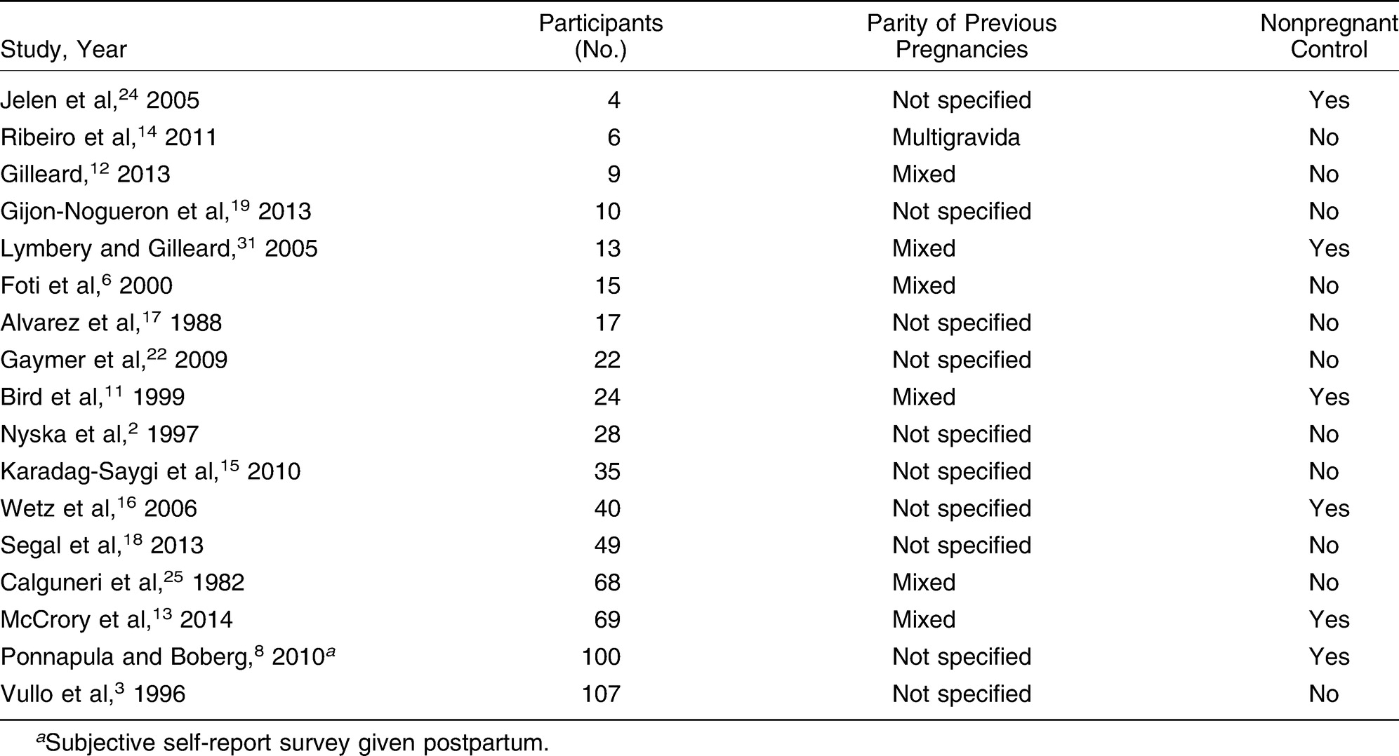
There were inconsistencies noted in the literature regarding speed of gait, foot length, width of steps, joint laxity, and decreased longitudinal arch height [24]. Most of the studies made no distinction regarding the number of previous pregnancies, and others had a mixture of primigravida and multigravida women or used nulliparous or nonpregnant controls, making it difficult to compare results and draw conclusions from the body of literature as a whole. For example, Calguneri et al [25] observed a 50% increase in joint laxity in second pregnancies compared with first pregnancies. According to their paradigm, multigravida women display greater change in laxity and dysfunction during and after pregnancy than first-time mothers [25].
There is, however, objective and subjective evidence suggesting better musculoskeletal outcomes throughout pregnancy for women who exercise before pregnancy and remain physically fit during pregnancy [6,26,27,28,29]. Pregnant women who are inactive or have low muscle strength are susceptible to straining certain muscle groups, including those associated with hip abduction, leg extension, and ankle plantarflexion [6]. Changes in kinetic patterns through pregnancy, including increased hip abductor, hip extensor, and ankle plantar flexor muscle groups, contribute to low-back pain, foot pain, and painful cramps in the calf muscles [15]. Therefore, initiation of exercise programs early in pregnancy would be helpful in adjusting to the kinetic changes and would increase forefoot loading and plantar pressures under the heel, midfoot, and metatarsal heads [15,30].
Conclusions
The results of this literature review demonstrate that expectant mothers typically experience significant alterations in musculoskeletal structure and function in the lower extremity. The most pronounced mass increase occurs in the sagittal plane of the abdominal region, causing anterior displacement in center of mass and biomechanical compensations, including an increase in stance width, an increase in step time, a decrease in stride length, and a longer stance phase.
By increasing understanding and strengthening current paradigms, we can achieve a more complete model of the changes that occur during pregnancy and establish a more uniform protocol for treatment of the parturient female. There are few treatment recommendations for musculoskeletal conditions of the lower extremity that are associated with pregnancy, and a lack of understanding of the pregnant foot may result in benign neglect [6]. Physicians often wait for musculoskeletal issues to resolve spontaneously postpartum for lack of a reliable treatment protocol [6]. Further research is needed to provide greater understanding and knowledge of the musculoskeletal changes that occur during pregnancy.
Based on the findings and interpretations of this literature review, we propose that the ideal study design on the lower-extremity biomechanical and musculoskeletal changes associated with pregnancy incorporate a prospective, longitudinal cohort design. Participant selection should consist of nulliparous women expecting to become pregnant in the next 3 months who demonstrate a moderate level of physical fitness and a normal body mass index [31]. Prepregnancy baseline measurements should be taken for comparison with patients' own measurements at rigidly defined time points during and after gestation. Future studies could explore how conservative interventions might treat or resolve the negative musculoskeletal effects of pregnancy.
Financial Disclosure:
None reported.
Conflicts of Interest:
None reported.
References
- Bureau of the Census for the Bureau of Labor Statistics: Current Population Survey, June 2010: Fertility Supplement, US Census Bureau, Washington, DC, 2011.
- Nyska M, Sofer D, Porat A, et al: Plantar foot pressures in pregnant women. Isr J Med Sci33: 139, 1997.
- Vullo VJ, Richardson JK, Hurvitz EA: Hip, knee, and foot pain during pregnancy and the postpartum period. J Fam Pract43: 63, 1996.
- Weiss G, Goldsmith LT, Sachdev R, et al: Elevated first-trimester serum relaxin concentrations in pregnant women following ovarian stimulation predict prematurity risk and preterm delivery. Obstet Gynecol82: 821, 1993.
- Marnach M: Characterization of the relationship between joint laxity and maternal hormones in pregnancy. Obstet Gynecol101: 331, 2003.
- Foti T, Davids JR, Bagley A: A biomechanical analysis of gait during pregnancy. J Bone Joint Surg Am82: 625, 2000.
- Borg-Stein J, Dugan SA, Gruber J: Musculoskeletal aspects of pregnancy. Am J Phys Med Rehabil84: 180, 2005.
- Ponnapula P, Boberg JS: Lower extremity changes experienced during pregnancy. J Foot Ankle Surg49: 452, 2010.
- Frost HM: Muscle, bone, and the Utah paradigm: a 1999 overview. Med Sci Sports Exerc: 2000;32: 911, 1999.
- Ritchie JR: Orthopedic considerations during pregnancy. Clin Obstet Gynecol46: 456, 2003.
- Bird AR, Menz HB, Hyde CC: The effect of pregnancy on footprint parameters: a prospective investigation. JAPMA89: 405, 1999.
- Gilleard WL: Trunk motion and gait characteristics of pregnant women when walking: report of a longitudinal study with a control group. BMC Pregnancy Childbirth13: 71, 2013.
- McCrory JL, Chambers AJ, Daftary A, et al: The pregnant “waddle”: an evaluation of torso kinematics in pregnancy. J Biomech47: 2964, 2014.
- Ribeiro AP, Trombini-Souza F, de Camargo NevesSacco I, et al: Changes in the plantar pressure distribution during gait throughout gestation. JAPMA101: 415, 2011.
- Karadag-Saygi E, Unlu-Ozkan F, Basgul A: Plantar pressure and foot pain in the last trimester of pregnancy. Foot Ankle Int31: 153, 2010.
- Wetz HH, Hentschel J, Drerup B, et al: Changes in shape and size of the foot during pregnancy. Orthopade35: 1124, 2006.
- Alvarez R, Stokes IA, Asprinio DE, et al: Dimensional changes of the feet in pregnancy. J Bone Joint Surg Am70: 271, 1988.
- Segal NA, Boyer ER, Teran-Yengle P, et al: Pregnancy leads to lasting changes in foot structure. Am J Phys Med Rehabil92: 232, 2013.
- Gijon-Nogueron GA, Gavilan-Diaz M, Valle-Funes V, et al: Anthropometric foot changes during pregnancy: a pilot study. JAPMA103: 314, 2013.
- Root M, Orien W, Weed J: Normal and Abnormal Function of the Foot,Clinical Biomechanics Corp, Los Angeles,1977.
- Block RA, Hess LA, Timpano EV, et al: Physiologic changes in the foot during pregnancy. JAPMA75: 297, 1985.
- Gaymer C, Whalley H, Achten J, et al: Midfoot plantar pressure significantly increases during late gestation. Foot (Edinb)19: 114, 2009.
- Bohemen EK, Gendi NS: Flatfeet in pregnancy. Br J Rheumatol35: 396, 1996.
- Jelen K, Tetkova Z, Halounova L, et al: Shape characteristics of the foot arch: dynamics in the pregnancy period. Neuro Endocrinol Lett26: 752, 2005.
- Calguneri M, Bird HA, Wright V: Changes in joint laxity occurring during pregnancy. Ann Rheum Dis41: 126, 1982.
- Clapp JF III: Long-term outcome after exercising throughout pregnancy: fitness and cardiovascular risk. Am J Obstet Gynecol199: 489, 2008.
- Fishbein EG, Phillips M: How safe is exercise during pregnancy?J Obstet Gynecol Neonatal Nurs19: 45, 1990.
- Hall DC, Kaufmann DA: Effects of aerobic and strength conditioning on pregnancy outcomes. Am J Obstet Gynecol157: 1199, 1987.
- Ostgaard HC, Zetherstrom G, Roos-Hansson E, et al: Reduction of back and posterior pelvic pain in pregnancy. Spine (Phila Pa 1976)19: 894, 1994.
- Gravante G, Russo G, Pomara F, et al: Comparison of ground reaction forces between obese and control young adults during quiet standing on a baropodometric platform. Clin Biomech (Bristol, Avon)18: 780, 2003.
- Lymbery JK, Gilleard W: The stance phase of walking during late pregnancy: temporospatial and ground reaction force variables. JAPMA95: 247, 2005.
© 2025 American Podiatric Medical Association