


Abstract
Background: Maintaining balance is a complex phenomenon that is influenced by a range of sensorimotor factors. Foot posture and mobility may also influence balance and postural sway. Recently, three-dimensional foot scanners have been used to assess foot posture. This tool allows many individuals to be scanned quickly and easily and helps eliminate patients' radiation exposure. The objective of this study was to determine whether static foot posture and mobility are independently associated with postural sway in a large community sample of older women using objective measures of balance status and the recently launched technology of three-dimensional foot scanning. Methods: This cross-sectional study included 140 community-dwelling elderly women (mean ± SD age, 73.9 ± 5.1 years) recruited in Kasama City, Japan. The postural sway variables were total path length and area and were measured by force plate. We measured static foot posture, sitting and standing navicular height, and mobility using a three-dimensional foot scanner. Foot mobility was determined as the amount of vertical navicular excursion between the positions of the subtalar joint, from neutral in sitting position to relaxed bilateral standing. Results: After adjusting for potential cofounders, analysis of covariance revealed that sitting navicular height was associated with total path length (P = .038) and area (P = .031). Foot mobility was associated with total path length (P = .018). Conclusions: These findings suggest that sitting navicular height and foot mobility are associated with postural sway in elderly women and might be an important factor in defining balance control in older adults.
Maintaining balance when performing functional tasks is a complex phenomenon that is influenced by a range of sensorimotor factors, including lower-extremity muscle strength, [1] peripheral sensation, [2] reaction time, and visual acuity. [1] Foot posture and mobility may also influence balance and postural sway. [3]
Foot and leg problems are a common condition reported by older adults, particularly in women, and, as such, they have a considerable effect on the daily lives of many older people. [4] Because foot posture influences lower-limb gait kinematics, [5] muscle activity, [6] functional ability, and balance, [3] measuring foot posture is widely considered to be an important component of musculoskeletal examination in clinical practice and research. [7] However, many measurement approaches are not suitable for routine use because they are time-consuming or require specialized equipment or clinical expertise. [7] The recent use of three-dimensional (3-D) foot scanners allows many individuals to be scanned quickly and easily and helps eliminate patients' exposure to x-rays. [8]
Unlike research on foot posture, assessment of foot mobility has received less attention in the literature. In 1982, Brody [9] first described the navicular drop test as an assessment of foot mobility. It is a measure of sagittal plane mobility of the midfoot as measured by the vertical change in the height of the navicular tuberosity. [9] In particular, the navicular drop test is an easy-to-perform technique and is more related to dynamic rearfoot function and lower-limb dysfunction. [10-13] It is widely used as a valid and reliable measure for determining foot mobility. [14-18]
On the whole, the available evidence indicates that foot and leg problems may be associated with functional impairment in people, but this information should be interpreted in light of certain study limitations, in particular, the way functional impairment and foot problems have been measured. [4] Foot problems have been clustered together with other pathologic abnormalities and labeled “lower-extremity problems,” [19] and participants' self-reported abilities and problems are often relied on to determine functional impairment. [4] Few studies have incorporated objective evaluations of functional performance and foot posture. [4] Although self-reporting of many instruments of functional ability and foot problems can be valid, [20] objective evaluations may be more sensitive to change and may provide better predictive validity. [21]
Furthermore, most studies have not explored the influence of other health-related factors on the relationship between foot posture and functional performance. It is important to consider the relative contribution of foot posture to postural sway after accounting for these potential confounders. [4] Moreover, previous studies have largely focused on radiographic or other less accessible measures of foot structure as potential predictors rather than tests that can be performed in a clinical setting. [22]
We found no published studies on the association of static foot posture and postural sway in an elderly population using a 3-D foot scanner. The objective was to provide additional insight into navicular height (foot posture) and mobility of the foot in older women.
Therefore, the main objective of this study was to determine whether static foot posture and mobility are independently associated with postural sway in a large community sample of older women after adjusting for the effects of medical factors using objective measures of balance status and the recently launched technology of 3-D foot scanning.
Methods
Participants and Data Collection
We conducted this cross-sectional study in August 2012 in Kasama City (population, 79,266; proportion of older adults, 24.0%), a rural region in Ibaraki prefecture, Japan. We mailed invitation letters to 831 women aged 65 to 85 years randomly drawn from the Basic Resident Register.
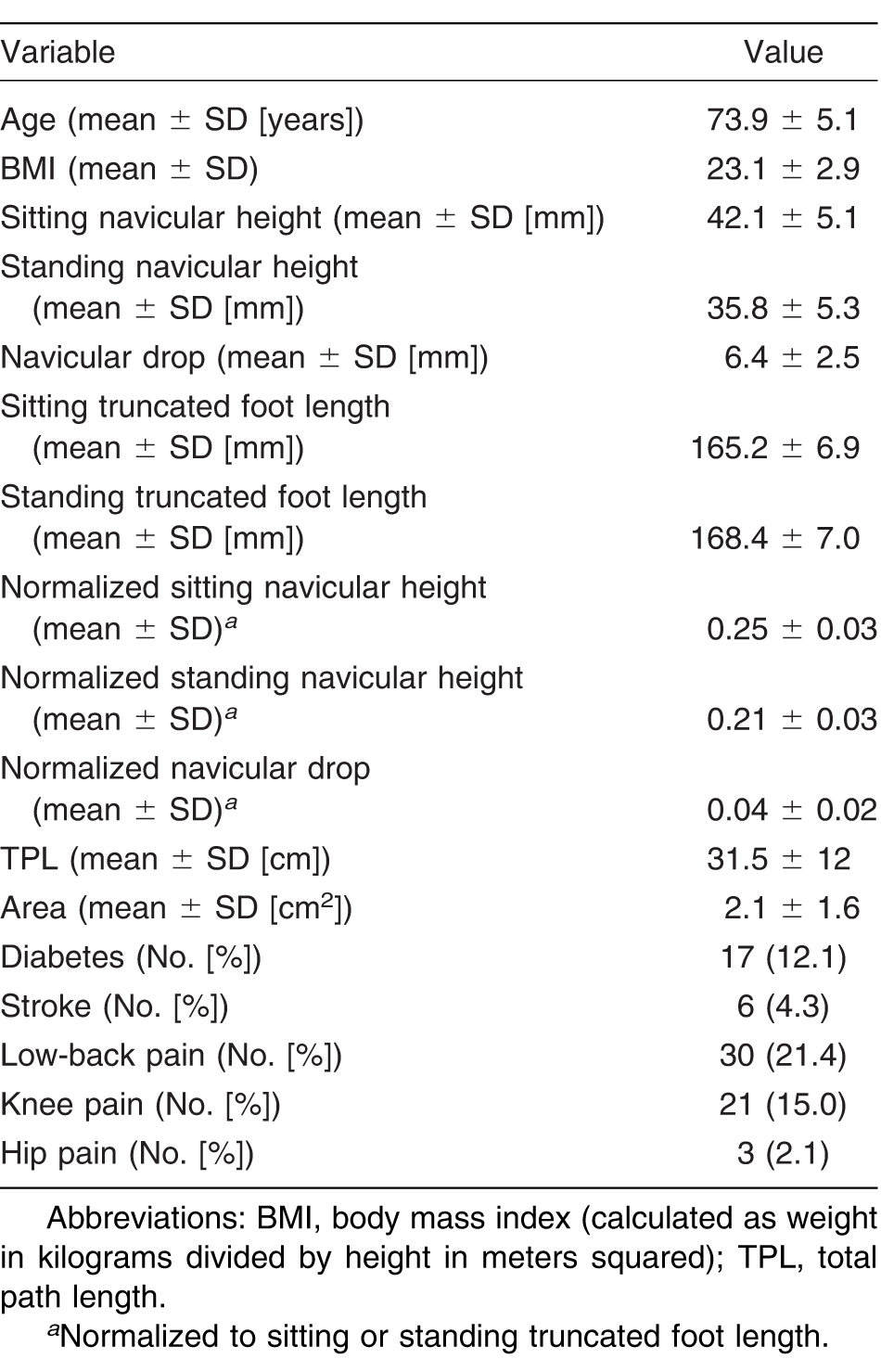
A total of 170 older women participated in this study, conducted in the health center in Kasama City. Of these participants, we excluded 30 due to their reliance on walking sticks or their unwillingness to be barefoot during the measurement, preventing us from collecting static foot posture or postural sway data. There were 140 participants (82.4%) for final data analysis. Medical history, demographic, static foot posture, and postural sway variables are shown in Table 1. All of the participants provided signed informed consent. This study was approved by the ethical committee of the University of Tsukuba, Tsukuba, Japan.

Table 1.
Medical History, Demographic, Static Foot Posture, and Postural Sway Variables in 140 Older Women
Measurement Variables
During the first 2 weeks of August 2012, we measured the postural sway, static foot posture, and body mass index of the participants, and we gathered medical histories via face-to-face interviews.
Williams and McClay [23] indicated that using truncated foot length—the perpendicular distance from the first metatarsophalangeal joint to the most posterior aspect of the heel—reduces the impact that toe deformities, such as claw toes and hallux valgus, may have on heel to longest toe foot length. Therefore, all of the data are normalized to truncated foot length after the measurement.
Postural Sway
Participants stood barefoot on a force plate (BM-101; Tanita Corp, Tokyo, Japan), separating their heels 10.6 cm from the sagittal-horizontal axis of the force plate. Toes were adjusted in a symmetrical and comfortable position. Participants' arms hung naturally at their sides as they looked at a fixed mark placed on a wall 1.5 m in front of them. [24] They were requested to stand as still as possible. Body sway was measured one time for 30 sec. [24] The force plate measures selected for this study were total path length traveled by the center of pressure in the allotted time and the 95% circular area for the center of pressure.
Static Foot Posture
We measured static foot posture using a recently launched 3-D foot scanner (Footstep PRO; Dream GP Company, Osaka, Japan). Modern 3-D surface scanning systems can obtain accurate and repeatable digital representations of the foot shape and have been used successfully in medical, ergonomic, and footwear development applications. [8]
For 3-D scanning, the barefoot participant sat on a chair. We measured navicular heights as described in the literature. [9] The most prominent portion of the navicular tuberosities on both feet were palpated and marked with a small, round, black sticky point while the participants maintained a relaxed sitting position. The 3-D scanner software located these black points as the navicular points. This black point is visible in Figure 1.
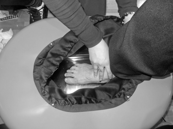
Figure 1.
Three-dimensional foot scanner by Dream GP Co.
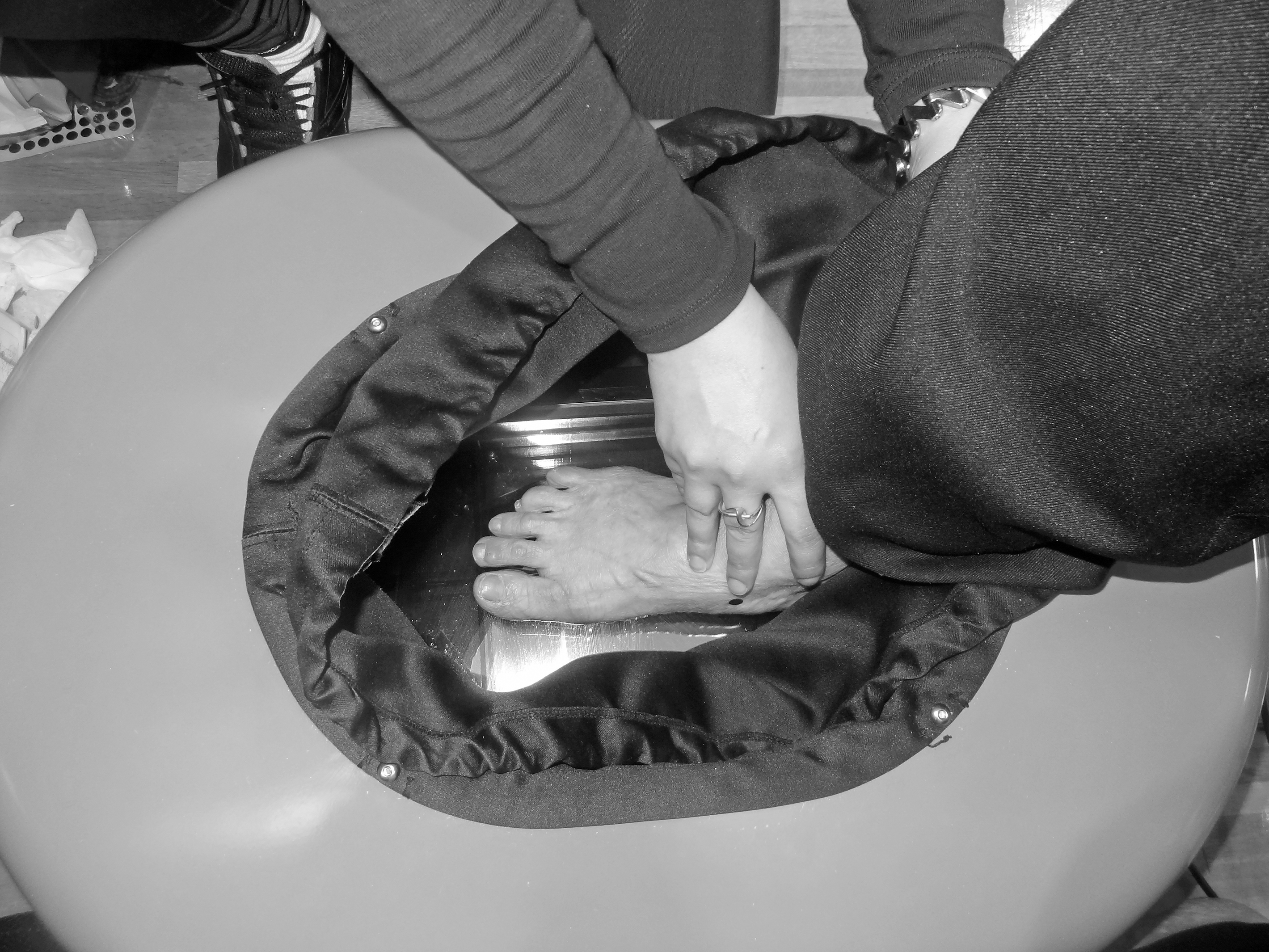
With the black point indicating the navicular tuberosity, participants individually sat on the end of a table so that their lower legs were nonweightbearing and their ankles were slightly plantarflexed. [25] They placed their right foot onto the factory-delineated center of the scanner as the measurer ensured proper positioning. To prevent ankle dorsiflexion, the participants were instructed not to forcibly push the platform of the 3-D machine. [25] Before starting the machine, light-blocking material attached to the rim of the scanner was secured to the participants' lower legs (Fig. 1).
When the scanner is started, a laser rotates on the rail around the foot measuring approximately 30,000 positions, including instep, heel, sole, and toe, which allows the software to reproduce exactly the shape of the foot. Each measurement is completed in approximately 13 sec.
After completing the measurements in a sitting position, the participants stood up without changing their foot position inside the machine, set their left foot on an adjacent wooden platform next to and level with the platform inside the scanner, and placed equal weight on each foot. This placed 50% of their body weight on the foot being assessed. The measurer checked the foot positioning in the scanner before starting the machine. Participants were also encouraged to use the handrail placed in front of them for balance, to relax their feet, and to ensure equal loading on each extremity. The handrail was placed at a level that they could easily reach without needing to raise or lower their arms too much. The participants looked straight ahead and stood as still as possible.
Once we obtained readings for the right foot in the sitting and standing positions, we repeated the measurements for the left foot. We collected four measurements (right and left leg in the sitting and standing positions) on each person and then sanitized the instruments with 70% alcohol before measuring the next person.
One investigator (M.S.) performed all of the markings of the navicular tuberosity. This investigator was a licensed athletic trainer with 2 years' experience in foot and posture assessment at the time of testing. In this study, navicular height was defined as the linear distance from the most medial prominence of the navicular tuberosity to the supporting surface while sitting and while standing with 50% body weight on each foot. [26] We defined foot mobility as the amount of vertical navicular excursion between the positions of the subtalar joint while neutral in sitting position and relaxed in bilateral standing (navicular drop). [18]
Statistical Analysis
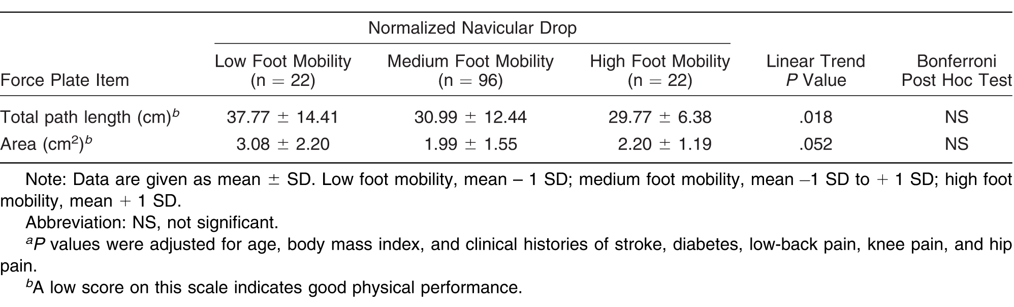
Because t test results indicated no significant differences between left and right feet data, we used mean data from both feet in the analyses. For analyses, we divided participants into three groups based on standard scores of navicular height and drop: low arch and low foot mobility (mean – 1 SD), medium arch and medium foot mobility (mean – 1 SD to + 1 SD), and high arch and high foot mobility (mean + 1 SD).
To examine the associations between static foot posture (navicular height and foot mobility) and postural sway, we used a one-way analysis of covariance (ANCOVA), which can adjust for potential confounders, because conditions such as age, body mass index, cardiovascular disease and stroke, low-back pain, knee pain, and hip pain are also likely to affect functional ability in older people. [4] Diabetes can also affect foot posture. Therefore, covariates included age, body mass index and clinical histories of stroke, diabetes, low-back pain, knee pain, and hip pain. We applied the Bonferroni post hoc test when the difference was significant (P < .05) according to the ANCOVA results. Statistical analyses were performed using a commercially available software program (SPSS version 18.0; SPSS Inc, Chicago, Illinois).
Results
After adjusting for potential cofounders, the linear trend in ANCOVA revealed that sitting navicular height was associated with total path length (P = .038) and area (P = .031) and that foot mobility was associated with total path length (P = .018). However, there was no association between standing navicular height and postural sway (Tables 2–4).
Table 2.
Associations Between Postural Sway and Sitting Foot Characteristics in Older Women
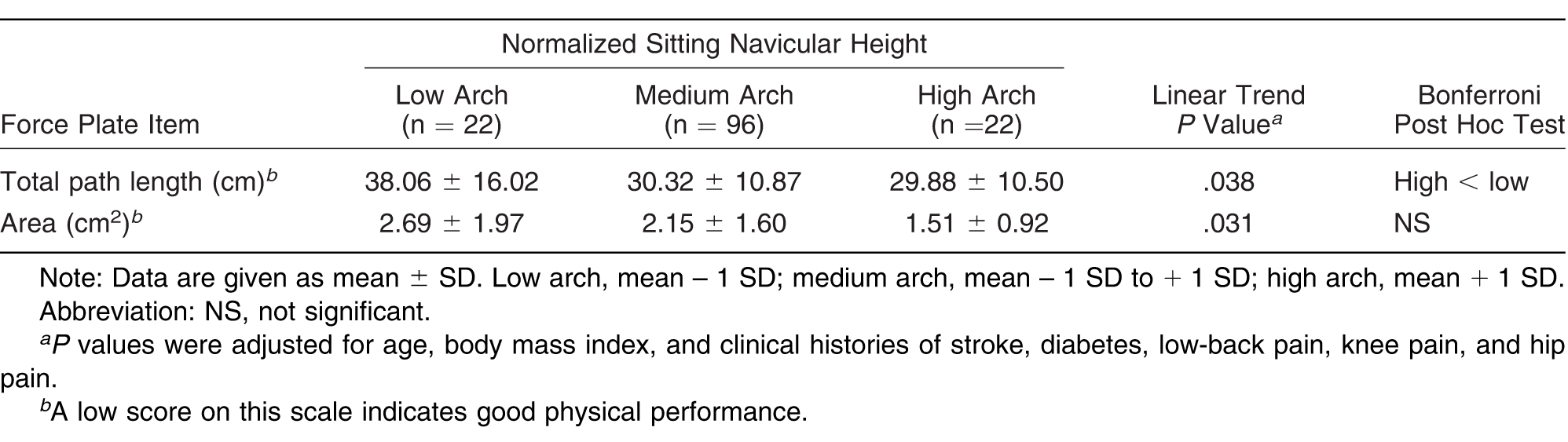
Table 3.
Associations Between Postural Sway and Standing Foot Characteristics in Older Women
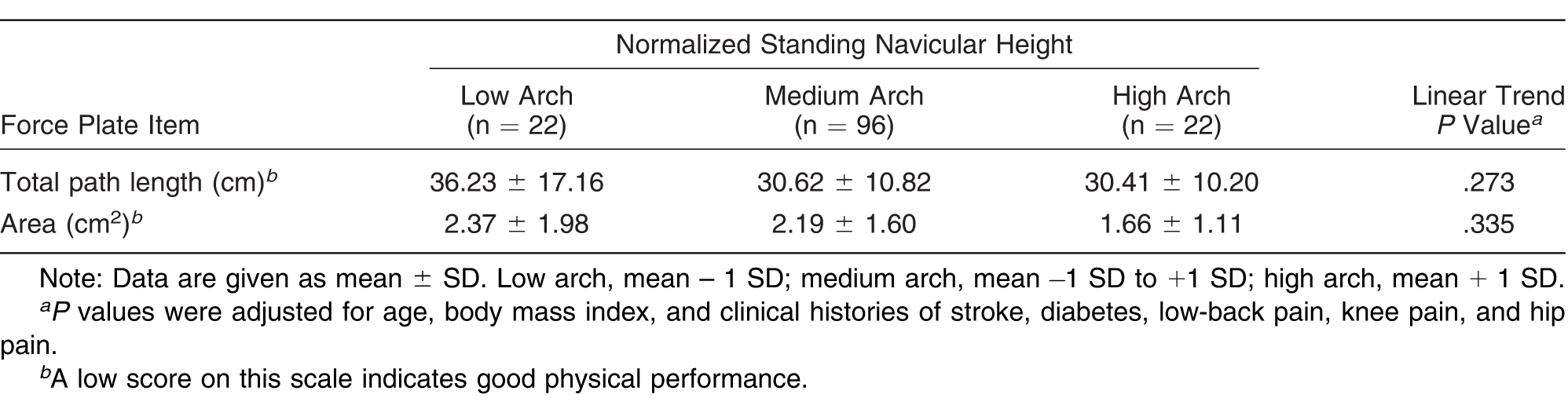
Table 4.
Associations Between Postural Sway and Foot Mobility in Older Women
Discussion
In this study, sitting navicular height and foot mobility were associated with postural sway. However, there was no association between standing navicular height and postural sway. The lack of standardization of data collection methods and approaches for measurements are limits of the study. Using the recently launched technology of the 3-D foot scanner makes it difficult to compare the results of this study with those of other studies. Overall, the present study provided evidence of some associations between sitting navicular height and foot mobility with postural sway in elderly women. Women with a low arch or a flatter foot and low foot mobility had more postural sway. The results of this study are consistent with those of previous studies that have reported that a flatter foot resulted in increased anteroposterior sway. [3,27] Cobb et al [27] believed that decreased joint congruity and, consequently, an increased reliance on soft-tissue structures for stability was the reason for decreased stability associated with increased forefoot varus. Spink et al [3] revealed that foot posture was an independent predictor of postural sway on foam, with a more pronated (flatter) foot corresponding to a poorer performance.
However, Menz et al [28] found that foot posture was not an independent predictor of performance on balance and function tests. This inconsistency with the present study results may be due to how we defined foot posture. In this study, we used navicular height to define foot posture, whereas Menz et al [28] used the Foot Posture Index, which is a multidimensional visual observation tool consisting of six criteria scored on a 5-point scale (range, −2 to +2). In the Foot Posture Index, the summed score indicates the degree of pronation or supination in the posture of the foot, with higher scores representing a more pronated (flatter) foot. This is different from measuring navicular height and categorizing the foot type based on that.
In this study, while in a standing position, people with low arches were also likely to have more postural sway than the groups with higher arches, but there was no significant association between standing navicular height and postural sway. Cornwall and McPoil [29] believed that this might be because the navicular bone not only moves in a vertical direction during the stance phase of gait but in the mediolateral direction as well, especially during the later portion of the stance phase. We considered only navicular height in this study, which is the vertical direction of movement only. If we could measure both lateral and vertical changes, the difference in postural sway may be statistically significant. Further study is needed to investigate this.
In addition, although participants were instructed to distribute their body weight equally when standing so that the assessed foot supported 50%, we could not control this percentage with accuracy. Therefore, there may be variations in the percentage of weightbearing and, as a result, different standing navicular heights in standing position. Tessem et al [30] previously reported that the amount of asymmetry in weight distribution between extremities during relaxed standing is 4% or less in healthy individuals. There are two other limitations of the present study: because of the cross-sectional design, we cannot prove a causal association, and we had only women as participants.
Despite these limitations, this study provides intriguing findings about associations of sitting navicular height and foot mobility with postural sway in elderly women. Women with a low arch or a flatter foot had more postural sway and poor balance. These findings suggest that sitting navicular height and foot mobility might be important factors in defining balance control in older adults. Further research investigating the effect of foot posture on falling is needed before making any generalizations about the potential risk of a fall. Other areas of research should include the effect of shoes or other forms of support on balance and postural sway in older adults with flat feet.
Acknowledgment
The Pigeon Company, especially Ms. Itagaki, who was so helpful with operation of the 3-D scanner. Also, all of Prof. Okura's laboratory assistants and staff, Kasama City's officers, and the participants for their great support and help.
Financial Disclosure
None reported.
Conflict of Interest
None reported.
References
- LordSR, ClarkRD, WebsterIW: Postural stability and associated physiological factors in a population of aged persons. J Gerontol46: M69, 1991.
- KristinsdottirEK, JarnloGB, MagnussonM: Aberrations in postural control, vibration sensation and some vestibular findings in healthy 64-92-year-old subjects. Scand J Rehabil Med29: 257, 1997.
- SpinkMJ, FotoohabadiMR, WeeE, et al: Foot and ankle strength, range of motion, posture, and deformity are associated with balance and functional ability in older adults. Arch Phys Med Rehabil92: 68, 2011.
- BarrEL, BrowningC, LordSR, et al: Foot and leg problems are important determinants of functional status in community dwelling older people. Disabil Rehabil27: 917, 2005.
- NawoczenskiDA, SaltzmanCL, CookTM: The effect of foot structure on the three-dimensional kinematic coupling behavior of the leg and rearfoot. Phys Ther78: 404, 1998.
- MurleyGS, MenzHB, LandorfKB: Foot posture influences the electromyographic activity of selected lower limb muscles during gait. J Foot Ankle Res2: 35, 2009.
- MenzHB, FotoohabadiMR, WeeE, et al: Visual categorisation of the arch index: a simplified measure of foot posture in older people. J Foot Ankle Res5: 10, 2012.
- TelferS, WoodburnJ: The use of 3D surface scanning for the measurement and assessment of the human foot. J Foot Ankle Res3: 19, 2010.
- BrodyDM: Techniques in the evaluation and treatment of the injured runner. Orthop Clin North Am13: 541, 1982.
- AllenMK, GlasoeWM: Metrocom meaurement of navicular drop in subjects with anterior cruciate ligament injury. J Athl Train35: 403, 2000.
- BennettJE, ReinkingMF, PluemerB, et al: Factors contributing to the development of medial tibial stress syndrome in high school runners. J Orthop Sports Phys Ther31: 504, 2001.
- LoudonJK, JenkinsW, LoudonKL: The relationship between static posture and ACL injury in female athletes. J Orthop Sports Phys Ther24: 91, 1996.
- MenzHB: Alternative techniques for the clinical assessment of foot pronation. JAPMA88: 119, 1998.
- EvansAM, CopperAW, ScharfbilligRW, et al: Reliability of the foot posture index and traditional measures of foot position. JAPMA93: 203, 2003.
- BuchananKR, DavisI: The relationship between forefoot, midfoot and rearfoot static alignment in pain-free individuals. J Orthop Sports Phys Ther35: 559, 2005.
- CoteKP, BrunetME, GansnederBM, et al: Effects of pronated and supinated foot postures on static and dynamic postural stabilityJ Athl Train40: 41, 2005.
- LangeB, ChipchaseL, EvansA: The effect of low-dye taping on plantar pressures during gait, in subjects with navicular drop exceeding 10mm. J Orthop Sports Phys Ther34: 201, 2004.
- MuellerMJ, HostJV, NortonBJ: Navicular drop as a composite measure of excessive pronation. JAPMA83: 198, 1993.
- MenzHB, LordSR: Foot problems, functional impairment, and falls in older people. JAPMA89: 458, 1999.
- Van SwearingenJM, BrachJS: Making geriatric assessment work: selecting useful measures. Phys Ther81: 1233, 2001.
- GuralnikJM, SimonsickEM, FerrucciL, et al: A short physical performance battery assessing lower extremity function: association with self-reported disability and prediction of mortality and nursing home admission. J Gerontol49: M85, 1994.
- MenzHB, MorrisME: Clinical determinants of plantar forces and pressures during walking in older people. Gait Posture24: 229, 2006.
- WilliamsDS, McClayIS: Measurements used to characterize the foot and the medial longitudinal arch: reliability and validity. Phys Ther80: 864, 2000.
- PellecchiaGL: Dual-task training reduces impact of cognitive task on postural sway. J Motor Behav37: 239, 2005.
- CornwallMW, McPoilTG: Relationship between static foot posture and foot mobility. J Foot Ankle Res4: 4, 2011.
- MuellerMJ, MenzHB, LandorfKB: A protocol for classifying normal- and flat-arched foot posture for research studies using clinical and radiographic measurments. J Foot Ankle Res2: 22, 2009.
- CobbSC, TisLL, JohnsonBF, et al: The effect of forefoot varus on postural stability. J Orthop Sports Phys Ther34: 79, 2004.
- MenzHB, MorrisME, LordSR: Foot and ankle characteristics associated with impaired balance and functional ability in older people. J Gerontol A Biol Sci Med Sci60: 1546, 2005.
- CornwallMW, McPoilTG: Relative movement of the navicular bone during normal walking. Foot Ankle Int20: 507, 1999.
- TessemS, HagstrømN, FallangB: Weight distribution in standing and sitting positions, and weight transfer during reaching tasks, in seated stroke subjects and healthy subjects. Physiother Res Int12: 82, 2007.
© 2015 American Podiatric Medical Association