


Abstract
Background: Vancomycin is a common treatment option for skin and skin structure infections caused by methicillin-resistant Staphylococcus aureus (MRSA). Given the increasing prevalence of MRSA, vancomycin is widely used as empirical therapy. In patients with lower-limb infections, antimicrobial penetration is often reduced because of decreased vascular perfusion. In this study, we evaluated the tissue concentrations of vancomycin in hospitalized patients with lower-limb infections. Methods: An in vivo microdialysis catheter was inserted near the margin of the wound and was perfused with lactated Ringer's solution. Tissue and serum samples were obtained after steady state for one dosing interval. Tissue concentrations were corrected for percentage of in vivo recovery using the retrodialysis technique. Results: Nine patients were enrolled (mean ± SD: age, 54 ± 19 years; weight, 105.6 ± 31.5 kg). Patients received a mean of 12.8 mg/kg of vancomycin every 12 hours (n = 7), every 8 hours (n = 1), or every 24 hours (n = 1). Mean ± SD steady-state trough vancomycin concentrations in serum and tissue were 11.1 ± 3.3 and 6.0 ± 2.6 μg/mL. The mean ± SD 24-hour free drug areas under the curve for serum and wound were 283.7 ± 89.4 and 232.8 ± 75.7 μg*h/mL, respectively. The mean ± SD tissue penetration ratio was 0.8 ± 0.2. Conclusions: These data suggest that against MRSA with minimum inhibitory concentrations of 1 μg/mL or less, vancomycin achieved blood pharmacodynamic targets required for the likelihood of success. Reduced concentrations may contribute to poor outcomes and the development of resistance. As other literature suggests, alternative agents may be needed when the pathogen of interest has a minimum inhibitory concentration greater than 1 μg/mL.
Staphylococcus aureus is a common pathogen in skin and skin structure infections, causing a major burden to health-care systems.[1] Of these S aureus infections, at least half are methicillin-resistant S aureus (MRSA), limiting the available options used to treat these infections.[1,2] Furthermore, the increasing prevalence of community-acquired MRSA has led to an increase in anti-MRSA antibiotic drugs being commonly and empirically prescribed for patients admitted to the hospital with a skin infection.[3-5]
Vancomycin has been the mainstay of treatment for the past few decades against MRSA infections requiring intravenous antibiotic agents.[1] In 2009, recommendations published by the American Society of Health-System Pharmacists, the Infectious Diseases Society of America, and the Society of Infectious Diseases Pharmacists advocated for achieving higher trough concentrations of 10 μg/mL or greater, and they should exceed 15 μg/mL if the vancomycin minimum inhibitory concentration (MIC) of the infecting pathogen is 1 μg/mL to achieve the pharmacodynamic target of area under the curve (AUC)/MIC of 400 for bloodstream infections and pneumonia.[6] To date, there are no data published to support a pharmacodynamic target in skin infections. As previous studies have described, increasing concentrations of vancomycin, although necessary, have also increased the toxicity associated with the drug, thereby limiting its use.[7,8]
Recently, there have been discussions questioning the utility of vancomycin for MRSA infections, citing increasing MICs, high-level resistance to vancomycin, and increased failures with vancomycin.[9-11] One rationale behind the decreased effectiveness of vancomycin could be impaired penetration into the site of infection. Multiple studies have demonstrated penetration into different sites.[12-15] It is further hypothesized that patients with lowerlimb infections, commonly those with diabetes and peripheral vascular disease, may have altered penetration of commonly used antibiotic agents. Therefore, understanding the pharmacokinetics of vancomycin in infected tissue may help us understand whether altered penetration is a contributing factor to the decreased effectiveness. To assess the pharmacokinetics of vancomycin at the site of infection, we used an in vivo microdialysis technique to describe wound and serum pharmacokinetics in patients with lower-limb infections receiving vancomycin.
Materials and Methods
Study Protocol
This study was designed as an open-label pharmacokinetic study in patients with lower-limb infections admitted to Hartford Hospital in Hartford, Connecticut, receiving intravenous vancomycin as part of their clinical management. The study protocol was reviewed and approved by the institutional review board at Hartford Hospital. Before any study-related activities took place, written informed consent was obtained from all of the patients.
Patients
Adult patients aged 18 years or older with lower-limb infections defined as mild or moderate by the Infectious Diseases Society of America or as grade 2 or 3 by the International Consensus on the Diabetic Foot who provided consent were enrolled.[16] Exclusion criteria included hypersensitivity to anesthetics (lidocaine or lidocaine derivatives), pregnancy or breastfeeding, no palpable pedal pulses, requiring multiple procedures during the study, or participating in another study of an investigational drug or device within the preceding 30 days.
Drug Administration
Vancomycin was dosed per protocol. Patients received a loading dose of vancomycin at 25 mg/kg and then were administered a regimen based on their weight and calculated creatinine clearance to target a trough concentration of 10 to 20 μg/mL. All of the patients received vancomycin intravenously via a peripheral catheter or a peripherally inserted central catheter as a 60- to 120-min infusion.
Microdialysis Procedure
The microdialysis procedure was performed as previously described.[17-19] The procedure began with a local injection of a 0.5% lidocaine solution if the patient still had sensation present in the extremity. A microdialysis probe (CMA 60 or 63 microdialysis catheter; CMA Microdialysis AB, Solna, Sweden), with a membrane length of 30 mm and a molecular weight cutoff value of 20 kDa, was inserted under sterile conditions within 10 cm of the wound margin into the subcutaneous tissue. This placed the semipermeable membrane within 5 cm of the wound. A microinfusion pump (CMA 107 microdialysis pump; CMA Microdialysis AB) was connected, and the catheter was flushed at a rate of 15 μL/min for 5 min and then was continuously perfused with lactated Ringer's solution at a rate of 2 μL/min during the entire study period. Catheters were placed in the subcutaneous tissue more than 4 hours before sampling for all of the patients.
Sample Collection
Venous blood was collected at various time points from either a peripheral intravenous catheter or a peripherally inserted central catheter after a minimum of four doses (ie, steady state). Some patients had samples drawn at 1 hour because the infusion was 60 min. The other patients' infusion time was 120 min, and as such they do not have the 1-hour sampling. Blood samples were collected using a 10-mL red top BD Vacutainer (Becton, Dickinson, and Co, Franklin Lakes, New Jersey). Blood samples were centrifuged (2,000g for 10 min), and the separated serum was immediately stored at −80°C until analysis.
Dialysate samples were collected over 1 hour corresponding to each blood sample time point. Dialysate samples were collected in 200-μL microvials (CMA Microdialysis AB), which were then stored in amber polypropylene tubes and frozen at −80°C until analysis.
In Vivo and In Vitro Retrodialysis
Microdialysis catheters were calibrated for each patient after sampling using the in vivo retrodialysis technique. A stock solution (Cperfusate) of vancomycin at 200 μg/mL was flushed through the catheter at 15 μL/min for 5 min; was allowed to equilibrate at 2 μL/min for approximately 15 min, followed by 2 μL/min for 60 min; and was collected into a microvial. The amount of recovery was calculated based on the concentration of the dialysate sample collected (Cdialysate) compared with the Cperfusate using the following equation: % recovery in vivo = 100 – (100 × Cdialysate/Cperfusate).
Retrodialysis by gain and loss was performed to assess relative recovery as previously described.[17] In brief, microdialysis probes (n = 3) were placed into a glass beaker of lactated Ringer's solution containing 200 μg/mL of vancomycin solution (Cmedium) and perfused with blank lactated Ringer's solution for recovery by gain. For recovery by loss, probes (n = 3) were placed into a glass beaker of blank Ringer's solution and perfused with a vancomycin solution at a concentration of 200 μg/mL. To mimic in vivo retrodialysis conditions, microdialysis probes were perfused at 2 μL/min at room temperature. Dialysate samples (Cdialysate) were collected every hour for 3 hours. During the switch from gain to loss, probes were placed in lactated Ringer's solution and perfused with lactated Ringer's solution for 1 hour. Then recovery by loss was performed, collecting dialysate samples every hour for 3 hours. The level of recovery by gain was calculated as follows: % recovery by gain = (Cdialysate/Cmedium) × 100. The level of recovery by loss was determined similarly to the level of in vivo recovery: % recovery by loss = 100 – (100 × Cdialysate/Cperfusate).
Protein Binding Studies
Protein binding studies were conducted in triplicate for each patient at various time points of the dosing interval. In brief, a blood sample was collected into a 10-mL red top BD Vacutainer (Becton, Dickinson, and Co) at each of the time points and was centrifuged (2,000g for 10 min) to obtain separated serum. Exactly 0.9 mL of serum was transferred into each regenerated cellulose 30-kDa molecular weight cutoff ultrafiltration device (Centrifree centrifugal filters; Millipore Corp, Billerica, Massachusetts) and were centrifuged at 2,000g using a fixed-angle rotor for 45 min at 10°C to obtain ultrafiltrate (Cultrafiltrate). An aliquot of serum was also retained at each corresponding time point for total drug concentration determination in the serum (Cserum). Protein binding was calculated using the following equation: % protein binding = 100 – (100 × Cultrafiltrate/Cserum).
Analytical Procedures
Vancomycin concentrations in plasma and dialysate were determined using a validated high-performance liquid chromatography assay that was developed using a previously published assay at the Center for Anti-infective Research and Development in Hartford, Connecticut.[20] The serum assay was linear over a range of 1 to 80 μg/mL (r2 ≥ 0.9989). The mean interday coefficients of variation for high (60 μg/mL) and low (2 μg/mL) check samples were 2.44% and 4.32%, respectively; the mean intraday coefficients of variation were 2.2% and 2.5%, respectively. The dialysate assay was linear over a range of 0.1 to 10 μg/mL (r2 ≥ 0.9983). The mean interday coefficients of variation for high (8 μg/mL) and low (0.2 μg/mL) check samples were 3.17% and 6.46%, respectively; the mean intraday coefficients of variation were 0.9% and 2.7%, respectively.
Pharmacokinetic Analysis
Serum
Total drug concentrations in serum were analyzed for each participant by noncompartmental analysis using WinNonlin software (version 5.3; Pharsight Corp, Mountain View, California). The maximum concentration (Cmax), area under the concentration-time profile for a 24-hour interval (AUC0-24), half-life, clearance, and volume of distribution were calculated for serum data. The Cmax was determined by visual inspection of the concentration-time profile. The area under the concentration-time profile of the dosing interval (AUC0→τ) was calculated using the linear-log trapezoidal method and multiplied by the number of doses in a 24-hour period. The half-life was calculated as ln(2)/λz, where λz is the terminal elimination rate constant. The terminal elimination rate constant was estimated by linear regression analysis of the terminal portion of the concentration-time curve. Clearance was calculated as dose/AUC0→τ. The volume of distribution was calculated as dose/(λz* AUC0→τ). A mean of each individual's protein binding percentages was applied to that individual's total drug concentrations to determine free drug concentrations.
Tissue
Dialysate samples were corrected using the in vivo recovery calculated for each patient's individual catheter using the following equation: Ctissue = 100 × (Cdialysate/% recovery in vivo), where Ctissue is the drug concentration in the interstitium. The Cmax of tissue concentrations was determined by visual inspection of the concentration-time profile. The AUC0→τ was calculated using the linear-log trapezoidal rule and then was extrapolated by the number of doses per 24-hour period to calculate AUC0-24. Each patient's penetration ratio was calculated using AUC0-24 from tissue (AUCtissue) and the free drug AUC0-24 in serum (fAUCserum) using the following equation: wound penetration ratio = AUCtissue/fAUCserum.
Results
Patients
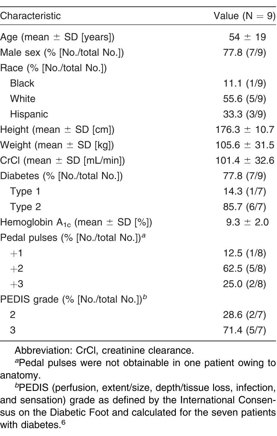
Nine patients provided consent and were enrolled. The baseline characteristics of these patients are listed in Table 1. The mean dose of vancomycin for the nine patients was 12.8 mg/kg, with frequency adjusted based on creatinine clearance. Seven of the patients received every-12-hour (q12h) dosing, and one patient each received q24h and q8h dosing.
Table 1.
Patients' Characteristics When Enrolled in the Study and When Sampling Occurred
Pharmacokinetic Analysis
Serum
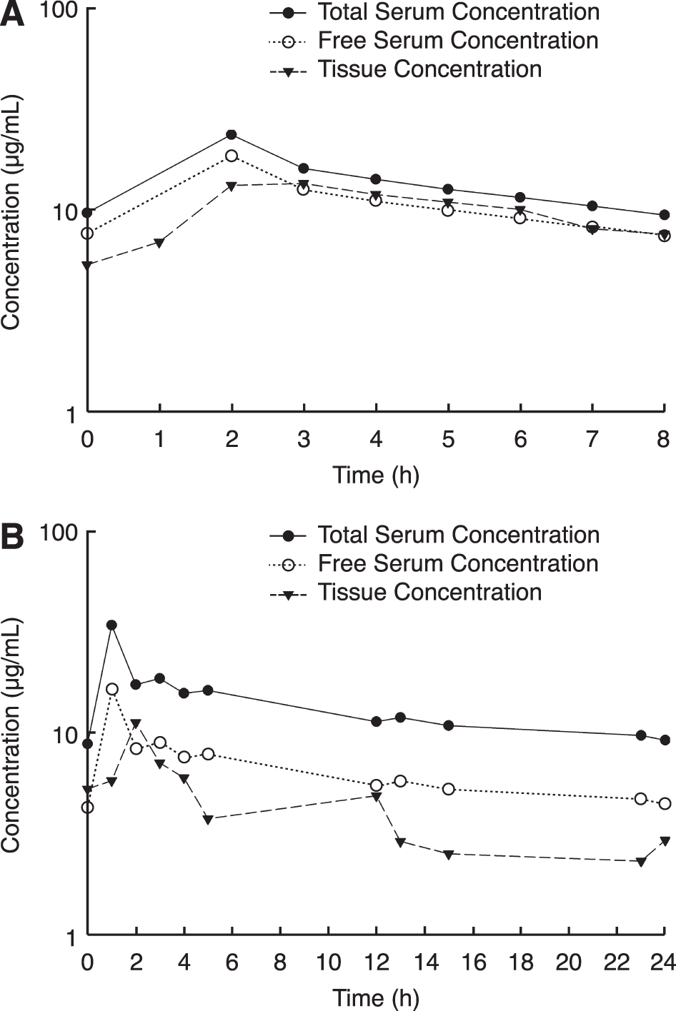
The mean pharmacokinetic parameters using total drug concentrations in serum at steady state are described in Table 2. One patient's Cmax and one patient's Cmin were excluded due to a contaminated sample and the next dose being administered before the end of sampling, respectively. The mean total and free drug concentrationtime profiles of the seven patients receiving q12h vancomycin is shown in Figure 1. The total and free drug concentration-time profiles of the two patients who received either the q8h or q24h regimens are depicted in Figure 2. Mean protein binding in the serum was 31.7% (range, 14.4%–51.7%). Protein binding was found to be linear over a range of concentrations. The mean ± SD total AUC0-24 for all nine patients was 417.2 ± 96.0 μg*h/mL. After correcting for protein binding, the mean ± SD free AUC0-24 calculated was 283.7 ± 89.4 μg*h/mL.
Table 2.
Steady-State Pharmacokinetics for Vancomycin in Serum and Tissue Samples Obtained from Patients with Lower-Limb Infections
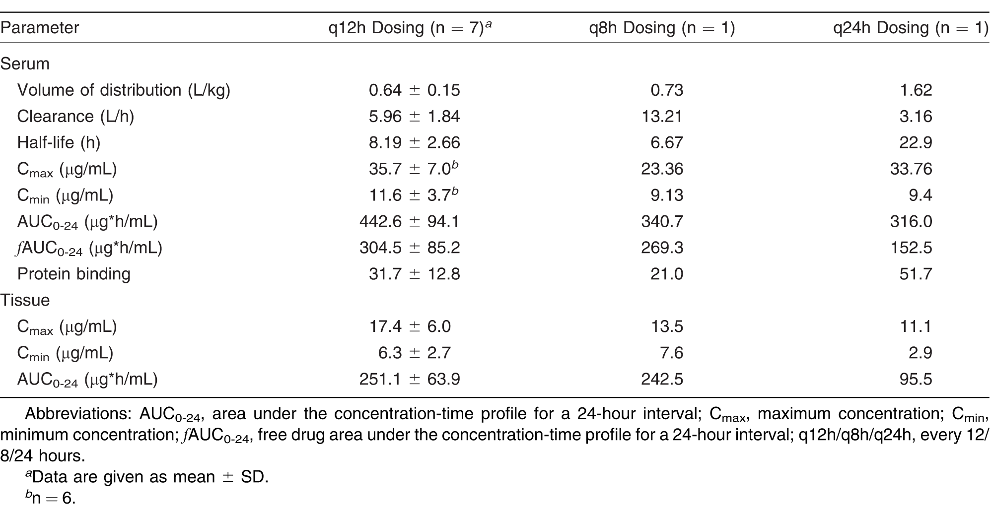
Figure 1.
Mean ± SD vancomycin concentrationtime profiles for the seven patients who received vancomycin every 12 hours.
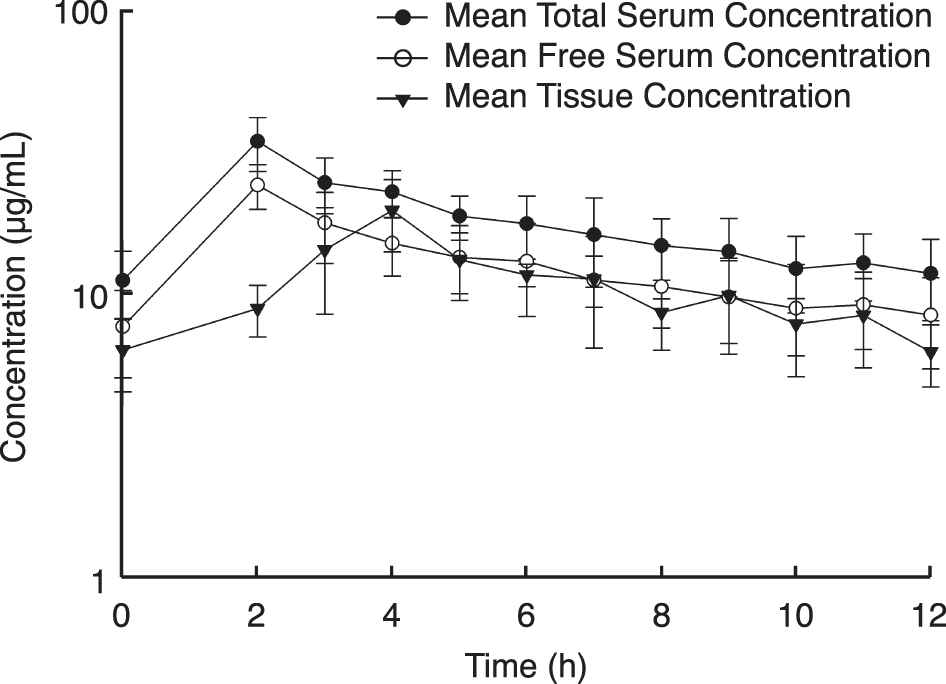
Figure 2.
Vancomycin concentration-time profiles for the two patients with alternative dosing schedules. One patient each received vancomycin every 8 hours (A) and every 24 hours (B).
Tissue
The mean ± SD recovery rate for the microdialysis catheters was 40.0% ± 12.5% (range, 26.5%–64.28%). In vitro retrodialysis experiments were similar between gain and loss, with mean ± SD relative recovery of 42.3% ± 6.0% and 51.0% ± 7.7%, respectively.[21] The steady-state tissue concentration-time profiles are shown in Figure 1 for patients receiving q12h dosing and in Figure 2 for the two patients receiving alternative dosing strategies. Assuming that only free drug will cross through the filter of the microdialysis catheter, the mean ± SD trough concentration in the tissue was 6.0 ± 2.6 μg/mL, which resulted in a mean ± SD AUC0-24 of 232.8 ± 75.7 μg*h/mL for all nine patients. The mean ± SD penetration of vancomycin into the infected tissue comparing the AUC in tissue with the free AUC in serum (AUCtissue/fAUCserum) from all patients was 0.8 ± 0.2.
Discussion
Vancomycin has been the mainstay of treatment against skin and skin structure infections caused by MRSA for the past few decades. Given increasing rates of MRSA and community-acquired MRSA,[1-5] the empirical use of vancomycin has increased. Current susceptibility studies also suggest that the MIC90 (the MIC required to inhibit the growth of 90% of organisms) for vancomycin is 1 μg/mL[22] and is likely higher in patients with previous treatment. The ability to achieve adequate pharmacodynamic exposures at the site of infection is important to obtain microbiologic and clinical success. As such, in vivo microdialysis is a technique that allows investigators to measure the concentration-time profile at a site of infection.[23] In our study, we assessed the tissue pharmacokinetics of vancomycin in hospitalized patients with lower-limb infections.
The patient population of this study consisted of primarily obese men who were poorly managing their diabetes. Most patients were admitted for surgical intervention. Of the two patients without diabetes, one had a history of trauma that left his left lower limb without sensation. The other patient was an otherwise healthy man who developed erythema and swelling with concurrent abscess of the fourth interspace and was admitted after failing oral outpatient therapy.
Mean ± SD serum trough concentrations of vancomycin were 11.1 ± 3.3 μg/mL and were deemed therapeutic with trough levels greater than 10 μg/mL. Protein binding was widely variable in patients, ranging from 14% to 52%, consistent with recent vancomycin protein binding estimates in the literature.[24] Correcting for protein binding, the mean ± SD total AUC0-24 in serum of 417.2 ± 96.0 μg*h/mL corresponds to a mean ± SD free AUC0-24 of 283.7 ± 89.4 μg*h/mL. In the tissue, a lower Cmin was observed below 10 μg/mL (mean ± SD, 6.0 ± 2.6 μg/mL). Although the trough concentration in the tissue was lower, the overall profile as characterized by AUC was only slightly lower than that in the serum, with a mean ± SD penetration ratio (AUCtissue/fAUCserum) of 0.8 ± 0.2. The tissue concentrations slowly increased, suggesting delayed distribution over the dosing interval to achieve a peak concentration at 4 hours versus the 2 hours in blood. Given no tissue pharmacodynamic target, we inferred the only pharmacodynamic target for vancomycin in blood of 400 for AUC/MIC. The current regimens in patients would achieve this AUC/MIC target against isolates with MICs of 1 μg/mL or less in serum and tissue. This supports the body of evidence emerging that suggests alternative agents for infections caused by MRSA pathogens with vancomycin MICs greater than 1 μg/mL.
Available information describing vancomycin penetration into infected tissue via in vivo microdialysis is limited. A single study described the tissue penetration of vancomycin in patients with diabetes and without diabetes after cardiac surgery.[13] In this study, the investigators inserted the catheter into healthy tissue in the thigh. The system was pumped with lactated Ringer's solution at a rate of 1.5 μL/min. Vancomycin was administered as a continuous infusion, and samples were obtained hourly for 6 hours. Plasma concentrations in patients with diabetes and those without were similar, approximately 37 μg/mL. However, the tissue concentrations were significantly lower in patients with diabetes (mean, 3.7 μg/mL) versus those without diabetes (mean, 11.9 μg/mL). The penetration ratio as compared by AUCtissue/AUCplasma ranged from 0.08 to 2.02 (mean, 0.44) for patients with diabetes and from 0.35 to 3.72 (mean, 1.45) for those without diabetes. Given the sample size, it was not possible to compare patients with diabetes and those without diabetes; however, grossly comparing penetration ratios of those with diabetes and those without shows similarity between the numbers, 0.7 ± 0.4 and 0.8, respectively. In the seven patients with diabetes, there was no apparent relationship between hemoglobin A1c level and penetration ratio.
One difference to note between our study and the study by Skhirtladze et al[13] is the patient population. In the study by Skhirtladze et al, the study group consisted of patients undergoing cardiac surgery and implantation of the microdialysis catheter in healthy tissue. This is in contrast to our study, in which patients were actively infected and sampling took place from the site of infection. These differences should be noted because the presence of infection has been noted to change antimicrobial pharmacokinetics in humans and in animal models.[25,26] In addition, patients in our study were located on surgical wards, whereas at least nine of 12 patients in the study by Skhirtladze et al were in a step-down or intensive care unit, with many of those receiving catecholamine support. These differences may also contribute to slight differences in the results observed between studies.
In conclusion, vancomycin penetrates into the infected tissue of patients with lower-limb infections at values similar to or slightly lower than in the blood based on AUC ratios. Reduced concentrations at the site of infection may contribute to the decreased effectiveness previously mentioned in the literature and possibly to the development of resistance. As other literature suggests, alternative agents may be needed when the pathogen of interest has an MIC greater than 1 μg/mL.
Acknowledgment
Lee J. Steere, RN, for his efforts with intravenous access and sample collection; Christina Sutherland, BS, for conducting all of the high-performance liquid chromatography analyses; and Rebecca Keel, PharmD, Shawn MacVane, PharmD, Jami Jain, PharmD, and Wonhee So, PharmD, for their assistance with sample collection and processing.
Financial Disclosure: None reported.
Conflict of Interest: None reported.
References
- National Nosocomial Infections Surveillance System: National Nosocomial Infections Surveillance (NNIS) System Report, data summary from January 1992 through June 2004, issued October 2004. Am J Infect Control32: 470, 2004.
- StreitJM, JonesRN, SaderHS, et al: Assessment of pathogen occurrences and resistance profiles among infected patients in the intensive care unit: report from the SENTRY antimicrobial surveillance program (North America, 2001). Int J Antimicrob Agents24: 111, 2004.
- NaimiTS, LeDellKH, Como-SabettiK, et al: Comparison of community- and health care–associated methicillin-resistant Staphylococcus aureus infection. JAMA290: 2976, 2003.
- OchoaTJ, MohrJ, WangerA, et al: Community-associated methicillin-resistant Staphylococcus aureus in pediatric patients. Emerg Infect Dis11: 966, 2005.
- MoranGJ, AmiiRN, AbrahamianFM, et al: Methicillin-resistant Staphylococcus aureus in community-acquired infections. Emerg Infect Dis11: 928, 2005.
- RybakM, LomaestroB, RotschaferJC, et al: Therapeutic monitoring of vancomycin in adult patients: a consensus review of the American Society of Health-System Pharmacists, the Infectious Diseases Society of America, and the Society of Infectious Diseases Pharmacists. Am J Health Syst Pharm66: 82, 2009.
- LodiseTP, PatelN, LomaestroBM, et al: Relationship between initial vancomycin concentration-time profile and nephrotoxicity among hospitalized patients. Clin Infect Dis49: 507, 2009.
- LodiseTP, LomaestroB, GravesJ, et al: Larger vancomycin doses (at least four grams per day) are associated with an increased incidence of nephrotoxicity. Antimicrob Agents Chemother52: 1330, 2008.
- WhitenerCJ, ParkSY, BrowneFA, et al: Vancomycin-resistant Staphylococcus aureus in the absence of vancomycin exposure. Clin Infect Dis38: 1049, 2004.
- AppelbaumPC: Reduced glycopeptide susceptibility in methicillin-resistant Staphylococcus aureus (MRSA). Int J Antimicrob Agents30: 398, 2007.
- LodiseTP, GravesJ, EvansA, et al: Relationship between vancomycin MIC and failure among patients with methicillin-resistant Staphylococcus aureus bacteremia treated with vancomycin. Antimicrob Agents Chemother52: 3315, 2008.
- PayneCJ, ThomsonAH, StearnsAT, et al: Pharmacokinetics and tissue penetration of vancomycin continuous infusion as prophylaxis for vascular surgery. J Antimicrob Chemother66: 2624, 2011.
- SkhirtladzeK, HutschalaD, FleckT, et al: Impaired target site penetration of vancomycin in diabetic patients following cardiac surgery. Antimicrob Agents Chemother50: 1372, 2006.
- LodiseTP, DrusanoGL, ButterfieldJM, et al: Penetration of vancomycin into epithelial lining fluid in healthy volunteers. Antimicrob Agents Chemother55: 5507, 2011.
- RicardJD, WolffM, LacheradeJC, et al: Levels of vancomycin in cerebrospinal fluid of adult patients receiving adjunctive corticosteroids to treat pneumococcal meningitis: a prospective multicenter observational study. Clin Infect Dis44: 250, 2007.
- LipskyBA, BerendtAR, CorniaPB, et al: 2012 Infectious Diseases Society of America clinical practice guideline for the diagnosis and treatment of diabetic foot infections. JAPMA103: 2, 2013.
- KimA, SuecofLA, SutherlandCA, et al: In vivo microdialysis study of the penetration of daptomycin into soft tissues in diabetic versus healthy volunteers. Antimicrob Agents Chemother52: 3941, 2008.
- BulikCC, WiskirchenDE, ShepardA, et al: Tissue penetration and pharmacokinetics of tigecycline in diabetic patients with chronic wound infections described by using in vivo microdialysis. Antimicrob Agents Chemother54: 5209, 2010.
- SahreM, SabarinathS, GrantM, et al: Skin and soft tissue concentrations of tedizolid (formerly torezolid), a novel oxazolidinone, following a single oral dose in healthy volunteers. Int J Antimicrob Agents40: 51, 2012.
- HagiharaM, SutherlandC, NicolauDP: Development of HPLC methods for the determination of vancomycin in human plasma, mouse serum and bronchoalveolar lavage fluid. J Chromatogr Sci51: 201, 2013.
- MacVaneSH, HousmanST, NicolauDP: Assessing the impact of combination antibiotic therapy on in vitro microdialysis recoveries. Paper presented at: 53rd Interscience Conference on Antimicrobial Agents and Chemotherapy; September 10-13, 2013; Denver, CO. Abstract F-1700.
- FarrellDJ, CastanheiraM, MendesRE, et al: In vitro activity of ceftaroline against multidrug-resistant Staphylococcus aureus and Streptococcus pneumoniae: a review of published studies and the AWARE Surveillance Program (2008-2010). Clin Infect Dis55 (suppl 3): S206, 2012.
- JoukhadarC, MullerM: Microdialysis: current applications in clinical pharmacokinetic studies and its potential role in the future. Clin Pharmacokinet44: 895, 2005.
- CrandonJL, MacVaneSH, NicolauDP: Clinical laboratory based assay methodologies may underestimate and increase variability of vancomycin protein binding in patients. Pharmacotherapy34: 203, 2014.
- KeelRA, CrandonJL, NicolauDP: Pharmacokinetics and pulmonary disposition of tedizolid and linezolid in a murine pneumonia model under variable conditions. Antimicrob Agents Chemother56: 3420, 2012.
- KutiJL, NicolauDP: Levofloxacin penetration into epithelial lining fluid differs between infected and uninfected patients. Paper presented at: 52nd Interscience Conference on Antimicrobial Agents and Chemotherapy; September 9-12, 2012; San Francisco, CA. Abstract A-1964.
© 2015 American Podiatric Medical Association