


Abstract
Background: We assessed the efficacy of customized foot orthotic therapy by comparing reulceration rates, minor amputation rates, and work and daily living activities before and after therapy. Peak plantar pressures and peak plantar impulses were compared with the patients not wearing and wearing their prescribed footwear. Methods: One hundred seventeen patients with diabetes were prescribed therapeutic insoles and footwear based on the results of a detailed biomechanical study and were followed for 2 years. All of the patients had a history of foot ulcers, but none had undergone previous orthotic therapy. Results: Before treatment, the reulceration rate was 79% and the amputation rate was 54%. Two years after the start of orthotic therapy, the reulceration rate was 15% and the amputation rate was 6%. Orthotic therapy reduced peak plantar pressures in patients with reulcerations and in those without (P < .05), although a significant decrease in peak plantar impulses was achieved only in patients not experiencing reulceration. Sick leave was reduced from 100% to 26%. Conclusions: Personalized orthotic therapy targeted at reducing plantar pressures by off-loading protects high-risk patients against reulceration. Treatment reduced the reulceration rate and peak plantar pressures, leading to patients’ return to work or other activities.
The long-term nature of the neuropathic diabetic foot and the irreversibility of its lesions makes it a high-risk foot. [1] Partial foot amputations are a risk factor for ulcer recurrence [2] because the resultant reduced surface area modifies the pressure distribution such that pressures increase over focal areas. [3,4] Reulceration rates in patients subjected to different treatments have been examined by several authors. [5–10] Protection of the high-risk diabetic foot is achieved through the use of orthopedic insoles and therapeutic shoes. This type of treatment is the most suitable for redistributing pressures and preventing ulcer recurrence. [11]
In a review by Bus et al [5] of 1,651 articles addressing ulcer prevention in the diabetic foot, the scientific evidence regarding orthotic treatment was found to be meager. Treatment is essentially based on clinical experience, and there are practically no studies or protocols to indicate the optimal features of interventions or the efficiency of the different orthotic devices designed to improve outcome measures, such as reulceration rates, peak plantar pressures, and peak plantar impulses. Among the reports that have provided useful data are those by Tyrrell, [12] Guldemond et al, [13] Munteanu and Bassed, [14] and Michaud and Nawoczenski. [15] Other authors report reulceration rates recorded in response to different treatments. [5–7,10,16]
The International Working Group on the Diabetic Foot [17] has published an international standard protocol for orthotic treatment. However, this protocol is rarely used in Spain, perhaps because the different treatment modes still lack evidence-based data.
Through orthotic treatment, we can more efficiently approach the preventive care of the neuropathic diabetic foot by prescribing insoles specially designed for each foot to help distribute pressures. [3] These insoles, used with footwear designed according to the biomechanics of the foot to accommodate deformities, [18,19] achieve adequate pressure distribution. In many patients, rocker-bottom outsoles are needed to reduce push-off forces and help with the dynamics of walking. [20–22]
To help with footwear prescription, the guidelines prepared by Dahmen et al [23] may be used. These guidelines try to match the therapeutic needs of each diabetic foot abnormality to the characteristics of the footwear.
Given the scarce scientific literature indicating the characteristics, design, and best methods for orthopedic treatments targeted at the diabetic neuropathic high-risk foot, the present orthotic treatment method is based on the biomechanics of the individual foot and on the concepts proposed for nondiabetic feet by the different authors. Among these, we could cite the work of Rose et al, [24] who described no significant benefit of using short wedges to off-load forefoot pressures, or of Milani and Hennig, [25] who managed to off-load pressures using a long heel pronation wedge, reducing forefoot pressure. Also, Tyrrell [26] indicated that insoles made of ethyl vinyl acetate work better than those made of foam. Studies such as those by Nawoczenski and Ludewig [27] and Nube et al [28] reveal that the use of a Morton’s extension offers no pressure relief. Michaud and Nawoczenski [29] reported that semirigid insoles give poor results in patients with limited mobility and increased pressures, and Guldemond et al [30] significantly reduced pressures using longitudinal arcs and metatarsal mounds.
At the Orthopedic Unit of the Universidad Complutense de Madrid (Madrid, Spain), we have experience with the use of different densities of heat-molded materials, such as ethyl vinyl acetate and 6-mm Poron (Rogers Corp, Rogers, Connecticut). In 2007, Paton et al [31] published the results of a study examining the physical properties of 15 materials used to prevent ulcers in diabetic patients with neuropathy. Of these materials, 6-mm Poron offered the best results, and ethyl vinyl acetate was also cited as a material providing good results. We use this material for the base insole layer and then overlay the base with further layers of decreasing density.
The present prospective study was designed to address whether a personalized orthopodologic regimen based on the plantar pressures and biomechanics of the individual foot could reduce the reulceration rate in patients with diabetic neuropathic foot ulcers.
Methods
Patients
Patients were recruited among those with diabetic neuropathy referred from the Diabetic Foot Unit to the Orthopedic Unit of the Podiatric Clinic of the Universidad Complutense de Madrid for personalized orthopodologic treatment. The inclusion criteria were a history of neuropathic ulcers or reulcerations, absence of an open ulcer at the study outset, and a capacity to walk (some of the patients did so with the help of felt insoles or postoperative footwear). To fulfill this last criterion, patients were required to indicate that they could perform routine activities around the house, climb a flight of stairs, and walk one block per day, similar to the requirements described by Reiber et al. [32] Patients were excluded if they had critical ischemia according to the Fontaine criteria. [33]
Before enrollment, all of the patients read and signed an informed consent form. The study protocol was approved by the review board of the Universidad Complutense de Madrid. Initially, 117 diabetic patients (79.5% men and 20.5% women) with high-risk foot factors and a history of ulcers were enrolled. Of these patients, three died (2.6%) and 14 abandoned the study (12.0%). This left a study population of 100 patients with a mean ± SD age of 59.4 ± 10.7 years who were observed for 2 years after the onset of a personalized orthotic treatment regimen.
Seventy-six patients had type 2 insulin-dependent diabetes, 17 had type 2 non–insulin-dependent diabetes, and 7 had type 1 diabetes. The mean ± SD diabetes duration was 16 ± 8 years. The mean ± SD levels of blood glucose and hemoglobin A were 168 ± 55.9 mg/dL and 7.8 ± 1.7 g/dL, respectively. Sixty-one percent of the patients had had neuroischemic ulcers, and the remaining 39% had experienced neuropathic ulcers.
Because of recent ulcers, none of the patients were working at the study outset: 48% were on sick leave, and the remaining 52% reported that their normal physical activity was somewhat reduced. Of these latter patients, 35% were retired, 15% were housewives, and 2% had a mental illness.
Procedure
Neuropathy was diagnosed by examining ten sites on the foot using a Semmes-Weinstein monofilament 5.07/10 g (Sensifil-Novalab Ibérica, Madrid, Spain) and a neurotensiometer (Horwell 997; Sensifil-Novalab Ibérica). [34,35] Vascular involvement was defined as an ankle-arm index lower than 0.8, [36] transcutaneous oxygen pressure of less than 30 mm Hg (using a TCM4 transcutaneous monitor; Radiometer Medical ApS, Brønshøj, Denmark), [37] and a lack of dorsal pedal and posterior tibial pulses. [36]
In each foot, the factors assessed were joint mobility, goniometry, and biomechanics. In addition, any plantar callus zones, lesions, or partial amputations were recorded, noting the localization and history of the different lesions.
For this study, we defined a reulceration as one or more plantar lesions (lack of skin integrity) appearing at the same site after a primary ulcer. In the initial examination, we assessed, through a full anamnesis and information in clinical records, the ulcers experienced by the patients before the study outset and determined whether these had been primary ulcers or reulcerations.
Plantar pressures while walking were recorded using a 2-m-long Footscan 7.x gait interface system (RSscan International, Lammerdries, Belgium) at a recording speed of 500 photograms per second. Barefoot pressures were determined for subsequent selective off-loading using the appropriate insoles. Three recordings were obtained first with the patients barefoot and then with the patients wearing their customized footwear, and mean peak plantar pressures and peak plantar impulses were determined.
The Footscan system, through a series of sensors, quantifies pressures and determines the distribution of such pressures across the plantar surface. The instrument’s software calculates peak plantar pressures and peak plantar impulses by dividing the foot sole into ten anatomical zones: medial and lateral rearfoot, midfoot, each of the five metatarsals, the hallux, and the remaining toes. When testing a patient with a minor foot amputation, the system permits adjustment of the plantar area. The Footscan ascribes each pressure measurement to a given anatomical zone. Using its software, it generates an image of the contact surface under the rearfoot, midfoot, and forefoot in which pressures are color coded.
When recording the patient’s gait using the Footscan, the different stages of gait when increased pressures are produced were determined to establish whether they corresponded to the push-off stage or the risk zone previously identified according to the patient’s history of ulcers.
For each foot, we prepared a radiophotopodogram by obtaining an imprint of the sole of the foot on radiographic paper and superimposing this footprint on a plantar radiograph to visualize bone prominences corresponding to the zones of highest pressure observed in the Footscan. A mold was then prepared using the impression of the sole made in a foam impression box (Fig. 1A).
The treatment design was based on the radiophotopodogram findings, peak plantar pressures, peak plantar impulses, and the stages of gait by considering the pressures caused by vertical push-off forces produced during gait or pressure exerted on a bone prominence during ambulation. The main goals of the method used were to give the foot good stability and to off-load zones of maximum pressure, sometimes by fenestrating the insole to off-load rigid joints that extend beyond the plantar level (Fig. 1B–D).
The idea was to eliminate pressures to reduce the risk of recurrent ulcers by applying the footwear modifications described in the literature. Our own contribution was the use of a fenestrated outsole covered with a rocker bottom in patients with a rigid plantarflexed metatarsal (Fig. 1E and F).
A shoe was prescribed according to the length and width of the foot, as measured using a Heider device (Langlaufschuhbedarf GmbH, Dortmund, Germany), and the given foot abnormality. The type of footwear (eg, extra-depth shoe or bottine) was selected according to the algorithm described by Dahmen et al. [23]
When pressure was mainly static, located over a bone prominence, and there was joint mobility, pressures were off-loaded using a heat-molded laminar (ethyl vinyl acetate) insole of varying density (25°–60° Shore). For metatarsal off-loading, we used different orthoses depending on the pathologic abnormality and the metatarsal head needing off-loading (metatarsal or central head mounds). A shock-absorbing material, 6-mm Poron (hereafter designated Poron), was used to off-load areas of bone protrusion and previous ulcer and wound sites.
When pressure coincided with a bone prominence and joints showed insufficient mobility for selective off-loading, we used a heat-molded laminar ethyl vinyl acetate insole with a maximum thickness of 1.5 to 2.0 cm and low to medium density (25°–33° Shore), with a high-density ethyl vinyl acetate (60° Shore) bottom layer. In this type of insole, we embedded a Poron longitudinal inner arch piece, in keeping with the anatomical configuration of the mold obtained during loading. Poron plugs were also introduced in selected forefoot zones showing elevated pressure. Use of this thickness enabled us to fenestrate the insole in the area of bone prominence and, thus, to avoid direct pressure. When fenestrations were large or when these gave rise to suction on soft tissues, the insole was cut according to the biomechanics of the foot. The use of pronation and supination wedges was sometimes necessary to redistribute pressures and offset the pressure on a hallux valgus or varus or limb dysmetria.
Figure 1.
A, The radiophotopodogram serves to identify the ulcer risk zone indicated (third metatarsal). The dashed-dotted line indicates where the metatarsal off-loading piece should be placed. B, The insole designed for the foot in A has a large fenestration in the ulcer zone to accommodate a plantarflexed third metatarsal. Note the 6-mm Poron lining of the fenestration. C, Short off-loading insole. D, Head-on view of the insole in C used to off-load a plantarflexed metatarsal. E, Fenestration in the outsole of the shoe in F filled with 6-mm Poron. F, Negative heel rocker used to compensate limb dysmetria. Note that the rocker is pivoted behind the zone of most pressure. G, U-shaped rocker used for the foot in A to off-load pressures at the central metatarsal heads.
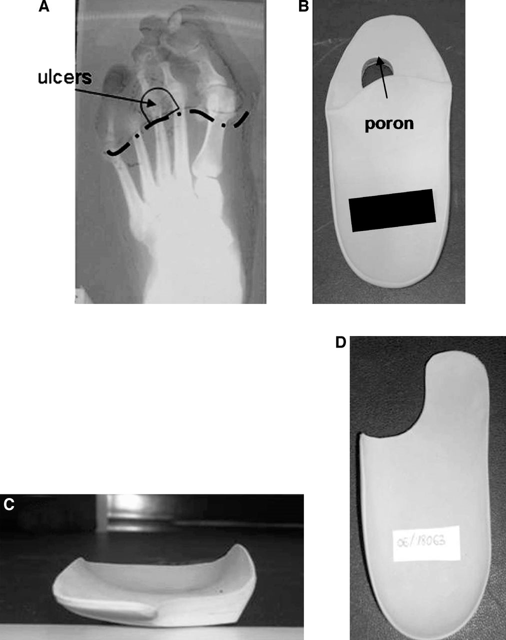
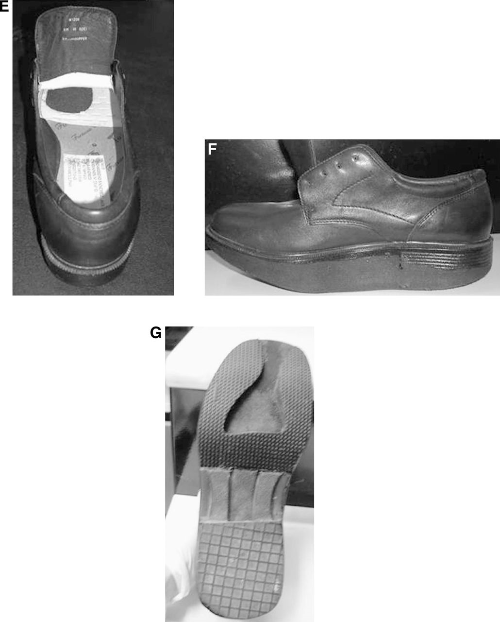
Customized outsoles were prescribed in cases of increased vertical pressure in the push-off stage of walking, hallux rigidus or limitus, and first-ray or digit amputations. The different outsoles incorporated several different customizations. A 1-cm U-shaped forefoot rocker was used when excess pressures affected the central metatarsals (Fig. 1G). A 1-cm forefoot rocker was used when excess loading affected the first or fifth metatarsals or both; the rocker was pivoted behind the zone of most pressure. The part of the outsole corresponding to the lesion site was cut out, and this area was covered with a half sole or rocker in cases of substantial metatarsal plantarflexion with a level difference of more than 1 cm. A rockered platform was prescribed when there was lower-limb dysmetria and pressures needed compensating.
Reulceration rates recorded in the 2 years before treatment were compared with those observed after the 2 years of orthotic treatment using the McNemar test. Once treatment was initiated, we determined any reulcerations produced at follow-up visits scheduled for 1, 3, 6, 12, 18, and 24 months. These data were recorded as cumulative reulceration rates (expressed per patient). Then, based on the number of plantar ulcers retrospectively recorded in patients’ clinical records, we also calculated cumulative reulceration rates by lesion at the different follow-up times.
Mean peak plantar pressures and peak plantar impulses recorded with the patient barefoot and with the patient wearing the prescribed orthotic devices were compared using a paired t test. This comparison enabled us to determine whether the prescribed footwear could significantly reduce pressures in all of the patients. Mean peak plantar pressure and peak plantar impulse measures were also compared in patients stratified according to whether they had experienced reulceration during orthotic treatment. All of the statistical tests were performed using a commercially available software program (SPSS, version 15.0; SPSS Inc, Chicago, Illinois). The level of significance was set at P < .05.
Results
The data obtained from the patients’ clinical records and anamnesis indicated that none of the patients had received previous orthotic treatment. All had experienced previous ulcers, with a reulceration rate of 79% and a partial amputation rate of 54% for the 2 years before the study outset.
The mean ± SD duration of the last ulcer was 66.7 ± 56.2 weeks. Seventy-nine patients experienced ulcer recurrence: 79.8% (63 patients) in the first year after the primary ulcer had resolved, 13.9% (11 patients) in the second year, 3.8% (three patients) in the third year, and 2.5% (two patients) in the fourth year.
During the 2 years leading up to the treatment intervention, the 100 patients included in the study had experienced 162 ulcerations. However, 15 of these ulcerations were not considered in these analyses because they did not affect plantar zones. This left 147 plantar ulcers retrospectively recorded in the patients.
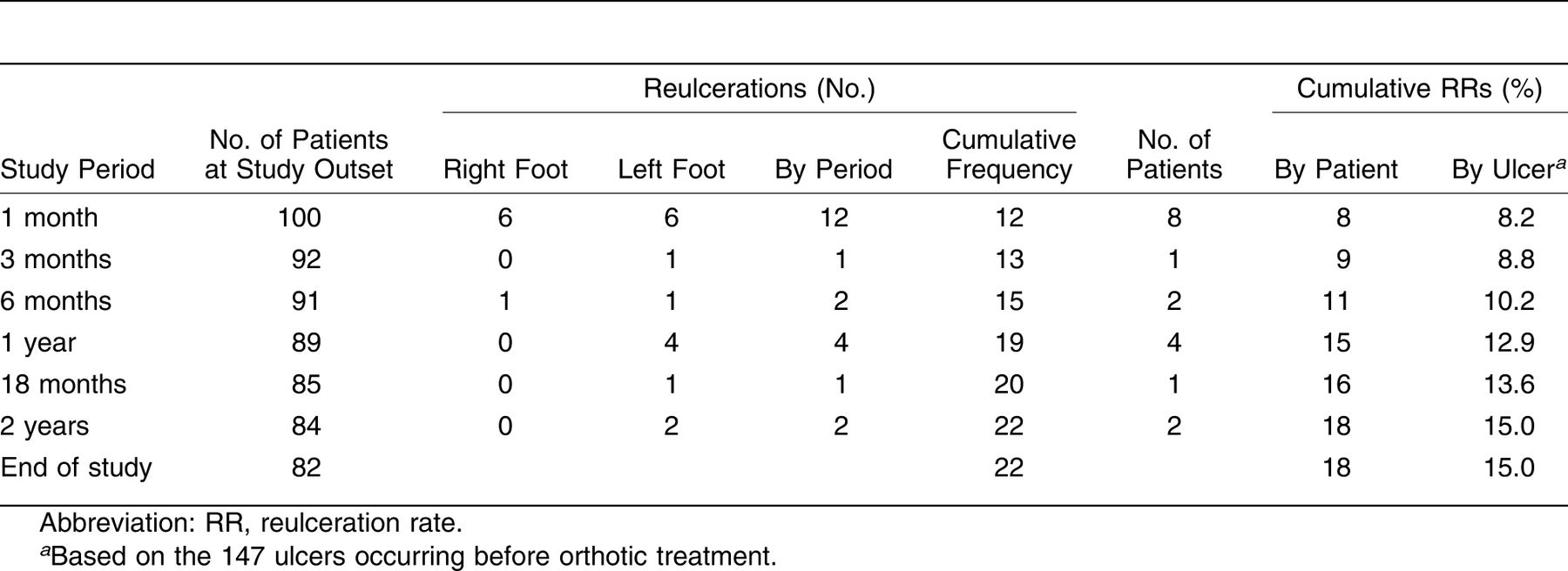
After orthotic treatment, cumulative reulceration rates were 8% at 1 month, 9% at 3 months, 11% at 6 months, 15% at 1 year, 16% at 18 months, and 18% at 2 years. Cumulative reulceration rates were also expressed by lesion based on the 147 ulcers occurring before orthotic treatment as 8.2% at 1 month, 8.8% at 3 months, 10.2% at 6 months, 12.9% at 1 year, 13.6% at 18 months, and 15.0% at 2 years (Table 1).
Using the McNemar test, we determined the efficacy of the orthotic treatment regimen by comparing the number of ulcerations recorded in the 2 years preceding treatment (147 ulcerations) with those recorded after treatment (22 reulcerations), obtaining a significant difference (P < .001; odds ratio, 0.016). Fifty-four patients had undergone previous partial amputations of their feet during their illness.
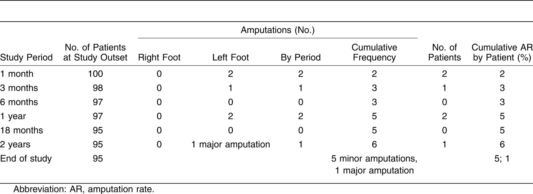
For the 2 years of orthotic treatment, the amputation rate was 6% (six patients), including minor foot amputations in five patients and one major foot amputation (Table 2).
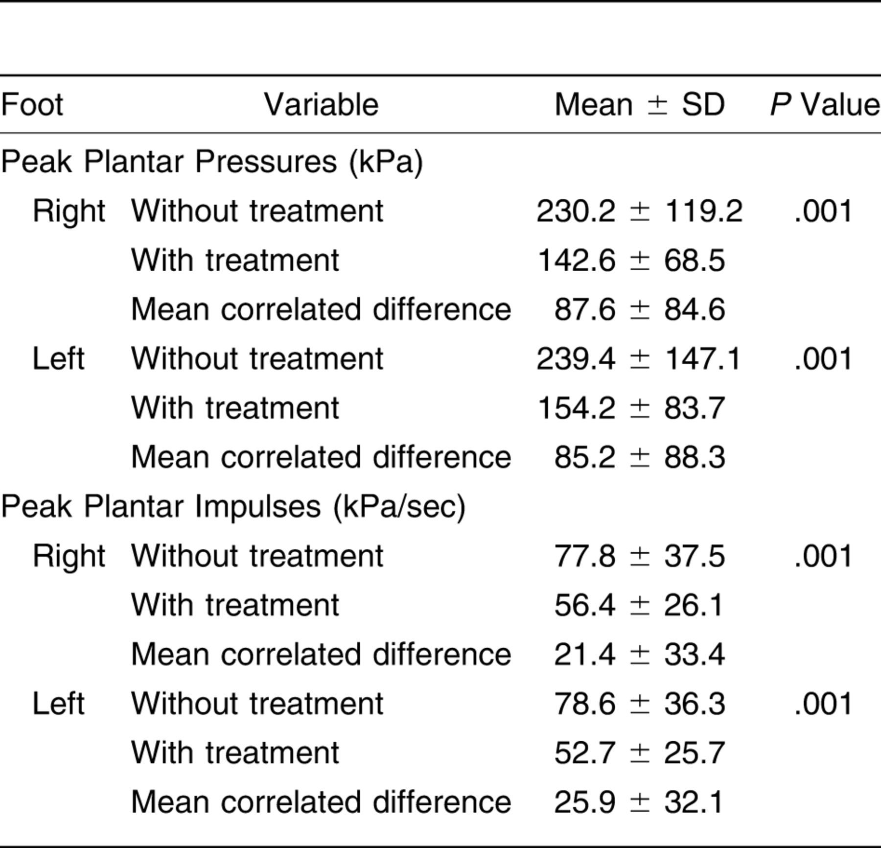
A paired t test was used to determine whether orthotic treatment was effective at reducing peak plantar pressures and peak plantar impulses as soon as the patients wore their prescribed orthotic devices. Table 3 shows the pressures recorded with and without customized orthoses.
Table 1.
Cumulative RRs Obtained at Each Time Point During the 2 Years by Number of Lesions and Number of Patients
The differences recorded in correlated means were significant (P < .001). Thus, we observed mean ± SD reductions in peak plantar pressures of 85.2 ± 88.3 kPa in the left feet and 87.6 ± 84.6 kPa in the right feet and mean ± SD reductions in peak plantar impulses of 25.9 ± 32.1 kPa/sec in the left feet and 21.4 ± 33.4 kPa/sec in the right feet.
Table 2.
Cumulative ARs Obtained at Each Time Point During the 2 Years by Number of Patients
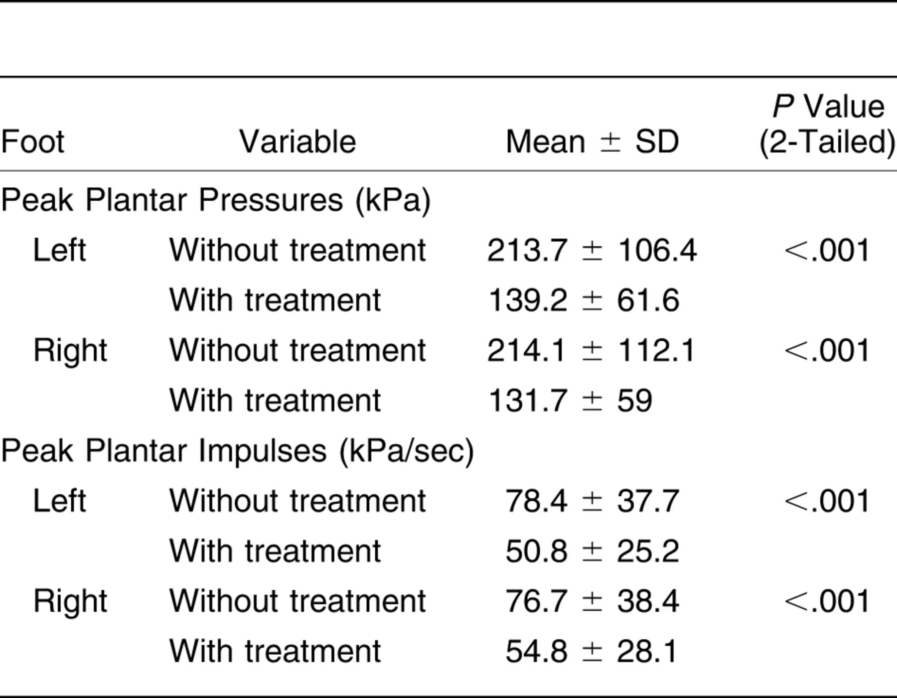
We repeated this analysis comparing mean peak plantar pressures and peak plantar impulses separately for patients who did not reulcerate (n = 82); these results are shown in Table 4.
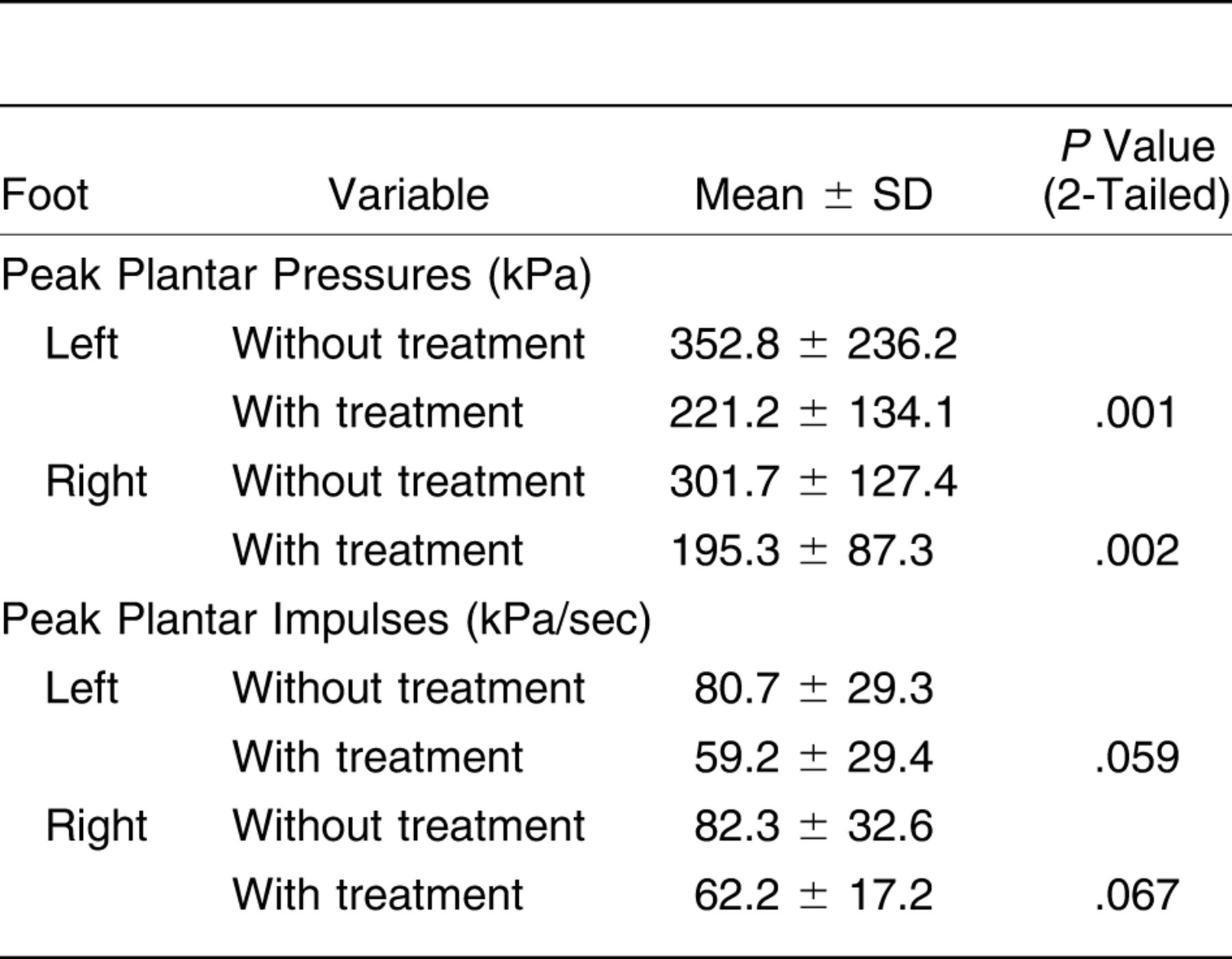
In patients reulcerating during orthotic treatment (n = 18), a significant reduction in mean peak plantar pressures was observed. However, when we compared mean peak plantar impulses with and without orthotic treatment, no significant differences emerged (Table 5).
Table 3.
Peak Plantar Pressures and Impulses Recorded with and Without Orthotic Footwear in 200 Right and Left Feet
At the study end, all of the working patients (48%) who were on sick leave had returned to work. Half of the remaining 52% of patients were able to resume their normal level of physical activity, and of the 26% remaining, 18% could not do so because of reulcerations. In a final 8% of patients, their state of continued dependency was attributed to other causes: 2% were mentally handicapped (able to walk) and 6% were retired, with a mean ± SD age of 83.50 ± 4.8 years. Although these elderly individuals could walk, when asked, they reported that they had stopped performing many of the daily living activities they had conducted before their last ulcer.
Discussion
Despite recommendations for orthopodologic treatment as a preventive measure to reduce pressures and the incidence of reulceration, [17] the high-risk foot patients included in this study had not received previous treatment when they were referred to the Orthopedic Unit of the Universidad Complutense de Madrid.
Table 4.
Analysis of Means (Paired t Test) of Peak Plantar Pressures and Impulses Recorded with and Without Orthotic Treatment in 82 Patients Who Did Not Reulcerate During the Study
Several studies [5–7,10,16] have addressed the issue of reulceration in the diabetic foot, although not always in high-risk patients with a history of ulcers and partial amputations. To examine the effects of treatment in terms of pressure relief, we compared the cumulative reulceration rate before and after orthotic treatment. The McNemar test revealed a significant reduction in this rate (P < .001; odds ratio, 0.016). This odds ratio is much higher than those reported by Zou et al [16] and Tyrrell [12] for the efficacy of orthopodologic treatments also measured in terms of lowering the risk of reulceration.
Table 5.
Analysis of Means (Paired t Test) of Peak Plantar Pressures and Impulses Recorded with and Without Orthotic Treatment in 18 Patients Reulcerating During the Study
After 1 year of orthotic treatment, the rate of reulceration was 12.9%, and the rate of patients experiencing reulceration was 15%. Both of these results are lower than the rates reported by Bus et al, [5] who achieved a reulceration rate of 16% after the use of customized footwear for 1 year, and by others. [5–7,10,16] We concur with the argument put forward by Bus et al [5] that it is within the first year, and mainly the first month, of orthotic treatment that most reulcerations occur such that we recommend more intense follow-up during this period to evaluate the factors contributing to reulceration.
We based our treatment designs on the biomechanics of the foot and on the results of studies performed in diabetic and nondiabetic patients. [12–15] We also separately assessed each foot of a patient as an independent biomechanical unit. The success of orthotic treatment could be related to the different pieces composing the insoles prescribed herein and confirms the hypothesis of the benefits of tailored treatment for each foot.
The present method consists of giving support to, but never increasing, the arch. In cases in which a greater amount of loading needs to be offset, we use the full arch height of the patient to increase the support surface and then distribute pressures by embedding Poron in the medial arch support so that reaction forces are optimally absorbed.
The study by Tyrrell [12] assessed the effects on the reulceration risk of the use of low-density ethyl vinyl acetate insoles shaped according to the foot’s physiologic arch. The work of Guldemond et al [13] on achieving metatarsal re-equilibrium is also interesting, although we used only metatarsal head mounds in patients capable of good movement who had not undergone metatarsal head amputation or had a rigid plantarflexed metatarsal.
The treatment of patients with metatarsal head amputations is a great challenge owing to joint rigidity and poor shock absorbency. When a lifting piece is used under a plantarflexed metatarsal, an increase in pressure is produced, and, consequently, ulcers recur. We believe that this is the great error of many orthoses and explains the high reulceration rates obtained in many patient series. [5–8] The present method consists of allowing the metatarsal to remain flexed and distributing pressures across the rest of the plantar surface using cutouts or half insoles.
Peak plantar pressures and impulses were analyzed before and immediately after fitting the feet with their personalized footwear. Some authors [38,39] claim that peak pressures achieved with an orthosis are important but are not the only predictor of treatment outcome. We also believe that other mechanisms are likely to intervene in increasing plantar pressures, as stated by Zou et al [16] and Yavuz et al. [40]
In these routine observations of the gait cycle, we noted that we could reduce peak plantar pressures through the use of complete orthotic treatment (insoles and footwear). In the active propulsion stage, we found that pressure increased across a smaller area as the support base of the foot was diminished. Time plays a critical role here; we observed that when impulse time increased, the Footscan system indicated the existence of high pressures in some patients.
During the active propulsion stage, horizontal and vertical forces are greater and are directed toward the forefoot across a relatively small area; pressures are higher and generally occur over the metatarsal heads. [41] Given these features of gait, besides examining peak pressure we decided to determine the moment of impulse in the propulsion stage with and without treatment. This variable is the time-force integral. To our knowledge, no study has related plantar impulse to the risk of reulceration, although some authors have mentioned this as a possible factor. [16,40]
Peak plantar pressures were reduced by orthotic treatment in patients who later experienced reulceration and in those who did not (P < .05). However, despite a reduction in mean peak plantar impulses recorded for the patient subset not reulcerating, treatment was unable to significantly mitigate peak impulses in patients with reulceration. It, thus, seems that a high peak plantar impulse promotes reulceration and should be controlled to prevent this problem. Future studies should focus on designing methods to reduce these peak plantar impulses.
At the study outset, all of the patients were incapacitated because of their chronic ulcers. Those who worked had to take sick leave, and those without jobs were unable to perform their routine living tasks, an observation made by others. [42] With treatment, this inactivity was improved in 100% of the working patients (52 patients) and in 50% of the remaining patients. In this non–professionally active subset of patients, only 26% maintained their situation of dependency despite having no foot ulcers.
The lack of a control group was the main limitation of this study. However, given the characteristics of these patients (they all had a history of ulcers and a high rate of reulceration, experienced a recent ulcer, and used standard orthotic footwear), we considered personalized preventive orthotic devices as the only ethical and available option for these patients.
These results indicate that personalized treatment based on a biomechanical study targeted at diminishing plantar pressures can reduce the risk of foot reulceration in high-risk patients with neuropathic ulcers and provide direction for future studies designed to reduce such pressures.
Acknowledgment
The staff of the University Podiatric Clinic, Universidad Complutense de Madrid, for their cooperation, and Ana Burton, BSc, for translating the original manuscript.
Financial Disclosure: None reported. | |
Conflict of Interest: None reported. |
References
- Boulton, AJ, LVileikyte, GRagnarson-Tennvall, et al: The global burden of diabetic foot disease. Lancet366:1719, 2005.
- Murdoch, DP, DGArmstrong, JBDacus, et al: The natural history of great toe amputations. J Foot Ankle Surg36:204, 1997.
- Giurini, JM and TELyons: Diabetic foot complications: diagnosis and management. Int J Low Extrem Wounds4:171, 2005.
- Lavery, LA, DCLavery, and TLQuebedeax-Farnham: Increased foot pressures after great toe amputation in diabetes. Diabetes Care18:1460, 1995.
- Bus, SA, GDValk, RWvan Deursen, et al: The effectiveness of footwear and offloading interventions to prevent and heal foot ulcers and reduce plantar pressure in diabetes: a systematic review. Diabetes Metab Res Rev24( suppl 1:):S162, 2008.
- Dalla, PL, EFaglia, MCaminiti, et al: Ulcer recurrence following first ray amputation in diabetic patients: a cohort prospective study. Diabetes Care26: 1874, 2003.
- Pham, H, DGArmstrong, CHarvey, et al: Screening techniques to identify people at high risk for diabetic foot ulceration: a prospective multicenter trial. Diabetes Care23:606, 2000.
- Tazi, O and CDebure: Preventing high-risk diabetic foot ulceration by a new method of custom-made shoes in high-risk patients: prospective study [in French]. J Mal Vasc33:191, 2008.
- Tyrrell, W: Orthotic intervention in patients with diabetic foot ulceration. J Wound Care8:530, 1999.
- Uccioli, L, EFaglia, GMonticone, et al: Manufactured shoes in the prevention of diabetic foot ulcers. Diabetes Care18:1376, 1995.
- Cavanagh, PR, AJBoulton, PSheehan, et al: Therapeutic footwear in patients with diabetes. JAMA288:1231, 2002.
- Tyrrell, W: Orthotic intervention in patients with diabetic foot ulceration. J Wound Care8:530, 1999.
- Guldemond, NA, PLeffers, APSanders, et al: Casting methods and plantar pressure: effects of custom-made foot orthoses on dynamic plantar pressure distribution. JAPMA96:9, 2006.
- Munteanu, SE and ADBassed: Effect of foot posture and inverted foot orthoses on hallux dorsiflexion. JAPMA96:32, 2006.
- Michaud, TC and DANawoczenski: The influence of two different types of foot orthoses on first metatarsophalangeal joint kinematics during gait in a single subject. J Manipulative Physiol Ther29:60, 2006.
- Zou, D, MJMueller, and DJLott: Effect of peak pressure and pressure gradient on subsurface shear stresses in the neuropathic foot. J Biomech40:883, 2007.
- Singh, N, DGArmstrong, and BALipsky: Preventing foot ulcers in patients with diabetes. JAMA293:217, 2005.
- Janisse, DJ: Prescription insoles and footwear. Clin Podiatr Med Surg12:41, 1995.
- Chantelau, E and AGede: Foot dimensions of elderly people with and without diabetes mellitus: a data basis for shoe design. Gerontology48:241, 2002.
- Myers, KA, JTLong, JPKlein, et al: Biomechanical implications of the negative heel rocker sole shoe: gait kinematics and kinetics. Gait Posture24:323, 2006.
- Chesnin, KJ, LSelby-Silverstein, and MPBesser: Comparison of an in-shoe pressure measurement device to a force plate: concurrent validity of center of pressure measurements. Gait Posture12:128, 2000.
- Mueller, MJ, DJLott, MKHastings, et al: Efficacy and mechanism of orthotic devices to unload metatarsal heads in people with diabetes and a history of plantar ulcers. Phys Ther86:833, 2006.
- Dahmen, R, RHaspels, BKoomen, et al: Therapeutic footwear for the neuropathic foot: an algorithm. Diabetes Care24:705, 2001.
- Rose, NE, LAFeiwell, and ACracchioloIII: A method for measuring foot pressures using a high resolution, computerized insole sensor: the effect of heel wedges on plantar pressure distribution and center of force. Foot Ankle13:263, 1992.
- Milani, TL and EMHennig: Measurements of rearfoot motion during running. Sportverletz Sportschaden14:115, 2000.
- Tyrrell, W: Orthotic intervention in patients with diabetic foot ulceration. J Wound Care8:530, 1999.
- Nawoczenski, DA and PMLudewig: The effect of forefoot and arch posting orthotic designs on first metatarsophalangeal joint kinematics during gait. J Orthop Sports Phys Ther34:317, 2004.
- Nube, VL, LMolyneaux, and DKYue: Biomechanical risk factors associated with neuropathic ulceration of the hallux in people with diabetes mellitus. JAPMA96:189, 2006.
- Michaud, TC and DANawoczenski: The influence of two different types of foot orthoses on first metatarsophalangeal joint kinematics during gait in a single subject. J Manipulative Physiol Ther29:60, 2006.
- Guldemond, NA, PLeffers, APSanders, et al: Casting methods and plantar pressure: effects of custom-made foot orthoses on dynamic plantar pressure distribution. JAPMA96:9, 2006.
- Paton, J, RBJones, EStenhouse, et al: The physical characteristics of materials used in the manufacture of orthoses for patients with diabetes. Foot Ankle Int28:1057, 2007.
- Reiber, GE, DGSmith, CWallace, et al: JAMA287:2552, 2002.
- Fontaine, R and JLFontaine: Arteritis and gangrene in diabetic patients in the surgical clinic. Rev Clin Esp112:571, 1969.
- Feng, Y, FJSchlosser, and BESumpio: The Semmes Weinstein monofilament examination as a screening tool for diabetic peripheral neuropathy. J Vasc Surg50:675, 2009.
- Lee, S, HKim, SChoi, et al: Clinical usefulness of the two-site Semmes-Weinstein monofilament test for detecting diabetic peripheral neuropathy. J Korean Med Sci18:103, 2003.
- McDermott, MM, RSufit, TNishida, et al: Lower extremity nerve function in patients with lower extremity ischemia. Arch Intern Med166: 1986, 2006.
- Kalani, M, KBrismar, BFagrell, et al: Transcutaneous oxygen tension and toe blood pressure as predictors for outcome of diabetic foot ulcers. Diabetes Care22:147, 1999.
- Mueller, MJ, DZou, and DJLott: “Pressure gradient” as an indicator of plantar skin injury. Diabetes Care28:2908, 2005.
- Yavuz, M, AErdemir, GBotek, et al: Peak plantar pressure and shear locations: relevance to diabetic patients. Diabetes Care30:2643, 2007.
- Yavuz, M, ATajaddini, GBotek, et al: Temporal characteristics of plantar shear distribution: relevance to diabetic patients. J Biomech41:556, 2008.
- van Schie, CH: A review of the biomechanics of the diabetic foot. Int J Low Extrem Wounds4:160, 2005.
- Ragnarson, TG and JApelqvist: Health-economic consequences of diabetic foot lesions. Clin Infect Dis39( suppl 2:):S132, 2004.
© 2013 American Podiatric Medical Association