


Abstract
Background: Integra bilayer wound matrix (IBWM) is a bilayer skin replacement system composed of a dermal regeneration layer and a temporary epidermal layer. It is used to treat various types of deep, large wounds via an inpatient procedure in an operating room. We sought to determine ease of use and effectiveness of IBWM in an outpatient clinical setting when treating diabetic foot ulcers. In addition, no epidermal autografting was performed in conjunction with the IBWM after silicone release, as is common in the inpatient setting. Methods: This 12-week, single-arm, prospective pilot study was conducted in three outpatient clinics. Weekly evaluations included monitoring the wound for signs of infection during the 12-week follow-up phase. Results: Eleven patients with diabetic foot ulcers who met the inclusion and exclusion criteria were enrolled. One patient was discontinued from the study owing to noncompliance leading to a serious adverse event. Therefore, ten patients who received the study intervention were included in the per-protocol population reported herein. The mean patient age was 60.6 years, with an average 11-year history of diabetes mellitus. Each ulcer was located on the plantar aspect of the foot. No infection was reported during the study. Patients treated with IBWM showed progressive wound healing over time: the greatest mean wound reduction was approximately 95% in week 12. Seven of ten patients (70%) achieved complete wound closure by week 12. No recurrent ulcers were reported during follow-up. Conclusions: These results are consistent with the hypothesis that IBWM is easy to use, safe, and effective when used on diabetic foot ulcers in an outpatient clinical setting without the secondary procedure of autografting for closure.
Yannas et al [1] developed the concept for Integra Dermal Regeneration Template (Integra LifeSciences Corp, Plainsboro, New Jersey) in the 1970s with the aim of developing a device that provides the immediate closure normally achieved with a thick skin graft and that confers the functional benefits of nonfibrotic healing. The initial design criteria were developed around physical properties, biological compatibility, and fibrosis.
Integra bilayer wound matrix (IBWM; Integra LifeSciences Corp) uses biocompatible cross-linked collagen/glycosaminoglycan with specific biochemistry composed of bovine collagen type I from tendon and the glycosaminoglycan chondroitin-6-sulfate. It suppresses inflammation by inhibiting the collagen-induced platelet aggregation driving the production of cytokines implicated in the inflammatory wound-healing process and the formation of granulation tissue. Furthermore, the collagen-glycosaminoglycan matrix is designed to have a significantly more open pore structure than collagen alone. This open porosity with controlled pore size and resorption rates permits dermal cellular ingrowth and remodeling without contraction and scarring. Epidermal function on the scaffold is achieved via a second, silicone layer of a defined thickness to prevent excess moisture loss. This silicone layer also prevents the formation of granulation tissue on the surface of the matrix. [2,3]
Originally introduced for use in the treatment of thermal injuries, IBWM has a long history of clinical use. [4,5] Applications include treating skull defects with exposed bone after burns or trauma, [6] degloving injuries, [7] after tumor excision, [8] and in patients with exposed bone after irradiation. [9] Yannas et al [1] detailed that in lower-extremity trauma, the dermal regeneration template has been particularly useful in covering small areas of bone and tendon, minimizing the need for local flaps or free tissue transfer. Barnett and Shilt [10] described use of the dermal regeneration template in open tibial fractures. Violas et al [11] described its use in the management of severe tissue defects, including bone exposure, in injured children.
In complex wounds, the dermal regeneration template has been used in covering avascular structures [12] and in restoring contour defects through sequential application to increase the thickness of the reconstruction. [13,14] In chronic wounds of the lower extremity, the dermal regeneration template has been used in reestablishing a dermal bed and reducing subsequent inflammation. [15,16] Given the ability to reduce scarring, IBWM has been used to resurface hypertrophic scars and keloids and to treat defects of the head and neck, including Mohs surgery. [17]
Lower-extremity ulcers impose significant clinical, humanistic, and economic burdens on society. One in five diabetic patients in the United States will be hospitalized with a foot complication at some time during their disease, and foot disorders such as ulceration, infection, and gangrene are the leading causes of hospitalization in these patients. [18] Annually, in the United States alone, 35,000 to 40,000 diabetic patients undergo a major amputation directly related to a nonhealing foot ulcer at a direct cost of $1.2 billion to the health-care system. This cost estimate does not include the cost of rehabilitation or prostheses, and it does not account for the financial impact of loss of time from work, loss of jobs, and welfare payments related to these wounds. [19–22]
Inpatient expenditures account for 70% to 80% of total costs related to lower-extremity ulcers. In contrast, outpatient service expenditures account for only 10% to 20% of total costs. [23] In current practice, application of IBWM on patients is performed in an operating room when patients are admitted to an inpatient service, which is costly and time consuming. Commonly, an epidermal autograft is placed on the wound after the silicone layer of IBWM is released, requiring an additional operating room procedure. We hypothesized that application of IBWM in the outpatient setting can easily, safely, and efficiently be performed, which could possibly save health-care dollars and make IBWM more affordable to more patients. This pilot study was conducted before the launch of a pivotal randomized clinical trial treating diabetic foot ulcers with IBWM in the outpatient setting.
Methods
Study Type
This was an institutional review board–approved prospective pilot study conducted in multiple outpatient clinical settings. Three sites participated in this study throughout the United States: Boston University Medical Center, Boston, Massachusetts (five patients enrolled); the Foot and Ankle Institute, St. George, Utah (three patients enrolled); and the Amputation Prevention Center at Broadlawns Medical Center, Des Moines, Iowa (three patients enrolled).
Enrollment Criteria
The inclusion criteria were as follows: men or women 18 years or older; type 1 or 2 diabetes with investigator-confirmed reasonable metabolic control, operationally defined as a hemoglobin A1c level of 12% or less (within the past 30 days); a full-thickness neuropathic diabetic foot ulcer (Semmes-Weinstein score >4 of 10 or no vibration sensation tested by biothesiometer at >25 V) measuring 1 to 10 cm2 with Wagner grade 1 or 2 (≤4 mm deep); and adequate vascular perfusion (ankle-brachial index of 0.65–1.2, toe pressure [plethysmography] >50 mm Hg, or transcutaneous oxygen pressure >30 mm Hg).
The exclusion criteria were as follows: suspected or confirmed signs/symptoms of wound infection; history of hypersensitivity to bovine collagen or chondroitin; previous entry into this study or participation in any study drug or medical device study within 30 days of enrollment; currently receiving a treatment regimen or medications that, in the opinion of the investigator, are known to interfere with wound healing (eg, cancer chemotherapy or equivalent immunosuppressants or systemic corticosteroids); excessive lymphedema that, in the opinion of the investigator, will interfere with wound healing; active Charcot’s foot or Charcot’s foot with a bony prominence that, in the opinion of the investigator, will inhibit wound healing; ulcers secondary to vasculitis, neoplasms, or hematologic disorders; a condition, other than diabetes, that, in the opinion of the investigator, would compromise the safety of the patient or the quality of the data; a condition, other than diabetes, that, in the opinion of the investigator, would seriously interfere negatively with the normal parameters of the wound-healing process; undergoing dialysis; and a history of irradiation in the treated foot.
Application of IBWM and Removal of the Silicone Layer
Integra bilayer wound matrix was applied according to the manufacturer’s instructions. Briefly, IBWM was applied to a viable wound bed in an aseptic field after sharp debridement of wounds. The wound bed was prepared using standard methods to ensure that the wound was free of debris and necrotic tissue and that the wound edges contained viable tissue; IBWM was cut to size and applied immediately after wound bed preparation. It is critical that the collagen layer be in direct contact with the prepared wound. Then, IBWM was firmly secured by staples in an interrupted manner under slight tension and adhered and conformed to the wound surface. Air bubbles were carefully removed by moving them to the edge of the sheet. The product was fenestrated when the treating physician determined that there was exudate that warranted fenestration. The silicone layer was removed when the collagen layer had been replaced by new wound tissue, generally 14 to 28 days after application of IBWM.
Study Procedure
This was a 12-week prospective pilot study to determine the safety and effectiveness of IBWM on patients with diabetic foot ulcers in the outpatient setting. The study included three phases: screening (weeks −2 and −1), treatment (week 0), and follow-up (weeks 1–12).
During the screening phase, patients were enrolled after the study was explained to them and a consent form was signed. Screening consisted of a history and physical examination, laboratory tests (hemoglobin A1c, glucose, prealbumin, creatinine, etc), and vascular noninvasive studies.
In the treatment phase, once the patient was screened into the trial, IBWM was applied according to the instructions provided by the manufacturer and after sharp debridement. Integra bilayer wound matrix was adhered to the wound and was firmly secured in place by staples. Then, a secondary dressing was applied to maintain dressing adherence and protect the wound area. Patients were instructed to keep the secondary dressing and wound area dry and off-loaded at all times using a walking boot.
During the follow-up phase, after initial IBWM application, patients returned to the clinic once a week for up to 12 consecutive weeks or until wound closure for dressing changes and wound measurements. Clinic visits were conducted a mean ± SD of every 7 ± 3 days for the duration of this phase, in which patients were monitored for safety and wound closure.
Study End Points and Safety Assessments
Wound Area
Each index ulcer was photographed at each visit. The surface area of each index ulcer was measured using the greatest length and the greatest width that is perpendicular to the greatest length measurement. The ulcer was then photographed immediately before and after debridement, if debridement was necessary.
Wound Closure
Complete wound closure was defined as 100% reepithelialization of a wound as determined by individual investigators. The investigators examined the wounds weekly and determined closure per definition.
Change in Wound Area
The percentage of patients with complete wound closure at the end of 12 weeks and the percentage of area reduction during the study were used to determine change in wound area.
Safety Assessments
We recorded the nature, intensity, and duration of adverse events and severe adverse events and their relationship to IBWM. In addition, clinical assessments were performed weekly to monitor for ulcer purulence, malodor, pain, erythema, tenderness, induration, swelling, soft-tissue infection (cellulitis), osteomyelitis, secondary amputation, and ulcer recurrence regardless of whether these assessments reached the level of an adverse event.
Statistical Analysis
The purpose of this pilot study was to determine ease of use and safety when applying IBWM in the outpatient clinical setting. We designed a single-arm prospective pilot study to test the hypothesis. Data analysis consisted of continuous variables that were summarized with descriptive statistics (number, mean, and standard deviation) and categorical variables that were summarized as proportions.
Results
Baseline Patient and Ulcer Characteristics
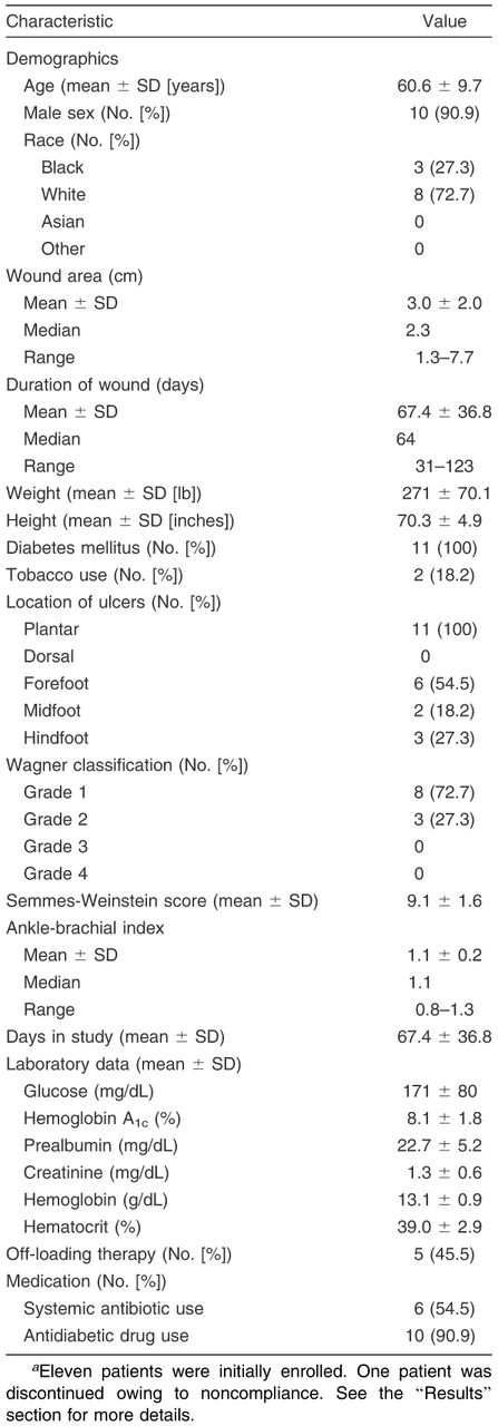
Eleven diabetic patients with chronic neuropathic foot ulcers were enrolled in this pilot study. Table 1 summarizes the baseline characteristics of these patients and their ulcers. The mean age of these patients is 60.6 years, with an average 11-year history of diabetes mellitus. All of the patients’ ulcers were located in the plantar aspect of the foot; 72.7% of the ulcers were Wagner grade 1, and 27.3% were Wagner grade 2. The laboratory data indicated that the study population’s mean ± SD glucose and hemoglobin A1c levels were 171 ± 80 mg/dL and 8.1% ± 1.8%, respectively. Ten patients (90.9%) were taking antidiabetic medications, and only 6 (54.5%) were taking antibiotics (Table 1).
IBWM on Wound Healing
The main objective of this pilot study was to determine the ease of use, safety, and effectiveness of IBWM in an outpatient clinical setting in patients with diabetic foot ulcers. Eleven patients were initially enrolled in this study after screening. However, one patient removed IBWM within 7 days of the first IBWM application, did not follow the offloading requirement, and missed a scheduled protocol visit. This patient’s ulcer was found to be larger and deeper and to have tendon exposure on the patient’s return to the clinic. The investigator then decided to discontinue this patient from the trial owing to noncompliance that lead to a serious adverse event. Therefore, ten patients who received the study intervention were included in the per-protocol population. Only one patient received a reapplication of IBWM, which was performed because the initial IBWM was displaced 3 days after application.
Patients were monitored for clinical signs of infection: purulence, malodor, pain, erythema, tenderness, induration, and swelling. Three patients developed malodor in their wounds in week 4, with the malodor resolving in week 8 without intervention. Pain and swelling were seen in one patient each at screening and week 1, and erythema occurred in 1 patient in week 4. No purulence, tenderness, induration, ulcer infection (cellulitis), osteomyelitis, secondary amputations, or recurrent ulcers were reported during the study.
IBWM on Wound Closure and Area Reduction
Complete wound closure was first seen in week 4 after IBWM treatment (n = 1). By the end of this pilot study, seven ulcers (70%) had achieved complete wound closure; mean ± SD time to closure was 7.43 ± 2.44 weeks (Table 2). Figure 1 demonstrates the change in average wound area over time during the 12-week study. Compared with baseline (week −1), the average wound area at weeks 4, 8, and 12 was changed by 44.2%, 10.1%, and 4.8% of baseline, respectively, which equates to approximately 56%, 90%, and 95% area reduction after treatment with IBWM at weeks 4, 8, and 12, respectively. A follow-up visit was conducted 1 week after initial closure, and complete closure was confirmed.
Discussion
In 2007, the treatment of diabetes and its complications in the United States generated at least $116 billion in direct costs; at least 33% of these costs were linked to the treatment of foot ulcers. Diabetic foot problems impose a major economic burden, and costs increase disproportionately to the severity of the condition. Compared with diabetic patients without foot ulcers, the cost of care for those with foot ulcers is 5.4 times higher in the year after the first ulcer episode and 2.8 times higher in the second year. Costs for treating the highest-grade ulcers are eight times higher than those for treating low-grade ulcers. [22–26] Patients with diabetic foot ulcers require more frequent emergency department visits and are more commonly admitted to the hospital, requiring longer lengths of stay. [19,24]
Medicare claims have shown that expenditures for diabetic patients with chronic neuropathic foot ulcers are three times higher than those for the general population. Lower-extremity ulcers account for 24% of the overall cost for the diabetic population with an ulcer. Length of stay in the hospital and intensive care drive the total cost of treatment. Inpatient stays account for 73% to 77% of total cost. Surgical intervention for and complications of foot ulcers, amputations, and peripheral vascular disease are also significant factors in the cost of limb salvage. [21,26,27] Therefore, reducing hospital procedures and length of hospital stay would result in a decrease in the total cost of diabetic foot care. As such, the application of IBWM in an outpatient setting may offer a lower-cost alternative when treating diabetic foot ulcers.
Table 1.
Baseline Characteristics of the 11 Patients Enrolled in the Studya
The Food and Drug Administration approved IBWM in 2002 for the management of wounds, including partial- and full-thickness wounds, pressure ulcers, venous ulcers, diabetic ulcers, chronic vascular ulcers, surgical wounds (donor sites/grafts, post–Mohs surgery, post–laser surgery, podiatric, and wound dehiscence), trauma wounds (abrasions, lacerations, second-degree burns, and skin tears), and draining wounds. Currently, the application of IBWM on patients with wounds is performed in the operating room. This may prove to be an unnecessary health-care expense and an unnecessary risk factor for patients who are insensate.
The original design of the study included a control arm and an intervention arm (standard of care only vs IBWM with standard of care). The study was stopped once it was shown that the application of IBWM in an outpatient clinical setting was easy, safe, and effective, even without the addition of an epidermal autograft after silicone release. That is, reduction in wound size was observed and no unexpected related adverse events occurred in the study population. When the study was halted, there were only two patients enrolled in the control arm. A control group of two patients was deemed insufficient to use in an analysis to compare the two arms, and they are not included herein. Therefore, this article provides the results from the ten patients in the IBWM arm without a major protocol violation (per-protocol population) who received the study intervention.
Table 2.
Complete Wound Closurea
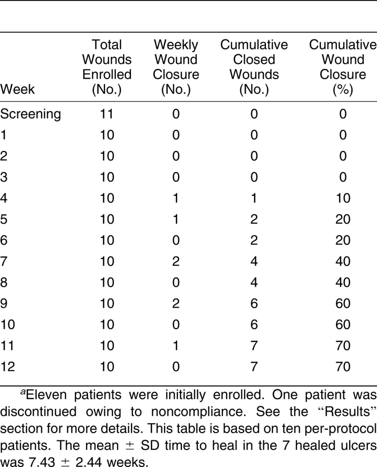
Figure 1.
Percentage of wound reduction during the study. Change in wound size is expressed as a percentage of wound reduction by comparing the wound area at each time point with the baseline wound area. Data are given as mean ± SD. Eleven patients were initially enrolled. One patient was discontinued owing to noncompliance. See the “Results” section for more details. The results shown in this figure are based on the ten per-protocol patients.
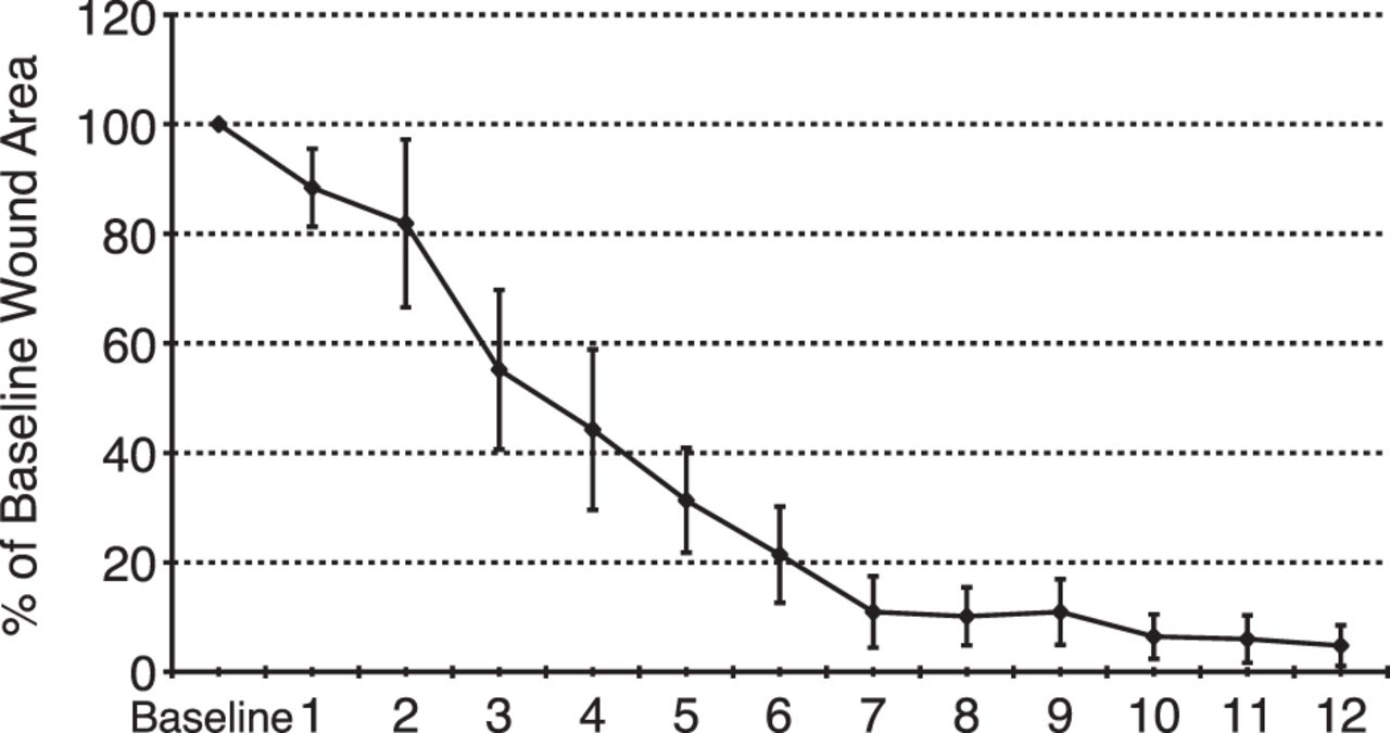
The data on wound characteristics in this pilot study demonstrate that three enrolled patients developed transient malodor in their wounds in week 4 that then resolved in week 8. One patient removed IBWM within 7 days of the first application, did not follow the off-loading requirement, and missed a scheduled protocol visit. This patient’s ulcer was found to be larger and deeper and to have tendon exposure. Thus, the investigator decided to discontinue this patient owing to noncompliance leading to a serious adverse event. There were no additional adverse events or severe adverse events or recurrent ulcers reported within 12 weeks. These data also demonstrate progressive wound healing in these patients over time: there were 56%, 90%, and 95% wound area reductions in weeks 4, 8, and 12, respectively. The study results show that the number of patients with complete wound closure in the active group was seven of ten (70%) (P = .37) during treatment weeks 1 through 12.
Conclusions
The results of this pilot study are consistent with the hypothesis that IBWM is easy to use, safe, and effective when used on diabetic foot ulcers in an outpatient clinical setting. There may, indeed, be cost-saving benefits associated with 1) not going into the operating room, 2) elimination of the additional procedure of epidermal autografting, and 3) reduced office visits.
Acknowledgment
Carl Van Gils, Foot and Ankle Institute, St. George, Utah, and Lee C. Rogers, Amputation Prevention Center at Broadlawns Medical Center, Des Moines, Iowa, for enrolling patients in the study.
Financial Disclosure: This study was funded by Integra Life Sciences Corporation. The sponsor funded the study’s execution, data collection, data analysis, and interpretation. Additionally, the role of the sponsor in this study includes manuscript editing. The Boston University research team is responsible for study execution, data analysis, data interpretation, and manuscript writing. | |
Conflict of Interest: None reported. |
References
- Yannas, IV, JFBurke, MWarpehoski, et al: Prompt, long-term functional replacement of skin. Trans Am Soc Artif Intern Organs27:19,1981.
- Baldwin, C, MPotter, EClayton, et al: Topical negative pressure stimulates endothelial migration and proliferation: a suggested mechanism for improved integration of Integra. Ann Plast Surg62:92,2009.
- Greenwood, J, MAmjadi, BDearman, et al: Real-time demonstration of split skin graft inosculation and Integra dermal matrix neovascularization using confocal laser scanning microscopy. Eplasty9:e33,2009.
- Heimbach, D, ALuterman, JBurke, et al: Artificial dermis for major burns: a multi-center randomized clinical trial. Ann Surg208:313,1988.
- Heimbach, DM, GDWarden, ALuterman, et al: Multicenter postapproval clinical trial of Integra dermal regeneration template for burn treatment. J Burn Care Rehabil24:42,2003.
- Yeong, EK, HFHuang, YBChen, et al: The use of artificial dermis for reconstruction of full thickness scalp burn involving the calvaria. Burns32:375,2006.
- Herlin, C, DLouhaem, MBigorre, et al: Use of Integra in a paediatric upper extremity degloving injury. J Hand Surg Eur32:179,2007.
- Komorowska-Timek, E, AGabriel, DCBennett, et al: Artificial dermis as an alternative for coverage of complex scalp defects following excision of malignant tumors. Plast Reconstr Surg115:1010,2005.
- GonyonJr, DL and MRZenn: Simple approach to the radiated scalp wound using INTEGRA skin substitute. Ann Plast Surg50:315,2003.
- Barnett, TM and JSShilt: Use of vacuum-assisted closure and a dermal regeneration template as an alternative to flap reconstruction in pediatric grade IIIB open lower-extremity injuries. Am J Orthop (Belle Mead NJ)38:301,2009.
- Violas, P, AAbid, PDarodes, et al: Integra artificial skin in the management of severe tissue defects, including bone exposure, in injured children. J Pediatr Orthop B14:381,2005.
- Muangman, P, LHEngrav, DMHeimbach, et al: Complex wound management utilizing an artificial dermal matrix. Ann Plast Surg57:199,2006.
- Jeng, JC, PEFidler, JCSokolich, et al: Seven years’ experience with Integra as a reconstructive tool. J Burn Care Res28:120,2007.
- Orgill, DP, FHStrausII, and RCLee: The use of collagen-GAG membranes in reconstructive surgery. Ann N Y Acad Sci888:233,1999.
- Campitiello, E, ADella Corte, AFattopace, et al: The use of artificial dermis in the treatment of chronic and acute wounds: regeneration of dermis and wound healing. Acta Biomed76( suppl 1:):69,2005.
- Clayman, MA, SMClayman, and DWMozingo: The use of collagen-glycosaminoglycan copolymer (Integra) for the repair of hypertrophic scars and keloids. J Burn Care Res27:404,2006.
- Ahmed, S, SSHussein, BPhilp, et al: Use of biologic dressing as a temporary wound dressing in reconstruction of a significant forehead Mohs defect. Dermatol Surg32:765,2006.
- American Diabetes Association :Economic costs of diabetes in the U.S. in 2007. Diabetes Care31:596,2008.
- Matricali, GA, GDereymaeker, EMuls, et al: Economic aspects of diabetic foot care in a multidisciplinary setting: a review. Diabetes Metab Res Rev23:339,2007.
- Gibbons, GW, EJMarcaccioJr, AMBurgess, et al: Improved quality of diabetic foot care, 1984 vs 1990: reduced length of stay and costs, insufficient reimbursement. Arch Surg128:576,1993.
- Harrington, C, MJZagari, JCorea, et al: A cost analysis of diabetic lower-extremity ulcers. Diabetes Care23:1333,2000.
- Driver, VR, MFabbi, LALavery, et al: The costs of diabetic foot: the economic case for the limb salvage team. JAPMA100:335,2010.
- Driver, VR and MYao: Discussion: the economics of limb salvage in diabetes. Plast Reconstr Surg127( suppl 1:):296S,2011.
- Davis, WA, PENorman, DGBruce, et al: Predictors, consequences and costs of diabetes-related lower extremity amputation complicating type 2 diabetes: the Fremantle Diabetes Study. Diabetologia49:2634,2006.
- Ortegon, MM, WKRedekop, and LWNiessen: Cost-effectiveness of prevention and treatment of the diabetic foot: a Markov analysis. Diabetes Care27:901,2004.
- Stockl, K, AVanderplas, ETafesse, et al: Costs of lower-extremity ulcers among patients with diabetes. Diabetes Care27:2129,2004.
- Carls, GS, TBGibson, VRDriver, et al: The economic value of specialized lower-extremity medical care by podiatric physicians in the treatment of diabetic foot ulcers. JAPMA101:93,2011.
© 2013 American Podiatric Medical Association