


Abstract
Background: Freiberg’s infraction is an osteochondrosis that is characterized by osteonecrosis of the metatarsal head, with pain and tenderness around the metatarso-phalangeal joint. We sought to evaluate the outcome of joint debridement and metatarsal remodeling for the treatment of advanced-stage Freiberg’s infraction. Methods: Between March 1, 2006, and April 30, 2011, 14 patients (eight females and six males) with symptomatic unilateral Freiberg’s disease refractory to conservative treatment (Smillie stages IV and V) underwent joint debridement with metatarsal head remodeling by two surgeons. To evaluate functional outcome, American Orthopaedic Foot and Ankle Society and 36-Item Short Form Health Survey forms were completed by the patients preoperatively and postoperatively at months 3, 6, and 12. Active-assisted range-of-motion exercise was allowed after 4 weeks of short-leg walking cast wear, and weightbearing on the forefoot was allowed as tolerated. Results: Mean patient age was 27.0 years (range, 16–53 years), and mean follow-up was 40.2 months (range, 14–54 months). Mean 6 SD American Orthopaedic Foot and Ankle Society and 36-Item Short Form Health Survey scores were 46.8 6 8.95 and 28.9 6 4.3 preoperatively and 76.2 6 9.5 and 45.6 6 7.7 1 year after surgery, respectively. There was a significant increase in both scores (P ≤ .001). Conclusions: In advanced-stage Freiberg’s infraction of the second metatarsal, joint debridement and metatarsal head remodeling is a safe and simple therapeutic option, and it provides better quality of life for patients.
Freiberg’s infraction, osteochondrosis of the metatarsal heads, was first described by Albert Freiberg in 1914.[1] This rare condition frequently occurs in the second metatarsal head in adolescents. Activity-related pain around the metatarsophalangeal joint, swelling, and limitation of motion of the affected joint are the main complaints of the patients. Diagnosis is confirmed by radiographic evaluation. Smillie [2] divided this disease into five stages according to radiographic findings.
In symptomatic patients, nonsurgical treatment should be attempted first. There are numerous surgical interventions described for symptomatic Freiberg’s infraction for those who do not respond to conservative treatment.[3,4] In the present study, we analyzed the outcomes of joint debridement with metatarsal head remodeling for the treatment of stages IV and V Freiberg’s infraction. The purpose of this study was to evaluate the results of joint debridement with metatarsal head remod-eling in the treatment of Freiberg’s disease and to show the effectiveness of this procedure.
Materials and Methods
Between March 1, 2006, and April 30, 2011, 19 patients who had joint debridement with metatarsal head remodeling at Bezmialem Vakif University, were evaluated retrospectively. All of the patients were operated on by two surgeons (M.E. and I.T.). Patients with Smillie stages IV and V who had refractory symptoms even after conservative treatment were included. Of the 19 patients, two with previous surgical intervention for hallux valgus were excluded, and three patients were lost to follow-up; the remaining 14 patients participated in the study. None of the patients underwent previous surgery for this problem. Nonsurgical treatment consisting of activity modification and the use of metatarsal pads in the shoes was attempted in all of the patients previously for a minimum of 6 months. Joint debridement with metatarsal head remodeling was performed with a dorsal longitudinal incision in all of the patients. Both surgeons used the same technique and the same postoperative rehabilitation program. Detailed information on the surgical interventions and the postoperative rehabilitation program were provided, and an informed consent form including the operative technique was signed by all of the patients; institutional review board approval was obtained from Bezmialem Vakif University. Active-assisted range-of-motion exercise was allowed after 4 weeks of short-leg walking cast wear, and weightbearing on the forefoot was allowed as tolerated. American Orthopaedic Foot and Ankle Society[5] and 36-Item Short Form Health Survey forms were completed by the patients preoperatively and postoperatively, and the results were compared.
Surgical Technique
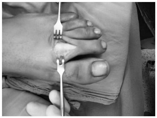
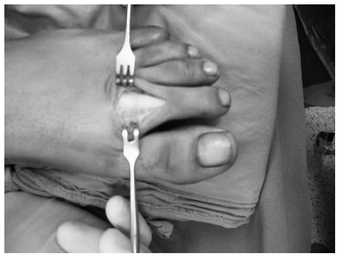
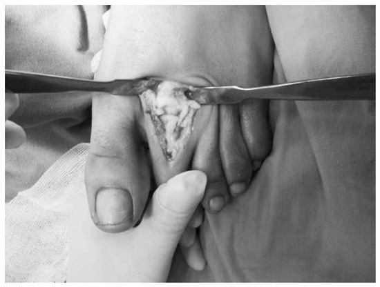
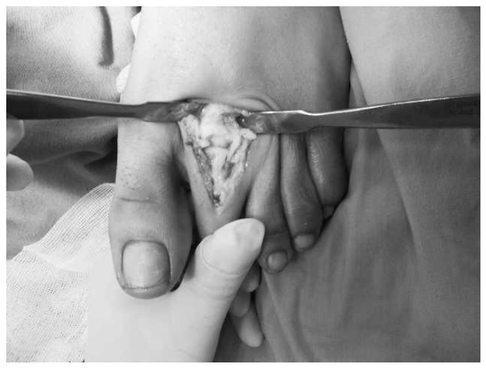
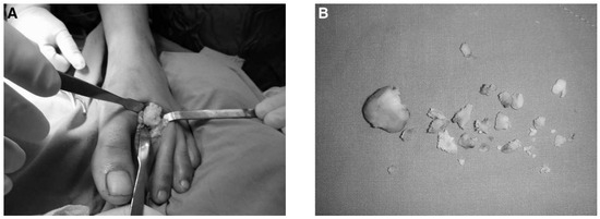
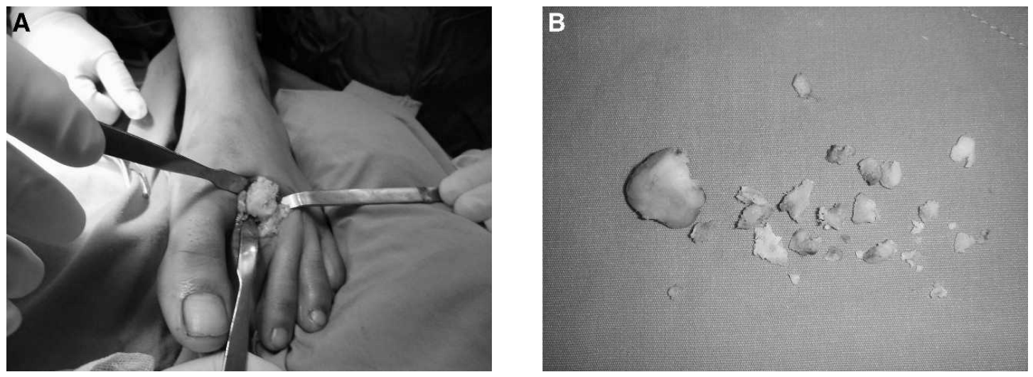
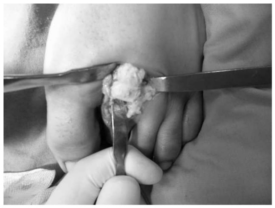
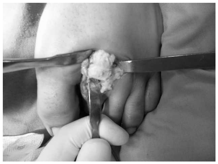
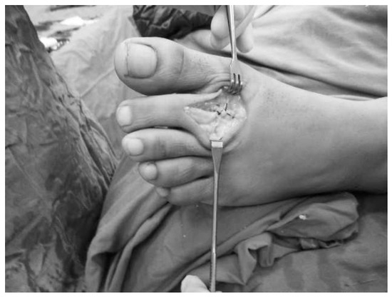
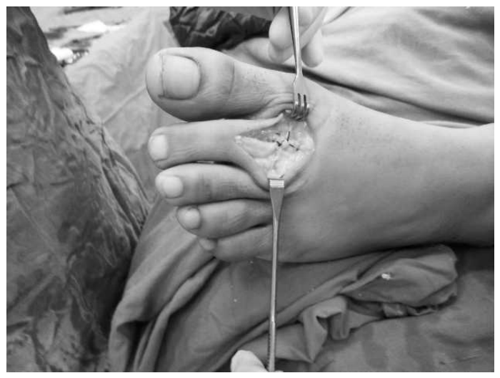
The patient is placed supine on a standard operating table with a well-padded tourniquet around the thigh. Through a dorsal longitudinal incision, the entire extensor mechanism is exposed over the metatarsophalangeal joint (Figure 1). After identifying the extensor digitorum brevis and extensor digitorum longus muscles, the extensor hood is incised just lateral to the extensor digitorum longus muscle and retracted medially. A longitudinal capsulotomy is performed with sharp dissection to expose the metatarsal head (Figure 2). All of the osteochondral fragments are debrided, and the toe is manually distracted and overflexed to see the entire metatar-sal head. All loose fragments, degenerated articular cartilage, and inflamed synovium are removed, and the head is remodeled and contoured with a rongeur (Figure 3). It is important not to skip any osteophytes in the plantar aspect of the metatarsophalangeal joint (Figure 4). Forceful irrigation is necessary to flush any remaining fragments while flexing and extending the toe. If there is extensor pressure on the joint, it is helpful to perform an approximately 1-cm Z-plasty lengthening of the extensor digitorum longus muscle, and the extensor pollicis brevis muscle can be tenotomized. After securing the hemostasis by direct pressure, the capsule is closed with fine absorbable sutures (Figure 5), and a dressing is applied to keep the joint in a reduced position because the metatarsal head is usually depressed dorsally and centrally after debridement.
Figure 1.
Dorsal longitudinal incision over the metatarsophalangeal joint.
Figure 2.
Capsulotomy to expose the metatarsal head.
Figure 3.
Operative view of the joint (A) and removal of the osteochondral fragments (B).
Figure 4.
Plantar aspect of the metatarsal head.
Figure 5.
Closure of the capsule with fine absorbable sutures.
Results
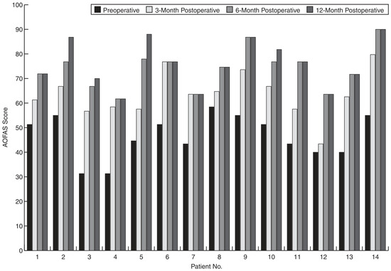
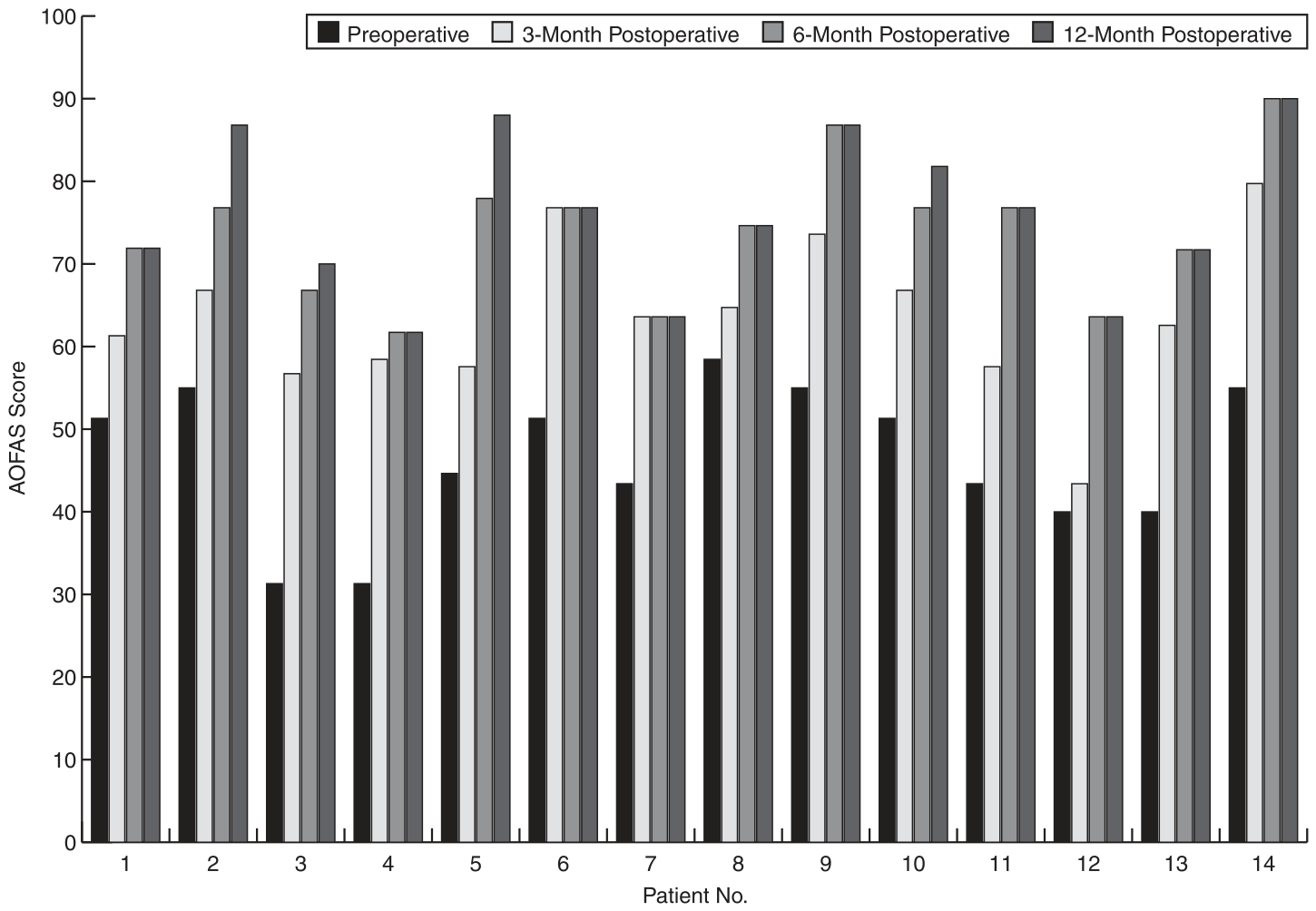
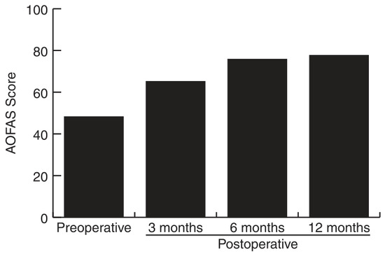
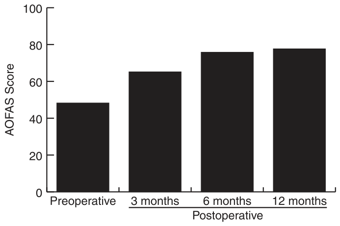
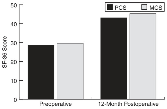
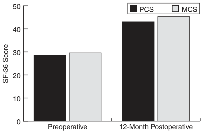
Fourteen metatarsophalangeal joints of 14 patients (eight females and six males) were all unilateral, and eight of the cases were right sided. The mean patient age at the time of the operation was 27.0 years (range, 16–53 years), and mean follow-up was 40.2 months (range, 14–54 months). Functional outcomes were evaluated by American Orthopaedic Foot and Ankle Society scores at 3-, 6-, and 12-month follow-up (Figure 6 and Figure 7) and by 36-Item Short Form Health Survey scores at 1-year follow-up (Figure 8). Preoperative American Orthopaedic Foot and Ankle Society scores were compared with scores 6 and 12 months postoperatively, and scores 3 and 12 months postoperatively were compared with the post hoc Dunn test. Mean 6 SD American Orthopaedic Foot and Ankle Society and 36-Item Short Form Health Survey scores were 46.8 ± 8.95 and 28.9 ± 4.3 preoperatively and 76.2 ± 9.5 and 45.6 ± 7.7 1 year after surgery, respectively. A significant increase was noted (Friedman v2 = 3.29, P < .01). Preoperatively, the physical and mental component summary values for the 36-Item Short Form Health Survey were noted and compared with the results 1 year after surgery (Figure 8). There was also a significant difference in 36-Item Short Form Health Survey values (Wilcoxon for the physical component summary: z = 3.296; P = .001 and for the mental component summary: z = 3.297; P = .001). One year after surgery, the range of motion of the second metatarsophalangeal joint increased by a mean of 27° (range, 6°–58°).
Figure 6.
Changes in American Orthopaedic Foot and Ankle Society (AOFAS) scores of patients preoperatively and 3, 6, and 12 months postoperatively.
Figure 7.
Mean American Orthopaedic Foot and Ankle Society (AOFAS) scores preoperatively and 3, 6, and 12 months postoperatively.
Figure 8.
Changes in 36-Item Short Form Health Survey (SF-36) scores of patients preoperatively and 12 months postoperatively. MSC indicates mental component summary; PCS, physical component summary.
Discussion
Freiberg’s infraction is an osteochondrosis that is characterized by osteonecrosis of the metatarsal head with pain at rest and on exercise and tenderness around the metatarsophalangeal joint. Moreover, swelling and stiffness of the metatarsophalangeal joint exacerbated by weightbearing activities are common findings. Freiberg’s infraction was first described by Freiberg[1] in 1914 and is also referred to as Kohler’s second disease in European countries.[6] The second metatarsal is the most frequently affected, and predominantly the dorsal aspect is involved. Freiberg’s infraction has also been reported in other metatarsals, but rarely in the first metatarsal. Although the condition is reported in males, it is predominantly seen in female teenagers.[7] In the radiographic examination, flattening of the metatarsal head with increased sclerosis and fragmentation is frequently detected. Hoskinson [7] indicated that early lesions of this disease can be most reliably demonstrated via 458 oblique views of the forefoot. A combination of clinical suspicion and plain films confirm the diagnosis.
The etiology of Freiberg’s disease is not clear yet. Acute or repetitive trauma, local plantar pressure, metatarsal length, and second-ray mobility may play a role in this condition. Crock [8] reported that radial arteries penetrate in both sides of the metatarsal head and unite to form a central arterial network that gives off branches to the subchondral area. Because the subchondral bone is enclosed by a convex hyaline cartilage, it is vulnerable just like the femoral head and the capitellum. The exact cause remains uncertain, and many cases are self-limited with revascularization of the metatarsal head. However, some of these cases result in significant deformity and secondary degenerative changes at the metatarsophalangeal joint.[9]
Because it is the longest and the least mobile, the second metatarsal is thought to be prone to repetitive traumatic stress, which may lead to excessive local plantar pressure on the metatarsal head. This repetitive traumatic stress may damage the vascularization of the subchondral bone and collapse cancellous bone and overlying cartilage. On the other hand, by using dynamic pedobarography, Belts et al [10] demonstrated that only 15% of their patients had higher pressures on the affected metatarsal head. Metatarsal length may also play a role in the disease process. Stanley et al [11] noted that the second metatarsal was the longest in 94% of 33 patients with unilateral Freiberg’s disease. However, there was no significant difference between the lengths of the second metatarsals of the affected and unaffected sides in the present study (P <. .005).
Smillie [2,12] categorized Freiberg’s infraction into five different stages according to radiographic views. Stage I is a subchondral fracture that can be detected on magnetic resonance imaging or bone scanning. In stage II, dorsal collapse of the articular surface can be seen on plain radiographs. Stage III is progressive collapse of the metatarsal head. In this stage, the plantar articular portion is intact. Early arthritic changes and joint space narrowing and collapse of the entire metatarsal head are specific findings of stage IV. Stage V reveals severe arthritic changes with joint space obliteration.
Nonsurgical treatment should be attempted first with activity modification and the use of metatarsal pads in the shoes.[3,13] In some acute cases, a short-leg cast extended to the toes for 4 to 6 weeks followed by a stiff-sole shoe with a metatarsal bar for several months may be beneficial. Surgical intervention is rarely indicated and should be reserved for patients who have symptoms that interfere with daily or sports activities in whom nonsurgical treatment fails.[3,4] Numerous surgical treatment options have been described to try to modify the disease process or as a salvage procedure for those with degenerative changes in the metatarsophalangeal joint. Surgical options include isolated joint debridement for mild-to-moderate symptomatic cases, metatarsal head resection, dorsal closing base wedge osteotomy of the metatarsal head for lesions localized to the dorsal aspect of the metatarsal head, elevation of the depressed fragments of the metatarsal head with bone grafting of the defect, resection of the base of the proximal phalanx with syndactylization of the second and third toes, DuVries arthroplasty of partial head resection for Smillie stage IV and V involvement, joint debridement with metatarsal head remodeling, arthroscopic debridement and drilling, prosthetic joint replacement, and osteochondral plug transplantation.[4,9]
There is still too much to be understood about the disease process, and current knowledge is not adequate to inform the long-term outcome of patients with Freiberg’s disease, regardless of the therapeutic option. Joint debridement with metatarsal head remodeling may be one of the most simple and safe methods in the treatment of advanced-stage Freiberg’s disease that is resistant to conservative treatment, and extensor tenoplasty may play a part, if necessary, in the therapeutic approach.
Funding
None reported.
Acknowledgments
Omer Uysal, PhD, head of the Medical Informatics and Biostatistics Department, Bezmialem Vakif University, Istanbul, Turkey, for his valuable contribution to the statistical analysis section of this study.
Conflicts of Interest
None reported.
References
- FREIBERG AH: Infraction of the second metatarsal bone. Surg Gynecol Obstet 19, 191, 1914.
- SMILLIE, I.S. Freiberg’s infraction (Kohler’s second dis-ease). J Bone Joint Surg Br 1957, 39, 580. [Google Scholar]
- Giannestras NJ, 2nd Ed ed; Lea & Febiger: Philadelphia, 1973.
- HAYASHI, K.; OCHI, M.; UCHIO, Y. ET AL: A new surgical technique for treating bilateral Freiberg disease. Ar-throscopy 2002, 18, 660. [Google Scholar]
- SOOHOO, N.F.; SAMIMI, D.; VYAS, R.; et al. Evaluation of the validity of the Foot Function Index. Paper presented at: 21st Annual Summer Meeting of the American Ortho-paedic Foot and Ankle Society (AOFAS), Boston, MA, July 15–17, 2005. [Google Scholar]
- KOHLER, A. Grenzen des Normalen und Anfange des Pathologischen,’’ in Rontgenbilde, p 77; LucasGrafe and Sillem: Hamburg, 1924. [Google Scholar]
- HOSKINSON, J. Freiberg’s disease: a review of the long term results. Proc R Soc Med 1974, 67, 10. [Google Scholar] [CrossRef] [Scilit] [PubMed]
- CROCK HV: The Blood Supply of the Lower Limb Bones in Man; Livingstone Ltd, Edinburgh and London, 1967.
- CANALE, S.T. Osteochondrosis or Epiphysitis and Other Miscellaneous Affections,’’ in Campbell’s Operative Orthopaedics, 11th Ed; Canale, S.T., Beaty, J.H., Eds.; Mosby Elsevier: Philadelphia, 2007; p. 1252. [Google Scholar]
- BELTS, R.P.; STANLEY, D. SMITH TWD: Foot pressure studies in Freiberg’s disease. Foot 1991, 1, 21. [Google Scholar]
- STANLEY, D.; BELTS, R.P.; ROWLEY, D.I. ET AL: Assessment of the etiological factors in the development of Freiberg’s disease. J Foot Surg 1990, 29, 444. [Google Scholar] [PubMed]
- SMILLIE, I.S. Treatment of Freiberg’s infraction. Proc R Soc Lond 1967, 60, 29. [Google Scholar] [CrossRef] [Scilit] [PubMed]
- TROTT AW: ‘‘Developmental Disorders,’’ in Disorders of the Foot; Jahss, M.H., Ed.; WB Saunders: Philadel-phia, 1982. [Google Scholar]
© 2023 American Podiatric Medical Association