


Simple Summary
Immunotherapy has become part of the main treatment for non-small cell lung cancer. However, it pertains with immunotherapy-related inflammation of the lungs, an uncommon but potentially life-threatening side effect. Identification of patients at risk, early detection, and treatment of this inflammation is crucial in improving symptoms and reducing death rates. In this article, we aim to find the real-life incidence rate of this side effect and review studies with risk factors and early detection methods to help in early recognition and treatment decision-making related to this uncommon but serious immunotherapy-related lung inflammation.
Abstract
It is well-known that immune checkpoint inhibitors (ICIs) can lead to uncommon but potentially life-threatening immune-related adverse events (irAEs) such as checkpoint-inhibitor pneumonitis (CIP). Early recognition, identification, and treatment are crucial with regard to decreasing toxicity from ICIs. However, there is a discrepancy in the incidence rates between the clinical trials and postmarketing studies. Several postmarketing studies have developed early detection methods and identified new risk factors in developing CIP. Thus, in this narrative review, we aim to review these incidence rates, early detection methods, and clinical risk factors for CIP in NSCLC patients, which could help to improve CIP diagnosis and management for enhanced NSCLC care. Major clinical trials and postmarketing studies of CIP incidence, early detection methods, and risk factors in NSCLC were reviewed. A wide array of potential risk factors has been implicated in the development of CIP in NSCLC, such as having preexisting interstitial lung disease, receiving PD-1 inhibitors, receiving combination ICI therapy instead of ICI monotherapy, lower pretreatment hemoglobin and albumin levels, increased baseline plasma IL-8 levels, and impaired baseline pulmonary function.
1. Introduction
Lung cancer is the most commonly diagnosed cancer worldwide and remains the leading cause of cancer mortality, making up 12.4% of all new cases and 18.7% of cancer deaths [1]. Non–small cell lung cancer (NSCLC) accounts for about 85% of all lung cancers [2]. The introduction of immune checkpoint inhibitors (ICIs) has significantly improved survival outcomes, including OS, progression-free survival (PFS), and event-free survival (EFS) in resectable, locally advanced, and metastatic NSCLC [3,4,5,6,7].
ICIs are monoclonal antibodies that enhance anti-tumor immune responses by blocking inhibitory immune checkpoint receptors on T-lymphocytes, thereby releasing the brakes on the immune system and revolutionizing lung cancer therapy [8]. Approved ICIs for NSCLC include inhibitors targeting programmed cell death-1 (PD-1, e.g., pembrolizumab), cytotoxic T-lymphocyte antigen-4 (CTLA-4, e.g., ipilimumab), and programmed death-ligand 1 (PD-L1, e.g., atezolizumab). However, by exciting immune activity, these agents can also disrupt normal immune tolerance mechanisms, leading to a spectrum of immune-related adverse events (irAEs) [9,10], including checkpoint-inhibitor pneumonitis (CIP) [11,12].
CIP is a rare but potentially life-threatening irAE characterized by immune-mediated focal or diffuse inflammation of the lung parenchyma following ICI therapy [13,14]. Clinically, CIP can present asymptomatically or with dyspnea and other respiratory symptoms (e.g., cough), accompanied by new inflammatory infiltrates on chest imaging after ICI initiation, in the absence of infection or tumor progression [15,16].
2. Incidence
According to clinical trial data, the incidence of CIP in NSCLC ranges from about 3% to 6.3%, with a reported mortality rate under 1% [17]. In real-world settings, however, higher pneumonitis rates have been observed, with approximately 5% to 19% of ICI-treated NSCLC patients developing CIP outside of trials [18,19] (Table 1). This discrepancy may be explained by broader patient inclusion in routine practice (e.g., patients with pre-existing lung conditions or poorer performance status who would be excluded from trials) [20]. Greater awareness and vigilance in recognizing CIP, as well as formal adjudication of pneumonitis cases in registries, have also likely contributed to higher reported incidences in recent years [20,21]. See Table 2 for the incidence of ICI-induced pneumonitis in NSCLC clinical trial data.
Table 1.
Incidence of pneumonitis in real-world practice.
Table 2.
Incidence of ICI-induced pneumonitis in NSCLC clinical trials.
3. Early Detection Methods
Among all irAEs in NSCLC, CIP is particularly concerning due to its potential lethality. Development of moderate-to-severe CIP often requires prompt high-dose corticosteroid therapy (e.g., methylprednisolone) and sometimes additional immunosuppressive treatments (such as tocilizumab, infliximab, or cyclophosphamide) to control inflammation [11,43,44]. Severe CIP typically mandates permanent discontinuation of ICI therapy, and can still lead to respiratory failure or death despite treatment [11,14]. Indeed, although relatively uncommon, CIP is one of the leading causes of treatment-related fatality in NSCLC patients receiving ICIs [12,22].
Early recognition of CIP is critical because timely intervention, primarily with corticosteroids, can prevent progression to respiratory failure. Oncologists and pulmonologists should maintain a high index of suspicion for pneumonitis in any ICI-treated patient who develops new respiratory symptoms. Baseline and periodic pulmonary function tests (PFTs) and imaging are useful tools for monitoring patients at risk.
3.1. PFTs
Several studies suggest that patients who develop CIP tend to have lower pre-treatment lung function. For instance, a retrospective analysis found that NSCLC patients who developed pneumonitis had a lower baseline forced expiratory volume in 1 s (FEV1) % predicted compared to those who did not, although baseline forced vital capacity (FVC) was similar between the groups, despite a comparable prevalence of COPD [45]. Similarly, a prospective multicenter study by Suzuki et al. [46]. reported that impaired baseline spirometry (reduced FVC and FEV1) and the presence of baseline dyspnea symptoms (as measured by a standardized dyspnea scale) were associated with a higher incidence of immune-related pneumonitis in patients receiving ICIs [46].
3.2. Radiologic Evaluation
Radiologic evaluation is the cornerstone of early detection of CIP. Routine CT scans in patients receiving immunotherapy can incidentally detect asymptomatic pneumonitis changes, such as ground-glass opacities or organizing pneumonia patterns, before clinical symptoms worsen. Prompt recognition of these imaging patterns, along with their correlation to clinical findings, can facilitate early management. In practice, when CIP is suspected, it is essential to rule out infectious etiology and tumor progression (often via bronchoscopy with lavage and, in some cases, lung biopsy) to confirm the diagnosis [15,16].
Once CIP is diagnosed, management follows established guidelines based on severity. Grade 1 (mild, asymptomatic) pneumonitis may be managed with close monitoring and temporarily suspending ICI treatment, whereas Grade 2 or higher (moderate-to-severe) pneumonitis warrants immediate immunosuppressive therapy, such as high-dose corticosteroids, and discontinuation of immunotherapy. If there is no improvement within 48–72 h of initiating steroids or in steroid-refractory cases, additional immunosuppressants such as infliximab (anti-TNF), mycophenolate mofetil, or intravenous immunoglobulin (IVIG) may be considered per guideline recommendations [11,44].
3.3. Early Consultation and Multidisciplinary Discussion
Early pulmonology consultation and a multidisciplinary approach, including oncology, pulmonology, radiology, and infectious disease specialists, is recommended to optimize CIP diagnosis and management. Rapid intervention is crucial, as CIP, although relatively rare, can progress quickly and has been identified as a major cause of ICI-related deaths [12].
4. Clinical Risk Factors for Checkpoint-Inhibitor Pneumonitis
4.1. Pre-Existing Interstitial Lung Disease
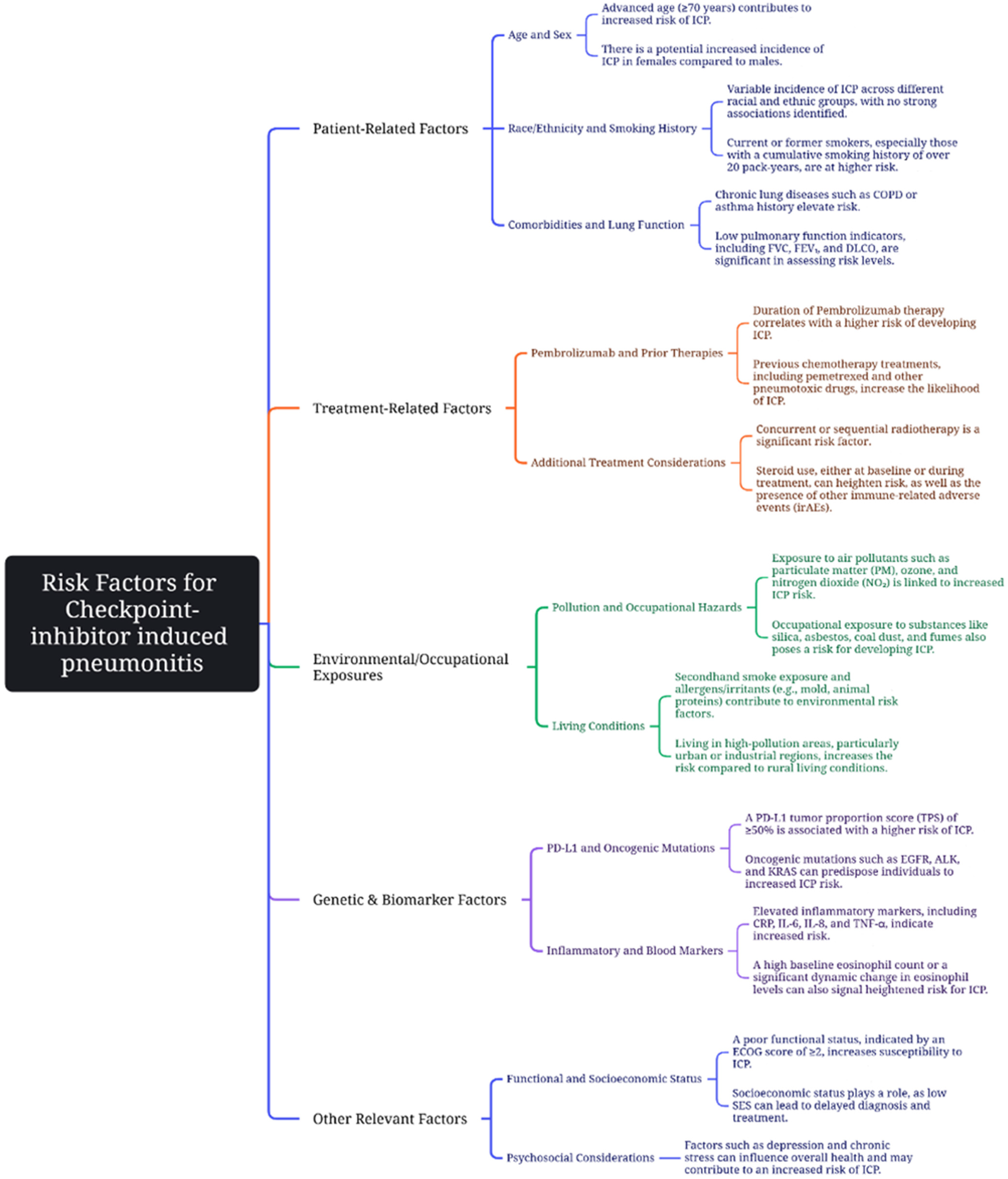
Identifying risk factors predisposing patients to checkpoint-inhibitor pneumonitis (CIP) is of great importance, as it may allow for preventive strategies or closer monitoring in high-risk individuals. Some of these risk factors can be found outlined in Figure 1. One consistently reported risk factor for ICI-related pneumonitis is pre-existing interstitial lung disease (ILD). ILD has been reported to exist in approximately 15% of lung cancer patients at the time of initial diagnosis and is associated with a poor prognosis [28,47,48].
Patients with underlying ILD, such as idiopathic pulmonary fibrosis, are far more likely to develop CIP when treated with ICIs. In a retrospective study of NSCLC patients receiving combination chemo-immunotherapy, those with pre-existing ILD had an odds ratio (OR) of approximately 19 for developing treatment-related pneumonitis compared to patients without ILD [21]. The same study also found that patients receiving pemetrexed-based chemotherapy in combination with ICIs had a higher risk of pneumonitis, suggesting that certain cytotoxic agents (e.g., pemetrexed) might potentiate lung inflammation [20]. In a retrospective study of NSCLC conducted by Kanai et al. [49], the incidences of nivolumab-related pneumonitis and severe nivolumab-related pneumonitis were significantly higher in patients with pre-existing ILD, although the treatment outcomes for pneumonitis were comparable between patients with and without pre-existing ILD [49]. In a retrospective cohort study by Sawa et al. [26], the risk of developing pneumonitis with ICIs in NSCLC patients with pre-existing ILD was similar to that observed with conventional chemotherapy [26].
Altan et al. [24] demonstrated that ILD in non-smokers and a longer duration of smoking, along with baseline symptoms of shortness of breath or cough in smokers, are associated with a higher risk of pneumonitis. However, the ability to evaluate the etiology of baseline symptoms was limited by the paucity of pre-treatment pulmonary function data [24].
4.2. History of Lung Irradiation
An earlier analysis by Barrón et al. [10] reported that a history of thoracic radiotherapy was associated with an increased likelihood of CIP and worse overall survival with subsequent immunotherapy, indicating that prior lung irradiation is another important risk factor to consider [10].
4.3. Chronic Obstructive Pulmonary Disease (COPD)
The influence of smoking history and chronic lung diseases such as COPD on CIP risk is less clear. Although COPD is common in lung cancer patients (in newly diagnosed lung cancer patients, it is estimated to be about 50%) and contributes to baseline pulmonary impairment, one analysis by Zeng et al. [28] showed that coexisting COPD (as defined by spirometry) did not significantly increase the risk of pneumonitis in NSCLC patients treated with ICIs. In contrast, another retrospective report observed that approximately 70% of patients who developed CIP had a history of COPD; however, this study relied on physician-diagnosed COPD rather than pulmonary function criteria, potentially confounding the results [28]. Overall, factors such as smoking status, older age, and common comorbidities (e.g., diabetes) have not consistently shown significant associations with CIP in most studies [22,28]. For example, a large single-institution study by Liu et al. [23] found that age, tumor histology, smoking history, pre-existing lung disease, performance status, concurrent chemotherapy, and baseline blood counts (neutrophil-to-lymphocyte ratio or monocyte counts) were not significantly different between patients who developed CIP and those who did not, suggesting that these factors were not strong predictors in that cohort [23].
In the study by Chao et al., the presence of COPD was associated with a higher incidence of CIP, and COPD patients experienced a lower grade of CIP, mainly Grade 2 [22].
4.4. PD-L1 Expression
In the study by Chao et al., PD-L1 expression of ≥50% was associated with a higher incidence of CIP [22].
In meta-analyses of controlled clinical trials conducted by Kong et al. [15], it was shown that ICI use was associated with a higher risk of both all-grade (1–5) and high-grade (3–5) pneumonia compared to chemotherapy, regardless of histology, treatment regimen, PD-L1 expression level, negative EGFR/ALK expression, or previous treatment history. Subgroup analyses revealed that the squamous group, the ICI versus combination chemotherapy group, the PD-L1 >50% group, and the previously untreated group had a higher risk of developing both all-grade and Grade 3–5 CIP [15]. However, in the study by Cho et al. [50], the proportion of tumors with positive PD-L1 expression did not differ between patients with and without ICI-related pneumonitis (i.e., CIP). In this study, extrathoracic metastases were less frequent (31.8% versus 58.6%) in patients with pneumonitis, whereas previous ILD (OR = 6.03, 95% CI: 1.19–30.45) and the absence of extrathoracic metastases (OR = 2.94, 95% CI: 1.08–7.69) were features independently associated with an increased risk of ICI-related pneumonitis (i.e., CIP) [50,51].
4.5. PD-1 Inhibitors
A meta-analysis of 19 trials conducted by Khunger M. et al. [52] demonstrated that PD-1 inhibitors were associated with a statistically significantly higher incidence of any-grade CIP compared to PD-L1 inhibitors (3.6% vs. 1.3%, p = 0.001). Furthermore, patients receiving PD-1 inhibitors experienced a higher incidence of Grade 3 or 4 pneumonitis (1.1% vs. 0.4%; p = 0.02) [52]. In a study by Fukihara et al. [25], the administration of pembrolizumab, as compared with nivolumab, was identified as an independent predictor of immune-related pneumonitis (i.e., CIP) [25].
4.6. Combination ICI Therapy
The incidence rate of CIP was higher in individuals receiving combination immune checkpoint inhibitor (ICI) therapy than in those receiving ICI monotherapy, regardless of tumor stage. Moreover, an inverse relationship was observed between cancer stage and CIP incidence in patients receiving combination ICI therapy; that is, patients with lower-stage cancer exhibited a higher rate of CIP. Tumor histologic type also influenced CIP development, with patients exhibiting the nonsquamous histologic type having a lower risk. Although female gender was associated with a higher incidence of CIP (incidence rate ratio [IRR] = 1.34; 95% CI: 0.67–2.66), this finding did not reach statistical significance [20].
4.7. Biomarkers
Hematological parameters and inflammatory markers have been identified as potential predictors of treatment response and prognosis in various cancers, including non-small-cell lung cancer (NSCLC), and they offer the advantages of being readily available, economical, and convenient [53]. Liu et al. [23] showed that lower pre-treatment hemoglobin and albumin levels were independent predictors of CIP [23]. Likewise, another study found that low serum albumin was associated with an increased risk of CIP and was an independent predictor of immune-related pneumonitis [25]. A study by Chao et al. [22] revealed that elevated baseline plasma IL-8 levels are associated with a decreased incidence of CIP, and previous ICI clinical trials demonstrated that greater pre-treatment IL-8 expression likely reflects an immunosuppressive, myeloid-enriched tumor microenvironment with limited adaptive T cell responses [22].
Figure 1.
Risk factors contributing to ICP in immunotherapy. These risk factors include: advanced age [23,24,54]; female sex [20,22]; smoking history [22,23,24]; chronic lung disease [22,23,24,54]; low pulmonary function indicators [29]; duration of pembrolizumab therapy [20,21,25]; previous chemotherapy treatments [20,21,24]; concurrent or sequential chemoradiotherapy [10,23,24]; exposure to air pollutants [10]; elevated PD-L1 tumor proportion score [21,22]; elevated inflammatory markers [22]; eosinophil levels [29]; poor functional status [10,55]. Incidence of ICP has no strong associations with race or ethnicity [20] (This figure was created using Microsoft Visio Professional 2019).
4.8. Baseline Pulmonary Function
Several analyses indicate that baseline pulmonary function may serve as a potential indicator of risk. Impaired lung function, as characterized by low FEV1 and reduced diffusion capacity prior to treatment, might predispose patients to CIP. Reuss et al. [45] reported that patients who developed CIP had a lower median FEV1% predicted at baseline than those without CIP, while baseline FVC% was similar [45]. In a prospective study by Suzuki et al. [46], poor baseline spirometry (reduced FVC and FEV1) and the presence of chronic dyspnea symptoms were significantly associated with the development of immune-related interstitial lung disease (i.e., CIP) during immunotherapy [46]. These findings suggest that underlying subclinical lung insufficiency or damage, such as from smoking or emphysema, may lower the threshold for triggering pneumonitis under ICI therapy [46].
Age ≥65 years and smoking history were associated with an increased risk of developing pneumonitis [26]. In an NSCLC-only series, the proportion of patients aged >70 years or those with prior ILD was higher in patients with ICI-related pneumonitis (i.e., CIP) than in those without (54.5% versus 30.3% and 18.2% versus 2.8%, respectively) [50,51].
Univariate analysis suggested that a history of chest radiotherapy was related to the incidence of CIP; however, multivariate logistic regression analysis showed that previous chest radiotherapy was not an independent risk factor for CIP [23].
Mao et al. [29], in a study of patients with NSCLC stages IIA–IIIB receiving neoadjuvant immunochemotherapy, it was revealed that post-neoadjuvant therapy fibrinogen level (post-FIB), post-neoadjuvant therapy diffusing capacity of the lungs for carbon monoxide expressed as a percentage of predicted (post-DLCO % pred), and d-value DLCO by alveolar volume (d-DLCO/VA % pred), as well as pre-neoadjuvant therapy BMI (pre-BMI), were independent risk factors for CIP. Thus, elevated BMI, increased fibrinogen levels, and decreased pulmonary diffusion function after neoadjuvant therapy are associated with an increased incidence of CIP [29].
4.9. Predictive Models
To improve risk stratification, researchers have proposed predictive models for CIP. Mao et al. [29], Chao et al. [22], and Lu et al. [56] developed predictive diagnostic models, or nomograms, that integrate various patient and disease factors to estimate an individual’s probability of developing pneumonitis during immunotherapy. These models typically incorporate factors such as lung function metrics, imaging findings, and patient demographics to generate a risk score. While these predictive models are promising, they require external validation and are not yet widely used in routine clinical practice.
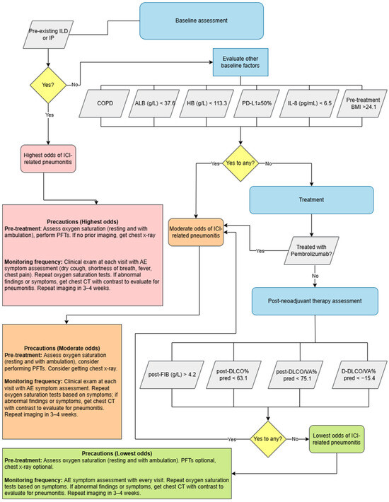
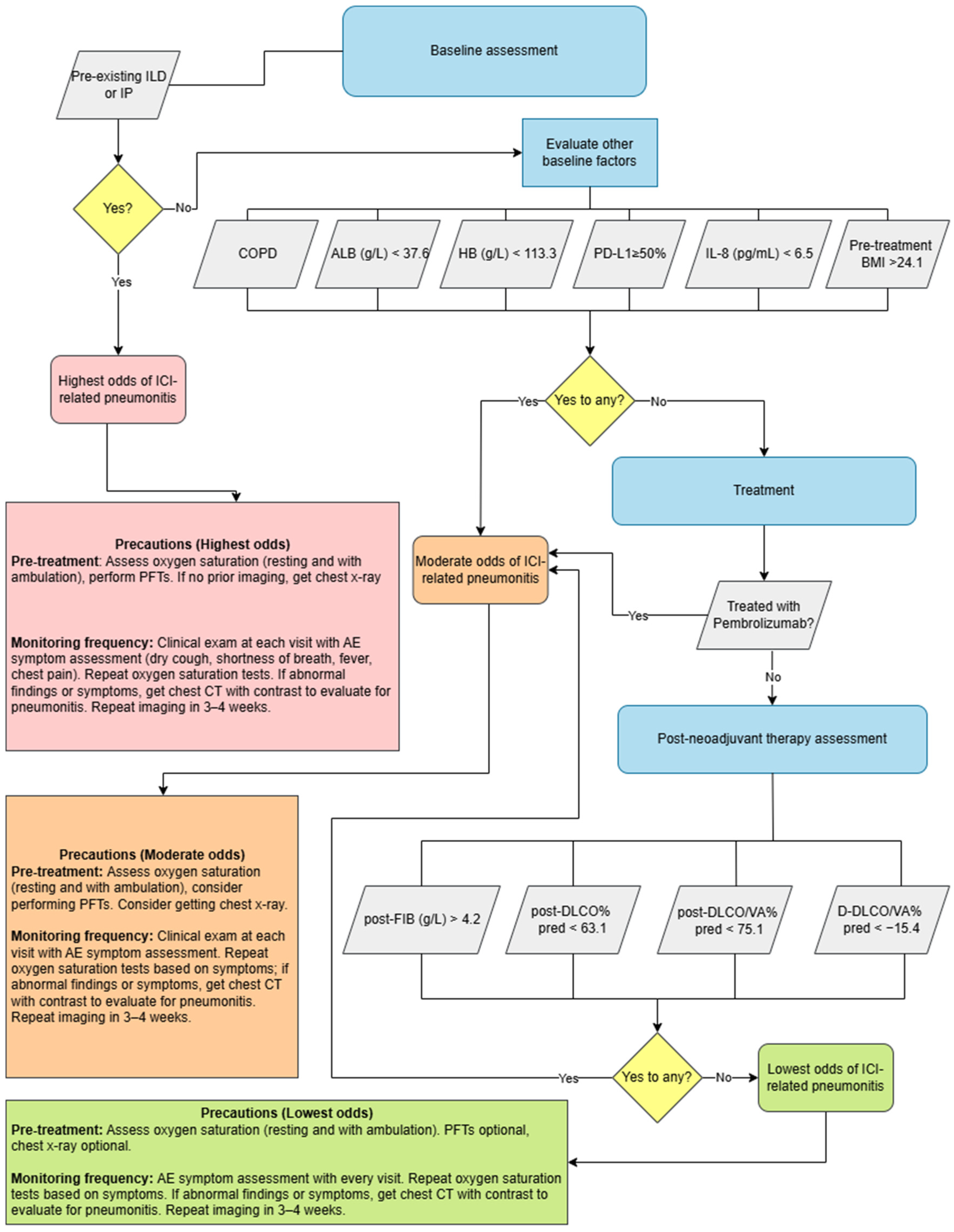
In summary, a multitude of factors have been evaluated for association with CIP, and key risk factors have emerged. As stated, pre-existing ILD (strongly predictive of CIP), prior thoracic radiation, and possibly baseline pulmonary impairment all seem to confer higher risk. Traditional patient factors (age, smoking) and basic tumor characteristics have not shown strong or consistent correlations. Ongoing prospective studies and pooled analyses are needed to definitively determine which patients are most susceptible to pneumonitis so that monitoring and prophylactic strategies can be tailored accordingly. Figure 2 represents a clinical algorithm combining the above factors with current clinical guidelines for management of CIP [57], and Table 3 summarizes the statistical data behind these risk factors.
Figure 2.
Clinical algorithm combining risk factors for immune checkpoint inhibitor (ICI) pneumonitis as identified in the literature with guidelines on management for immunotherapy-related pneumonitis from the NCCN [57]. Risk factors from Table 3b that have a p < 0.05 were included in this clinical algorithm. These include: pre-existing interstitial pneumonia (IP) [21]; pre-existing interstitial lung disease (ILD) [54]; chronic obstructive pulmonary disease (COPD) [22]; serum albumin level (ALB) [23,25]; hemoglobin (HB) [23]; PD-L1 expression [22]; IL-8 expression [22]; pre-treatment body mass index (BMI) [29]; treatment with pembrolizumab [25]; post-treatment fibrinogen levels (FIB) [29]; post-treatment diffusing capacity of the lung for carbon monoxide, expressed as a percentage of the predicted value (post-DLCO % pred) [29]; post-treatment diffusing capacity corrected for alveolar volume, expressed as a percentage of the predicted value (post-DLCO/VA % pred) [29]; and change in diffusing capacity per unit alveolar volume, expressed as a percentage of the predicted value (D-DLCO/VA% pred) [29]. ILD and IP have been combined into the same box to represent their overlapping definitions, though they are reported separately in the literature. Benchmarks for specific risk factors are the mean CIP group values as published in their respective studies. This figure was made using draw.io version 29.6.6; access link [https://app.diagrams.net], accessed on 24 March 2026.
Table 3.
(a) Risk factors in the development of CIP and their respective hazard ratios. (b) Risk factors in the development of CIP and their respective odds ratios.
5. Discussion
A wide array of potential risk factors has been implicated in the development of ICI-related pneumonitis in NSCLC; however, data regarding their impact on CIP incidence remain somewhat inconsistent across studies. This inconsistency can be attributed to limitations inherent in many available studies, including small patient cohorts, retrospective designs with inherent biases, heterogeneous patient criteria, a lack of standardized and detailed pulmonary function assessments, and limited longitudinal follow-up. In particular, the precise role of pulmonary function tests (PFTs) in the early diagnosis and prediction of CIP remains unclear.
It is crucial to identify patients at elevated risk for CIP to enable vigilant monitoring and prompt intervention as soon as pneumonitis is suspected. Recent efforts have focused on developing nomograms that integrate multiple risk factors into an estimated probability of CIP, such as those proposed by Mao et al. [29], Chao et al. [22] and Lu et al. [56]. These tools, once validated, could assist clinicians in stratifying risk and personalizing immunotherapy approaches, such as implementing closer imaging surveillance or prophylactic measures for high-risk patients.
Clinicians should be particularly attentive to patients who exhibit one or multiple risk factors for CIP development, and should consider more frequent and attentive imaging, with more attention paid to telltale symptoms of CIP during treatment such as dry cough, shortness of breath, fever, and chest pain. In particular, clinicians should tend toward the more conservative and proactive end of published monitoring guidelines when considering high-risk patients, given the importance of early identification and treatment of this dangerous side-effect. At this time, there is no definitive evidence as to whether pembrolizumab should be foregone altogether for patients with significant risk factors for CIP; the data described in this review should instead prompt clinicians to proceed with caution throughout ICI treatment of high-risk patients. In such cases, an interdisciplinary approach involving the patient’s full circle of care should be used, including close following of the patient via nurses and family practitioners, and consultation with pulmonologists and thoracic radiologists as necessary [24]. Additional imaging and pulmonary function tests may serve to provide further insight into disease development or progression. The development of any symptoms of CIP by high-risk patients should be treated with the utmost gravity, as early detection is crucial in proper care for this life-threatening complication; as such, a team of vigilant practitioners closely following high-risk patients receiving ICIs is key to ensuring improved clinical outcomes over time. While official guidelines do exist for the management of CIP depending on patient risk factors and history [57], more specific guidance must be developed regarding appropriate monitoring protocols for patients identified as being at-risk for later development of CIP, with particular courses of action recommended based on patient risk factors. In particular, the role of PFTs in the prediction and identification of CIP must be elucidated, as they are a simple and non-invasive test that could prove invaluable in the monitoring of patients at risk of developing CIP if a correlation is found. As more is understood about the risk stratification of CIP, researchers must also be prepared and responsive in developing more granular and specific clinical guidelines for the monitoring and prophylaxis of at-risk patients, as well as risk-benefit stratification to support clinical decision-making around choice of ICIs.
Ultimately, further research is needed to refine our understanding of CIP. Future prospective studies with larger patient populations and standardized data collection are essential to confirm risk factors, to identify reliable biomarkers for early detection, including clarifying the role of PFTs, and to improve management strategies. Such studies will help ensure that the life-saving benefits of immunotherapy in NSCLC can be realized while minimizing the impact of serious toxicities like pneumonitis.
Author Contributions
Conceptualization, O.A., L.Z., B.B. and C.S.; methodology, O.A., L.Z. and B.B.; software, O.A., L.Z. and B.B.; validation, O.A., L.Z. and B.B.; formal analysis, O.A., L.Z. and B.B.; investigation, O.A., L.Z. and B.B.; resources, O.A., L.Z. and B.B.; data curation, O.A., L.Z., B.B., M.S., R.S. and C.S.; writing—original draft preparation, O.A., L.Z., B.B., K.F., M.S., R.S. and C.S.; writing—review and editing, K.F., M.S., R.S. and C.S.; visualization, O.A., L.Z., B.B. and C.S.; supervision, O.A.; project administration, O.A., L.Z. and B.B. All authors have read and agreed to the published version of the manuscript.
Funding
This research received no external funding.
Data Availability Statement
No data were created or analyzed in this study. Data sharing is not applicable to this article.
Conflicts of Interest
The authors declare no conflicts of interest.
Abbreviations
The following abbreviations are used in this manuscript:
| NSCLC | Non-Small Cell Lung Cancer |
| HR | Hazard Ratio |
| irAEs | Immune-related Adverse Events |
| OR | Odds Ratio |
| CIP | Checkpoint Inhibitor Pneumonitis |
| ICP | Immune Checkpoint Pneumonitis |
| ALB | Albumin |
| HB | Hemoglobin |
| ILD | Interstitial Lung Disease |
| IP | Interstitial Pneumonia |
| ICI | Immune Checkpoint Inhibitor |
| PD-L1 | Programmed Death-Ligand 1 |
| PEM | Pemetrexed |
| PTX | Paclitaxel |
| nab-PTX | Nanoparticle Albumin-Bound Paclitaxel |
| COPD | Chronic Obstructive Pulmonary Disease |
| IL-8 | Interleukin-8 |
| pre-BMI | Pre-treatment Body Mass Index |
| post-FIB | Post-treatment Fibrinogen Level |
| post-DLCO % pred | Post-treatment Diffusing Capacity of the Lung for Carbon Monoxide expressed as a percentage of the predicted value. |
| post-DLCO/VA % pred | Post-treatment DLCO (diffusing capacity) corrected for alveolar volume, expressed as a percentage of the predicted value. |
| D-DLCO/VA % pred | Change in DLCO/VA (diffusing capacity per unit alveolar volume) expressed as a percentage of the predicted value. |
| Mod.1 | Model 1 |
| Mod.2 | Model 2 |
References
- Bray, F.; Ferlay, J.; Laversanne, M. Global cancer statistics 2022: GLOBOCAN estimates of incidence and mortality worldwide for 36 cancers in 185 countries. CA Cancer J. Clin. 2024, 74, 229–263. [Google Scholar] [CrossRef] [Scilit] [PubMed]
- Inamura, K. Lung Cancer: Understanding Its Molecular Pathology and the 2015 WHO Classification. Front. Oncol. 2017, 7, 193. [Google Scholar] [CrossRef] [Scilit]
- Spicer, J.D.; Garassino, M.C.; Wakelee, H. Neoadjuvant pembrolizumab plus chemotherapy followed by adjuvant pembrolizumab versus neoadjuvant chemotherapy alone in early-stage NSCLC (KEYNOTE-671): A randomized, double-blind, placebo-controlled, phase 3 trial. Lancet 2024, 404, 1240–1252. [Google Scholar] [CrossRef] [Scilit]
- Cascone, T.; Awad, M.M.; Spicer, J.D. CheckMate 77T Investigators. Perioperative Nivolumab in Resectable Lung Cancer. N. Engl. J. Med. 2024, 16, 1756–1769. [Google Scholar] [CrossRef] [Scilit]
- Spigel, D.R.; Faivre-Finn, C.; Gray, J.E. Five-year survival outcomes from the PACIFIC trial: Durvalumab after chemoradio-therapy in stage III non–small-cell lung cancer. J. Clin. Oncol. 2022, 40, 1301–1311. [Google Scholar] [CrossRef] [Scilit] [PubMed]
- Garassino, M.C.; Cho, B.C.; Kim, J.H. Pembrolizumab plus pemetrexed and platinum in nonsquamous NSCLC: 5-year out-comes from the phase 3 KEYNOTE-189 study. J. Clin. Oncol. 2023, 41, 1992–1998. [Google Scholar] [CrossRef] [Scilit]
- Socinski, M.A.; Nishio, M.; Jotte, R.M. Impower150 final overall survival analyses for atezolizumab plus bevacizumab and chemotherapy in first-line metastatic nonsquamous NSCLC. J. Thorac. Oncol. 2021, 16, 1909–1924. [Google Scholar] [CrossRef] [Scilit]
- Shiravand, Y.; Khodadadi, F.; Kashani, S.M.A. Immune checkpoint inhibitors in cancer therapy. Curr. Oncol. 2022, 29, 3044–3060. [Google Scholar] [CrossRef] [Scilit]
- Lin, M.X.; Zang, D.; Liu, C.G.; Han, X.; Chen, J. Immune checkpoint inhibitor-related pneumonitis: Research advances in prediction and management. Front. Immunol. 2024, 15, 1266850. [Google Scholar] [CrossRef] [Scilit]
- Barrón, F.; Sánchez, R.; Arroyo-Hernández, M. Risk of developing checkpoint inhibitor pneumonitis and its effect on over-all survival in non-small cell lung cancer patients previously treated with radiotherapy. Front. Oncol. 2020, 10, 570233. [Google Scholar] [CrossRef] [Scilit] [PubMed]
- Choi, J.; Lee, S.Y. Clinical characteristics and treatment of immune-related adverse events of immune checkpoint inhibitors. Immune Netw. 2020, 20, e9. [Google Scholar] [CrossRef] [Scilit]
- Wang, D.Y.; Salem, J.E.; Cohen, J.V. Fatal toxic effects associated with immune checkpoint inhibitors: A systematic review and meta-analysis. JAMA Oncol. 2018, 4, 1721–1728. [Google Scholar] [CrossRef] [Scilit]
- Tay, R.Y.; Califano, R. Checkpoint inhibitor pneumonitis—Real-world incidence and risk. J. Thorac. Oncol. 2018, 13, 1812–1814. [Google Scholar] [CrossRef] [Scilit]
- Marrone, K.A.; Naidoo, J.; Brahmer, J. Immunotherapy for lung cancer: No longer an abstract concept. Semin. Respir. Crit. Care Med. 2016, 37, 771–782. [Google Scholar] [CrossRef] [Scilit]
- Kong, Y.; Hong, L.; Xu, X.C.; Chen, Y.F.; Xu, J. The relative risk of immune checkpoint inhibitor pneumonitis in advanced non-small-cell lung cancer: Meta-analyses of controlled clinical trials. PLoS ONE 2024, 19, e0301931. [Google Scholar] [CrossRef] [Scilit] [PubMed]
- Nishino, M.; Giobbie-Hurder, A.; Hatabu, H.; Ramaiya, N.H.; Hodi, F.S. Incidence of programmed cell death 1 inhibitor–related pneumonitis in patients with advanced cancer: A systematic review and meta-analysis. JAMA Oncol. 2016, 2, 1607–1616. [Google Scholar] [CrossRef] [Scilit] [PubMed]
- Hao, Y.; Zhang, X.; Yu, L. Immune checkpoint inhibitor-related pneumonitis in non-small cell lung cancer: A review. Front. Oncol. 2022, 12, 911906. [Google Scholar] [CrossRef] [Scilit] [PubMed]
- Tao, H.; Gong, Y.; Wang, Y. Rate and risk factors of recurrent immune checkpoint inhibitor-related pneumonitis in patients with lung cancer. Transl. Lung Cancer Res. 2022, 11, 381–392. [Google Scholar] [CrossRef] [Scilit]
- Cathcart-Rake, E.J.; Sangaralingham, L.R.; Henk, H.J. A population-based study of immunotherapy-related toxicities in lung cancer. Clin. Lung Cancer 2020, 21, 421–427. [Google Scholar] [CrossRef] [Scilit]
- Suresh, K.; Voong, K.R.; Shankar, B. Pneumonitis in non–small cell lung cancer patients receiving immune checkpoint immunotherapy: Incidence and risk factors. J. Thorac. Oncol. 2018, 13, 1930–1939. [Google Scholar] [CrossRef] [Scilit] [PubMed]
- Yamaguchi, T.; Shimizu, J.; Oya, Y. Risk factors for pneumonitis in patients with non-small cell lung cancer treated with immune checkpoint inhibitors plus chemotherapy: A retrospective analysis. Thorac. Cancer 2022, 13, 724–731. [Google Scholar] [CrossRef] [Scilit]
- Chao, Y.; Zhou, J.; Hsu, S.; Ding, N.; Li, J.; Zhang, Y.; Xu, X.; Tang, X.; Wei, T.; Zhu, Z.; et al. Risk factors for immune checkpoint inhibitor-related pneumonitis in non-small cell lung cancer. Transl. Lung Cancer Res. 2022, 11, 295–306. [Google Scholar] [CrossRef] [Scilit]
- Liu, X.; Hao, N.; Yang, S.; Li, J.; Wang, L. Predictive factors and prognosis of immune checkpoint inhibitor-related pneumonitis in non-small cell lung cancer patients. Front. Oncol. 2023, 13, 1145143. [Google Scholar] [CrossRef] [Scilit]
- Altan, M.; Soto, F.; Zhong, L.L. Incidence and risk factors for pneumonitis associated with checkpoint inhibitors in advanced non-small cell lung cancer: A single center experience. Oncologist 2023, 28, e1065–e1074. [Google Scholar] [CrossRef] [Scilit]
- Fukihara, J.; Sakamoto, K.; Koyama, J. Prognostic impact and risk factors of immune-related pneumonitis in patients with non-small-cell lung cancer who received programmed death 1 inhibitors. Clin. Lung Cancer 2019, 20, 442–450.e4. [Google Scholar] [CrossRef] [Scilit]
- Sawa, K.; Sato, I.; Takeuchi, M.; Kawakami, K. Risk of pneumonitis in non-small cell lung cancer patients with preexisting inter-stitial lung diseases treated with immune checkpoint inhibitors: A nationwide retrospective cohort study. Cancer Immunol. Immunother. 2023, 72, 591–598. [Google Scholar] [CrossRef] [Scilit] [PubMed]
- Sumi, T.; Sekikawa, M.; Koshino, Y. Risk factors for severe immune-related pneumonitis after nivolumab plus ipilimumab therapy for non-small cell lung cancer. Thorac. Cancer 2024, 15, 1572–1581. [Google Scholar] [CrossRef] [Scilit] [PubMed]
- Zeng, Z.; Qu, J.; Yao, Y. Clinical outcomes and risk factor of immune checkpoint inhibitors-related pneumonitis in non-small cell lung cancer patients with chronic obstructive pulmonary disease. BMC Pulm. Med. 2022, 22, 458. [Google Scholar] [CrossRef] [Scilit] [PubMed]
- Mao, Z.; Pang, G.; Huang, X. Risk factors of immune checkpoint inhibitor-related pneumonitis after neoadjuvant immu-nochemotherapy for resectable NSCLC. BMC Pulm. Med. 2024, 24, 253. [Google Scholar] [CrossRef] [Scilit]
- Wakelee, H.; Liberman, M.; Kato, T. KEYNOTE-671 Investigators. Perioperative pembrolizumab for early-stage non-small-cell lung cancer. N. Engl. J. Med. 2023, 389, 491–503. [Google Scholar] [CrossRef] [Scilit]
- Heymach, J.V.; Harpole, D.; Mitsudomi, T. Perioperative durvalumab for resectable non-small-cell lung cancer. N. Engl. J. Med. 2023, 389, 1672–1684. [Google Scholar] [CrossRef] [Scilit] [PubMed]
- Reck, M.; Rodríguez-Abreu, D.; Robinson, A.G. KEYNOTE-024 Investigators. Pembrolizumab versus chemotherapy for PD-L1-positive non-small-cell lung cancer. N. Engl. J. Med. 2016, 375, 1823–1833. [Google Scholar] [CrossRef] [Scilit] [PubMed]
- Herbst, R.S.; Giaccone, G.; Marinis, F. Atezolizumab for first-line treatment of PD-L1-selected patients with NSCLC. N. Engl. J. Med. 2020, 383, 1328–1339. [Google Scholar] [CrossRef] [Scilit] [PubMed]
- Gandhi, L.; Rodríguez-Abreu, D.; Gadgeel, S. KEYNOTE-189 Investigators. Pembrolizumab plus chemotherapy in meta-static non-small-cell lung cancer. N. Engl. J. Med. 2018, 378, 2078–2092. [Google Scholar] [CrossRef] [Scilit]
- Socinski, M.A.; Jotte, R.M.; Cappuzzo, F. Impower150 Study Group. Atezolizumab for first-line treatment of metastatic nonsquamous NSCLC. N. Engl. J. Med. 2018, 378, 2288–2301. [Google Scholar] [CrossRef] [Scilit]
- Nishio, M.; Barlesi, F.; West, H. Atezolizumab plus chemotherapy for first-line treatment of nonsquamous NSCLC: Results from the randomized phase 3 IMpower132 trial. J. Thorac. Oncol. 2021, 16, 653–664. [Google Scholar] [CrossRef] [Scilit]
- Gogishvili, M.; Melkadze, T.; Makharadze, T. Cemiplimab plus chemotherapy versus chemotherapy alone in non-small cell lung cancer: A randomized, controlled, double-blind phase 3 trial. Nat. Med. 2022, 28, 2374–2380. [Google Scholar] [CrossRef] [Scilit]
- Paz-Ares, L.G.; Ciuleanu, T.E.; Cobo, M. First-line nivolumab plus ipilimumab with chemotherapy versus chemotherapy alone for metastatic NSCLC in CheckMate 9LA: 3-year clinical update and outcomes in patients with brain metastases or select somatic mutations. J. Thorac. Oncol. 2023, 18, 204–222. [Google Scholar] [CrossRef] [Scilit]
- Forde, P.M.; Spicer, J.; Lu, S. CheckMate 816 Investigators. Neoadjuvant nivolumab plus chemotherapy in resectable lung cancer. N. Engl. J. Med. 2022, 386, 1973–1985. [Google Scholar] [CrossRef] [Scilit]
- Garassino, M.C.; Khalifa, J.; Reck, M.; Chouaid, C.; Bischoff, H.; Reinmuth, N.; Cove-Smith, L.; Mansy, T.; Cortinovis, D.L.; Migliorino, M.R.; et al. Durvalumab after sequential chemoradiotherapy in unresectable stage III non-small-cell lung cancer—Final analysis from phase II PACIFIC-6 trial. ESMO Open 2025, 10, 105071. [Google Scholar] [CrossRef] [Scilit]
- Paz-Ares, L.; Luft, A.; Vicente, D.; Tafreshi, A.; Gümüş, M.; Mazières, J.; Hermes, B.; Şenler, F.Ç.; Csőszi, T.; Fülöp, A.; et al. Pembrolizumab plus Chemotherapy for Squamous Non-Small-Cell Lung Cancer. N. Engl. J. Med. 2018, 379, 2040–2051. [Google Scholar] [CrossRef] [Scilit]
- Antonia, S.J.; Villegas, A.; Daniel, D.; Vicente, D.; Murakami, S.; Hui, R.; Yokoi, T.; Chiappori, A.; Lee, K.H.; de Wit, M.; et al. Durvalumab after Chemoradiotherapy in Stage III Non-Small-Cell Lung Cancer. N. Engl. J. Med. 2017, 377, 1919–1929. [Google Scholar] [CrossRef] [Scilit]
- Khoja, L.; Day, D.; Wei-Wu Chen, T.; Siu, L.L.; Hansen, A.R. Tumour- and class-specific patterns of immune-related adverse events of immune checkpoint inhibitors: A systematic review. Ann. Oncol. 2017, 28, 2377–2385. [Google Scholar] [CrossRef] [Scilit]
- Haanen, J.; Obeid, M.; Spain, L. Management of toxicities from immunotherapy: ESMO Clinical Practice Guideline for diagno-sis, treatment and follow-up. Ann. Oncol. 2022, 33, 1217–1238. [Google Scholar] [CrossRef] [Scilit]
- Reuss, J.E.; Brigham, E.; Psoter, K.J. Pretreatment lung function and checkpoint inhibitor pneumonitis in NSCLC. JTO Clin. Res. Rep. 2021, 2, 100220. [Google Scholar] [CrossRef] [Scilit]
- Suzuki, Y.; Karayama, M.; Uto, T. Assessment of immune-related interstitial lung disease in patients with NSCLC treated with immune checkpoint inhibitors: A multicenter prospective study. J. Thorac. Oncol. 2020, 15, 1317–1327. [Google Scholar] [CrossRef] [Scilit]
- Fujimoto, D.; Kato, R.; Morimoto, T. Characteristics and prognostic impact of pneumonitis during systemic anti-cancer therapy in patients with advanced non-small-cell lung cancer. PLoS ONE 2016, 11, e0168465. [Google Scholar] [CrossRef] [Scilit] [PubMed]
- Omori, T.; Tajiri, M.; Baba, T. Pulmonary resection for lung cancer in patients with idiopathic interstitial pneumonia. Ann. Thorac. Surg. 2015, 100, 954–960. [Google Scholar] [CrossRef] [Scilit] [PubMed]
- Kanai, O.; Kim, Y.H.; Demura, Y. Efficacy and safety of nivolumab in non-small cell lung cancer with preexisting interstitial lung disease. Thorac. Cancer 2018, 9, 847–855. [Google Scholar] [CrossRef] [Scilit] [PubMed]
- Cho, J.Y.; Kim, J.; Lee, J.S. Characteristics, incidence, and risk factors of immune checkpoint inhibitor-related pneumonitis in patients with non-small cell lung cancer. Lung Cancer 2018, 125, 150–156. [Google Scholar] [CrossRef] [Scilit]
- Cadranel, J.; Canellas, A.; Matton, L. Pulmonary complications of immune checkpoint inhibitors in patients with nonsmall cell lung cancer. Eur. Respir. Rev. 2019, 28, 190058. [Google Scholar] [CrossRef] [Scilit]
- Khunger, M.; Rakshit, S.; Pasupuleti, V. Incidence of pneumonitis with use of programmed death 1 and programmed death-ligand 1 inhibitors in non-small cell lung cancer: A systematic review and meta-analysis of trials. Chest 2017, 152, 271–281. [Google Scholar] [CrossRef] [Scilit] [PubMed]
- Svaton, M.; Blazek, J.; Krakorova, G. Laboratory parameters are possible prognostic markers in patients with advanced-stage NSCLC treated with bevacizumab plus chemotherapy. J. Cancer 2021, 12, 5753–5759. [Google Scholar] [CrossRef] [Scilit] [PubMed]
- Sugano, T.; Seike, M.; Saito, Y. Immune checkpoint inhibitor-associated interstitial lung diseases correlate with better prognosis in patients with advanced non-small-cell lung cancer. Thorac. Cancer 2020, 11, 1052–1060. [Google Scholar] [CrossRef] [Scilit] [PubMed]
- Lin, X.; Deng, H.; Chu, T.; Chen, L.; Yang, Y.; Qiu, G.; Xie, X.; Qin, Y.; Lio, M.; Xie, Z.; et al. Safety and efficacy of immunotherapy rechallenge following checkpoint inhibitor-related pneumonitis in advanced lung cancer patients: A retrospective multi-center cohort study. Transl. Lung. Cancer Res. 2022, 11, 2289–2305. [Google Scholar] [CrossRef] [Scilit]
- Lu, H.; Jiang, A.; Yuan, Z.; Chen, D. Integrated machine learning risk model for predicting radiation pneumonitis in lung cancer patients with interstitial lung disease. Ann. Med. 2026, 58, 2634444. [Google Scholar] [CrossRef] [Scilit]
- National Comprehensive Cancer Network. Management of Immune Checkpoint Inhibitor-Related Toxicities; NCCN Guidelines; Version 1.2026; Dove Medical Press: London, UK, 2025. [Google Scholar]

Disclaimer/Publisher’s Note: The statements, opinions and data contained in all publications are solely those of the individual author(s) and contributor(s) and not of MDPI and/or the editor(s). MDPI and/or the editor(s) disclaim responsibility for any injury to people or property resulting from any ideas, methods, instructions or products referred to in the content. |
© 2026 by the authors. Licensee MDPI, Basel, Switzerland. This article is an open access article distributed under the terms and conditions of the Creative Commons Attribution (CC BY) license.