


Simple Summary
Considering the availability of human epidermal growth factor receptor 2 (ERBB2/HER2)-targeting therapy, the status of HER2 amplification in patients with all types of solid tumors needs to be clarified. We analyzed 89,374 eligible patients with solid tumors using comprehensive genomic profiling data from Japanese nationwide database, and HER2 amplification was detected in 5119 patients (5.7%): highest rates of HER2 amplification were observed in patients with tumors of the esophagus/stomach (12.9%), followed by tumors of the bladder/urinary tract (10.6%), breast (9.5%), biliary tract (8.4%), and uterus (8.4%). The detection of HER2 amplification was highest with FoundationOne CDx assay platform (7.4%) compared to other platforms. We report that HER2 amplification occurs in several tumors, including rare tumors, and is not limited to conventional tumor types. The results suggest that there is a need to expand the evaluation of HER2 amplification and consequently HER2-targeting therapy such as trastuzumab deruxtecan in Japan.
Abstract
Limited data exist on the prevalence of human epidermal growth factor receptor 2 (ERBB2/HER2) amplification in patients with all types of solid tumors. This retrospective, observational study (UMIN ID: UMIN000057382) analyzed the prevalence of HER2 amplification across all solid tumors using comprehensive genomic profiling (CGP) data from the Center for Cancer Genomics and Advanced Therapeutics database in Japan. We analyzed 89,374 eligible patients with solid tumors: HER2 amplification was detected in 5119 patients (5.7%). The highest rates of HER2 amplification were observed in patients with tumors of the esophagus/stomach (12.9%), followed by tumors of the bladder/urinary tract (10.6%), breast (9.5%), biliary tract (8.4%), and uterus (8.4%). Among the five assay platforms, FoundationOne CDx accounted for 69.7% of all CGP tests and had an HER2 amplification detection rate of 7.4%, compared to the other four platforms (range: 1.9–15.4% of all CGP tests [detection rates: 1.5–2.3%]). Substantial differences were observed in the mutation frequencies of multiple genes between HER2 amplified and HER2 non-amplified tumors. The results highlight that HER2 amplification extends beyond conventional tumor types and enables the identification, via CGP testing, of non-traditional tumor subsets (cancers other than breast and gastric cancer), including rare cancers, that could be candidates for HER2-targeting therapy such as trastuzumab deruxtecan in Japan.
1. Introduction
Human epidermal growth factor receptor 2 (HER2, also known as ERBB2) is a proto-oncogene that encodes a transmembrane tyrosine kinase receptor involved in the regulation of cell growth, differentiation, and survival [1]. HER2-positive tumors are identified by either protein overexpression (using immunohistochemistry with a score of 3+ [IHC 3+], which is the current standard) or IHC 2+ with concurrent gene amplification, confirmed by in situ hybridization (ISH) [2]. Thus, both amplification and overexpression of HER2 are established therapeutic targets, particularly in breast and gastric cancers [1,3].
In breast cancer, HER2 amplification occurs in approximately 20% of cases, while 15–20% of cases show HER2 overexpression, which is linked to aggressive tumor characteristics [3,4]. However, this subset now has effective treatment options due to HER2-targeted therapies (such as trastuzumab, pertuzumab) and newer drugs (such as trastuzumab deruxtecan [T-DXd]) [1]. Similarly, HER2 amplification is observed in 11–16% of gastric/gastroesophageal cancers, while HER2 overexpression in about 20% of cases, and anti-HER2 therapies have demonstrated clinical benefits [1,4].
In addition to these tumor types, HER2 amplification has also been increasingly recognized in various other solid tumors, including colorectal cancer, non-small cell lung cancer (NSCLC), biliary tract cancer, and bladder cancer, although at lower frequencies [4,5]. However, the overall landscape of HER2 amplification across the full spectrum of solid malignancies remains unclear.
For the treatment of patients with advanced HER2-amplified solid tumors, the HERALD trial conducted in Japan demonstrated the efficacy of T-DXd [6]. However, the prevalence of HER2 amplification detected by comprehensive genomic profiling (CGP) testing in Japan, as well as differences among assay platforms, have not yet been clearly defined. Understanding these aspects will become increasingly important in clinical practice for precision oncology.
CGP, based on next-generation sequencing (NGS), offers a broader approach for detecting multiple genomic alterations, including HER2 amplification, across tumor types and patient populations. In Japan, the Center for Cancer Genomics and Advanced Therapeutics (C-CAT) serves as a centralized database that integrates genomic and clinical information from patients who have undergone CGP under the National Cancer Genomic Medicine Program [7,8].
Large-scale genomic databases such as AACR Project GENIE have reported HER2 amplification frequencies of about 12% for esophagogastric cancer, 10% for breast cancer, and <4% for all other cancers. However, such prevalence estimates are derived largely from Western populations and may not reflect the genomic landscape in Asian patients, particularly in Japan [9,10]. Additionally, estimates can vary depending on the differences in the assay platforms used.
Overall, there is a need for genomically matched treatment with awareness of the prevalence of HER2 amplification in patients with all types of solid tumors without standard treatment or in patients with locally advanced or metastatic tumors who have completed standard treatment (including those who are expected to complete treatment). In Japan, there is a lack of Japan-specific real-world data regarding HER2 amplification using CGP testing.
Therefore, the primary objective of this study was to determine the prevalence of HER2 amplification across all solid tumors using CGP data from the C-CAT database in Japan. In addition, background information, including treatment histories and genetic mutations, was compared between patients with and without HER2 amplification.
2. Materials and Methods
2.1. Study Design and Data Source
This retrospective observational study (UMIN ID: UMIN000057382) evaluated the CGP data of patients with solid tumors from the C-CAT database, Japan’s national repository integrating genomic and clinical data under the National Cancer Genomic Medicine Program. Patient data were extracted and analyzed based on CGP test results recorded between 1 June 2019 and 31 December 2024 (database lock: May 2025). This large database (111,560 patients registered at the end of August 2025) includes 289 medical institutions certified by the Ministry of Health, Labour and Welfare as core cancer genome medical centers, core hospitals, and affiliated hospitals (as of 1 October 2025).
The observation period initiation date (index date) was based on the “expert panel (EP) held” date in the C-CAT database. Since the response to treatment at each facility is likely to differ based on the results of each CGP investigation, the information before the index date was regarded as background information, and the information after the index date was regarded as information from the follow-up period.
Informed consent for secondary data use by other parties was obtained from each patient prior to data transfer from their respective hospitals. Of note, >99% of patients in the C-CAT database have agreed to the secondary use of their genomic and clinical information, and all data extracted for this study were from patients who provided informed consent for secondary data use [11,12]. The CGP database included individual patient data and information on gene alterations, drug therapy (before and after EP), CGP specimens, and outcomes. CGP data were obtained using five platforms: tissue-based CGP (tissue CGP using tissue DNA) included the Oncoguide™ NCC OncoPanel System (NOP), FoundationOne®® CDx (F1CDx), and GenMineTOP®® (GMTOP), whereas plasma-based CGP using liquid biopsy (plasma CGP using circulating tumor DNA/ctDNA) included FoundationOne®® Liquid CDx (F1LCDx) and Guardant360®® CDx (G360CDx).
The study was approved by the Ethical Research Practice Committee for Research on Human Tissue, Information, and Other Human Material (Approval No. 000790) and reviewed by the C-CAT Data Utilization Review Board (Approval ID: CDU2024-026N) [13].
Individual patient data in this study were provided to the C-CAT database vendors as anonymously processed information by individual medical institutions and did not include personally identifiable information. Of note, in the C-CAT database, a genomic sequence falls under the personal identification code, and it is classified into the “personal information requiring consideration” category because it contains disease information. Patient consent, including consent for secondary data use prior to CGP investigation, was obtained from all facilities.
2.2. Patients
Eligible patients were required to be ≥18 years of age, enrolled in the C-CAT database during the study period (1 June 2019 to 31 December 2024), and having solid tumors. Patients were excluded if they had a record of a previous registration to avoid duplication of data, an unknown previous registration status, and CGP results including HER2 amplification that could not be judged or were indeterminate, i.e., not all alterations and co-alterations were detected, and the presence or absence of HER2 amplification was unknown. Eligible patients, with or without ERBB2 amplification, enrolled in the C-CAT database during the study period were categorized into the HER2 amplification and HER2 no-amplification groups, respectively. In this study, HER2 amplification was defined as the presence or absence of records in the C-CAT database and was not based on the copy number, because the copy number was not recorded in this database. Additionally, other HER2 alterations (mutations [such as single-nucleotide variants] and fusions) apart from amplification might have been included in both groups. Tumor types were classified according to OncoTree, which includes different histological and genomic subclassifications [14].
2.3. Endpoints
The primary endpoint was the prevalence of HER2 amplification using CGP test results. The secondary endpoints were the detection rates of HER2 amplification by CGP assay modality, patient background characteristics based on HER2 amplification (e.g., age, sex, tumor type, metastasis site, number of treatment lines before EP, and CGP platform type), and co-occurring gene alterations using CGP. The exploratory endpoint was to assess the implication of CGP results on the direction of treatment after EP, i.e., application of genomically matched treatment.
2.4. Statistical Analyses
Descriptive statistics were used to summarize categorical and continuous data, which are presented as mean and standard deviation (SD), and categorical data are presented as frequency and percentage. Chi-square tests were performed as necessary. All analyses were performed in accordance with the Strengthening the Reporting of Observational Studies in Epidemiology guidelines [15] and relevant sections of the Consolidated Standards of Reporting Trials guidelines [16]. Tumor types were categorized according to the OncoTree hierarchical classification system into the primary level (Level 1), secondary level (Level 2), tertiary level (Level 3), and quaternary level (Level 4), based on histological and genomic subclassifications. This allows meaningful comparisons of prevalence data across tumor subtypes for real-world interpretation and subsequent targeted therapies [14].
Patient characteristics were also evaluated, and patients were categorized into the HER2 amplification and HER2 no-amplification groups for all tumor types, with data presented using descriptive statistics. The percentages of tumor types with genetic mutations, HER2 amplification, and the frequency of co-occurring gene mutations [8] were visualized using a heat map. Missing data imputation was not performed. All analyses were performed using the SAS software package (version 9.4; SAS Institute Inc., Cary, NC, USA) and Python (version 3.11, Python Software Foundation, Wilmington, DE, USA).
3. Results
3.1. Patient Disposition and Baseline Characteristics
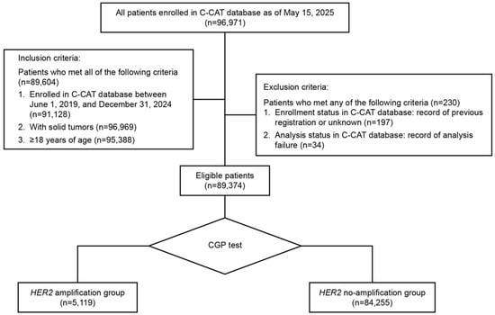
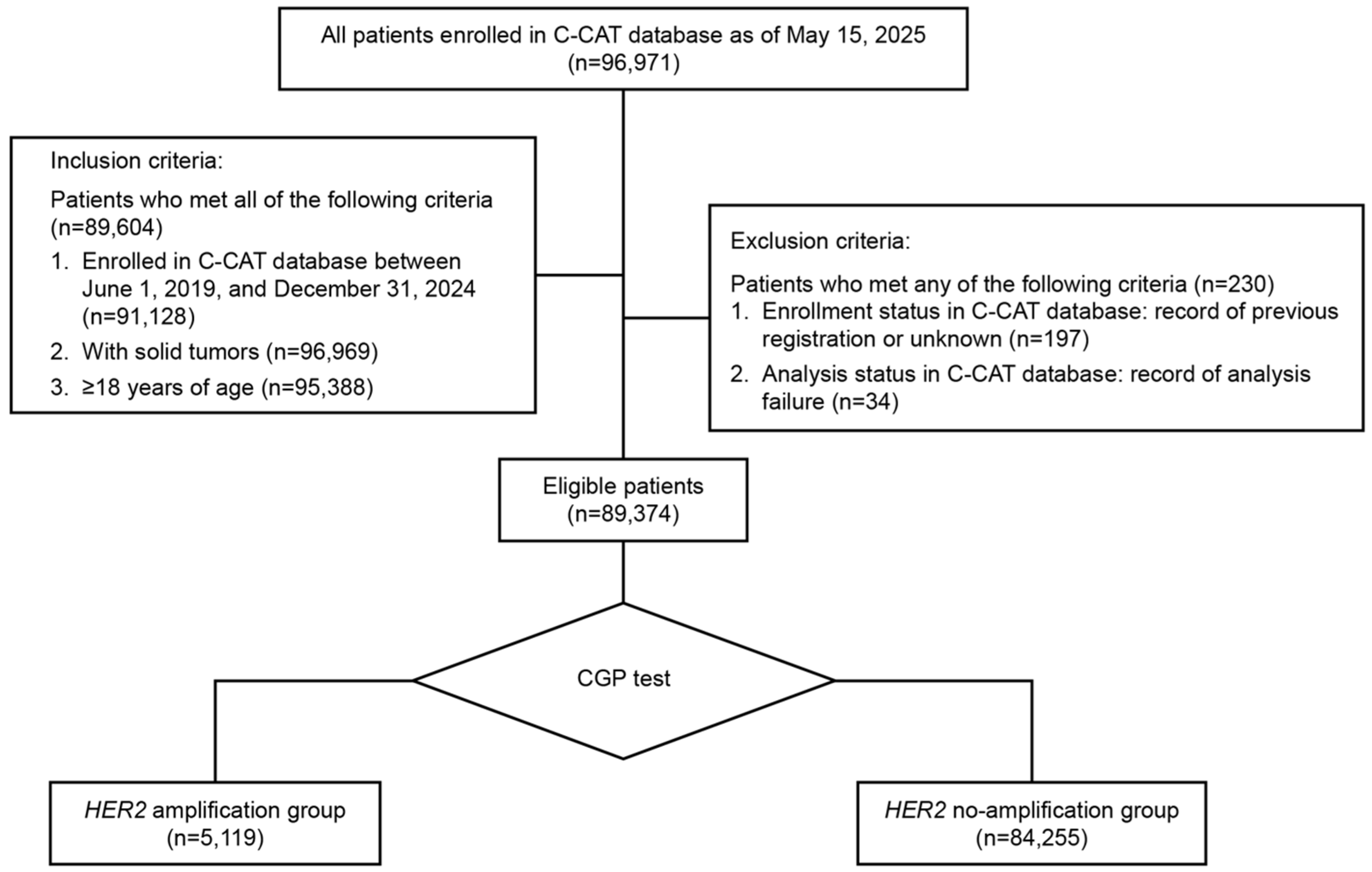
As of 15 May 2025 (C-CAT database ver.20250416), the records of 96,971 patients enrolled in the C-CAT database were evaluated. The analysis set comprised 89,374 eligible patients with solid tumors categorized into the HER2 amplification (n = 5119) and HER2 no-amplification (n = 84,255) groups, using evaluable CGP results (Figure 1).
Figure 1.
Patient flow diagram. C-CAT: Center for Cancer Genomics and Advanced Therapeutics; CGP: comprehensive genomic profiling; HER2: human epidermal growth factor receptor 2.
The commonest tumors (Level 1) identified in the database were tumors of the bowel (16.7% [14,888/89,374]), followed by those of the pancreas (15.6% [13,976/89,374]), biliary tract (8.9% [7966/89,374]), and breast (7.5% [6720/89,374]) (Table S1). Most of the patients presented with metastasis (89.5% [79,962/89,374]) before the index date (HER2 no-amplification group [89.2%] vs. HER2 amplification group [93.6%]), and the commonest site of metastasis was lymph nodes/lymph vessels (35.6% [31,774/89,374]), followed by the liver (32.9% [29,410/89,374]), lung (28.9% [25,792/89,374]), peritoneum (19.7% [17,595/89,374]), and bone (17.7% [15,838/89,374]) (Table 1). Most samples used in tissue CGP were collected surgically (61.0% [45,137/73,960]), followed by biopsies (38.6% [28,566/73,960]). A biopsy was performed at a higher rate in patients with prostate cancer (78.6% [2627/3341]), followed by those with other tumors (62.0% [1223/1971]) and lung cancer (61.7% [2408/3903]; Table S1). Biopsy specimens were collected from the primary lesion site in 68.2% (50,458/73,960) of patients and metastatic lesions in 31.3% (23,169/73,960) of patients (Table 1). The tumor type with the highest rate of plasma CGP was prostate cancer (39.4% [2169/5510]), followed by pancreatic cancer (28.9% [4033/13,976]) and lung cancer (27.9% [1514/5417]). When categorized by line of treatment (1st to ≥5th lines), the proportion of patients was almost evenly distributed among all treatment lines for overall tumor types. A higher proportion of patients with rare tumors underwent CGP testing after receiving first-line and second-line treatments compared with patients having other tumors. On the other hand, CGP testing was commonly performed after third-line treatment for tumor types with several treatment options (Table S1).
Table 1.
Patient characteristics by the HER2 amplification status.
3.2. Prevalence of HER2 Amplification
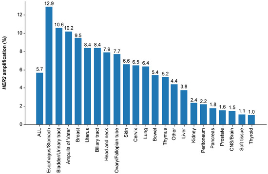
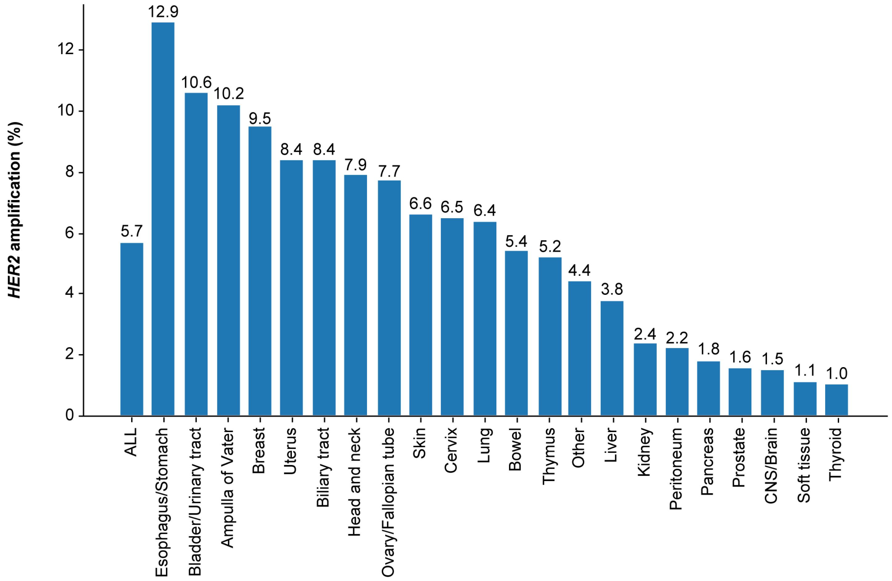
HER2 amplification was detected in 5.7% (5119/89,374) of eligible patients with solid tumors.
The HER2 amplification rate was highest in patients with tumors of the esophagus/stomach (12.9% [709/5499]), followed by those of the bladder/urinary tract (10.6% [157/1484]), ampulla of Vater (10.2% [54/531]), breast (9.5% [639/6720]), biliary tract (8.4% [673/7966]), and uterus (8.4% [258/3068]) (Figure 2 and Table S2).
Figure 2.
Prevalence of HER2 amplification in solid tumors. CNS, central nervous system; HER2: human epidermal growth factor receptor 2.
3.3. Prevalence of HER2 Amplification by the OncoTree Hierarchical Classification System
The prevalence of HER2 amplification in tumors of the esophagus/stomach (Level 1) was 12.9% (n = 709/5499), with Level 2 prevalence in esophagogastric adenocarcinoma of 16.1% (n = 491/3041) and esophageal squamous cell carcinoma of 6.6% (n = 95/1440), and Level 3 prevalence in gastroesophageal junction adenocarcinoma of 21.5% (n = 73/339) and stomach adenocarcinoma of 17.0% (n = 353/2074).
The prevalence of HER2 amplification in tumors of the bowel (Level 1) was 5.4% (n = 803/14,888), with Level 2 prevalence in colorectal adenocarcinoma of 5.7% (657/11,451) and small bowel cancer of 5.3% (28/529), and Level 3 prevalence in colon adenocarcinoma of 5.6% (338/6064) and rectal adenocarcinoma of 6.1% (258/4228).
The prevalence of HER2 amplification in tumors of the ampulla of Vater (Level 1) was 10.2% (54/531), with Level 2 prevalence in ampullary carcinoma of 10.0% (45/451).
The prevalence of HER2 amplification in tumors of the biliary tract (Level 1) was 8.4% (673/7966), with Level 2 prevalence in intraductal papillary neoplasm of the bile duct of 6.0% (325/5455); Level 3 prevalence in gallbladder cancer of 15.6% (291/1868) and cholangiocarcinoma of 5.9% (322/5419); and Level 4 prevalence in extrahepatic cholangiocarcinoma of 9.3% (93/995), intrahepatic cholangiocarcinoma of 4.8% (136/2842), and perihilar cholangiocarcinoma of 5.4% (60/1107).
The prevalence of HER2 amplification in tumors of the bladder/urinary tract (Level 1) was 10.6% (157/1484), with Level 2 prevalence in bladder urothelial carcinoma of 16.0% (91/568) and upper tract urothelial carcinoma of 7.9% (34/431).
The prevalence of HER2 amplification in tumors of the cervix (Level 1) was 6.5% (141/2177), with Level 2 prevalence in cervical adenocarcinoma of 10.2% (63/617) and cervical squamous cell carcinoma of 4.7% (44/941).
The prevalence of HER2 amplification in tumors of the uterus (Level 1) was 8.4% (258/3068), with Level 2 prevalence in endometrial carcinoma of 11.8% (201/1697) and uterine sarcoma/mesenchymal tumors of 2.3% (19/828) and Level 3 prevalence in uterine carcinosarcoma/uterine malignant mixed Müllerian tumor of 14.5% (40/276), uterine clear cell carcinoma of 18.6% (13/70), uterine endometrioid carcinoma of 6.2% (44/715), uterine serous carcinoma/uterine papillary serous carcinoma of 25.2% (70/278), and uterine smooth muscle tumor of 2.3% (12/512).
The prevalence of HER2 amplification in tumors of the ovarian/fallopian tube (Level 1) was 7.7% (379/4941), with Level 3 prevalence in clear cell ovarian cancer of 21.4% (187/875), mucinous ovarian cancer of 13.5% (28/207), serous ovarian cancer of 4.0% (74/1856), and endometrioid ovarian cancer of 4.1% (11/270). Its Level 4 prevalence was higher in high-grade serous ovarian cancer (4.2% [61/1436]) than in low-grade serous ovarian cancer (2.4% [2/85]).
The prevalence of HER2 amplification in tumors of the breast (Level 1) was 9.5% (639/6720), with Level 2 prevalence in invasive breast carcinoma of 9.5% (543/5704) and Level 3 prevalence in breast invasive ductal carcinoma of 9.7% (430/4422) and breast invasive lobular carcinoma of 3.1% (11/354).
The prevalence of HER2 amplification in tumors of the lung (Level 1) was 6.4% (348/5417), with Level 2 prevalence in NSCLC of 6.8% (289/4269) and Level 3 prevalence in lung adenocarcinoma of 7.2% (228/3177), lung squamous cell carcinoma of 4.6% (24/517), large cell lung carcinoma of 14.3% (3/21), and small cell lung cancer of 2.3% (7/308).
The prevalence of HER2 amplification in tumors of the head and neck (Level 1) was 7.9% (231/2911), with Level 2 prevalence in salivary carcinoma of 13.7% (133/972) and head and neck squamous cell carcinoma of 4.1% (37/908) and Level 3 prevalence in adenoid cystic carcinoma of 2.8% (12/425) and salivary duct carcinoma of 40.2% (86/214).
The prevalence of HER2 amplification in tumors of the skin (Level 1) was 6.6% (91/1381), with Level 2 prevalence in melanoma of 3.3% (20/614) and extramammary Paget disease of 27.7% (41/148) (Table S2).
3.4. Detection Rate of HER2 Amplification by CGP Assay Modality
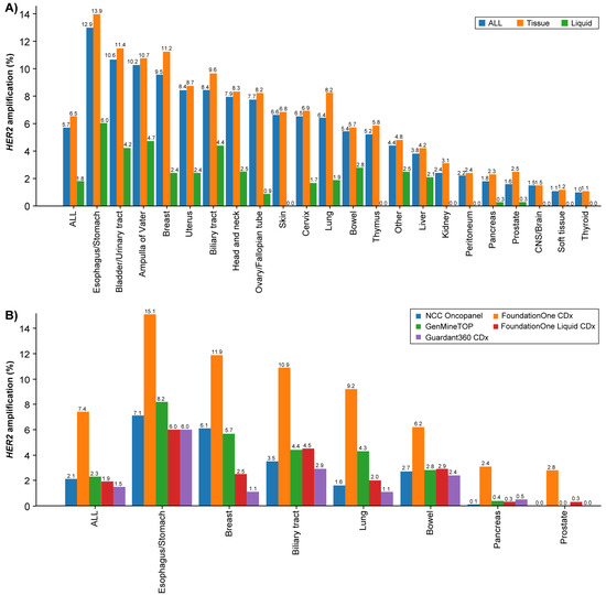
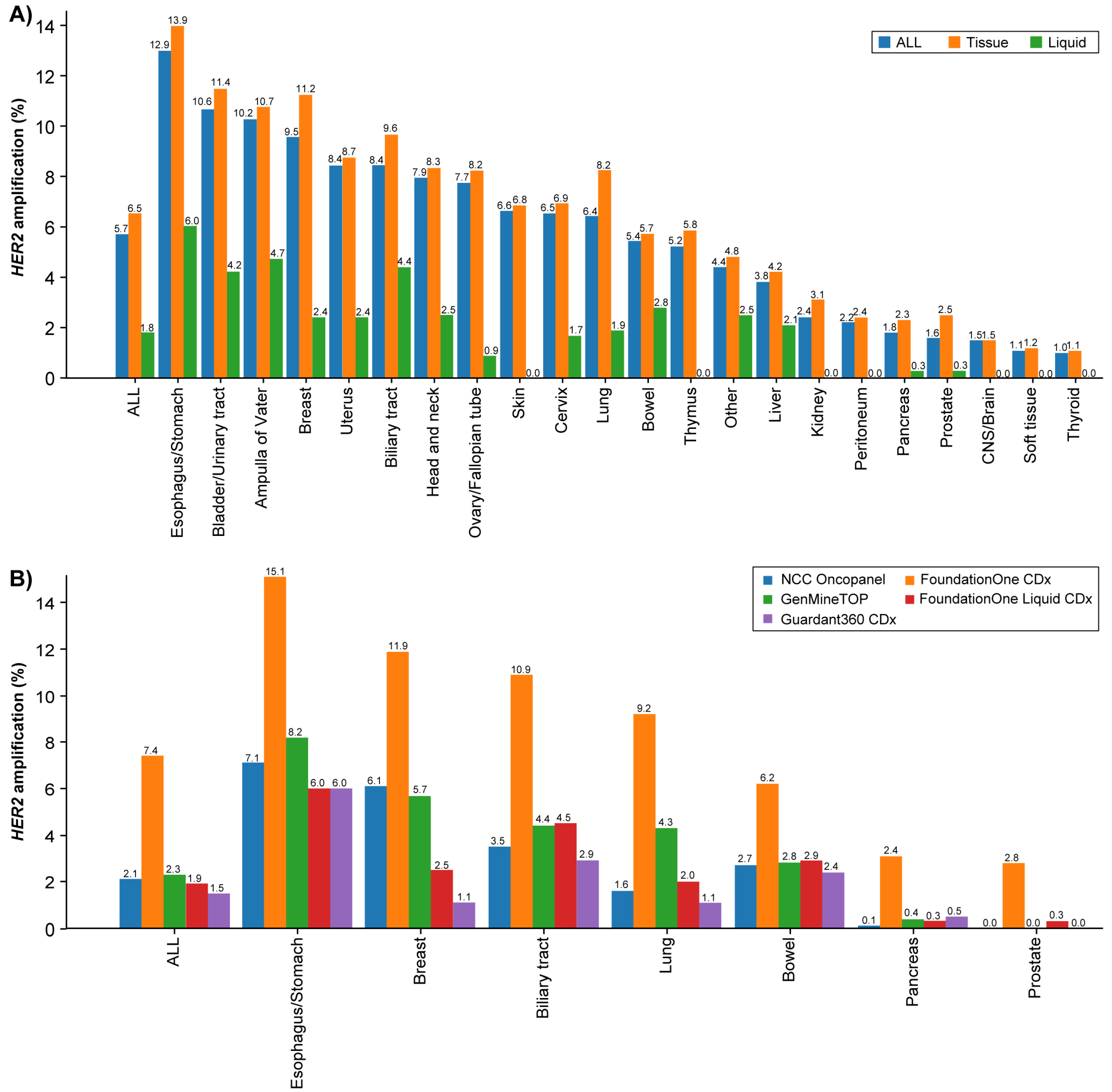
Tissue CGP testing was predominant (82.8% [73,960/89,374]), whereas plasma CGP testing accounted for 17.2% (15,414/89,374) of all tests. HER2 amplification was detected in 6.5% (4839/73,960) of tissue CGP, which was higher than 1.8% (280/15,414) for plasma CGP. When stratified by assay platforms, F1CDx accounted for 84.2% (62,299/73,960) of tissue CGP and F1LCDx for 89.0% (13,719/15,414) of plasma CGP. In tissue CGP, the highest rates of HER2 amplification were observed in the esophagus/stomach (13.9% [667/4801]), bladder/urinary tract (11.4% [150/1319]), breast (11.2% [609/5461]), and ampulla of Vater (10.7% [52/488]) (Figure 3). In plasma CGP, detection rates were highest in the esophagus/stomach (6.0% [42/698]), and subsequently in the ampulla of Vater (4.7% [2/43]), biliary tract (4.4% [78/1788]), and bladder/urinary tract (4.2% [7/165]) (Figure 3A and Table S3). The HER2 amplification detection rates by assay platform were 7.4% for F1CDx, 2.3% for GMTOP, 2.1% for NOP, 1.9% for F1LCDx, and 1.5% for G360CDx (Figure 3B and Table S3).
Figure 3.
Prevalence of HER2 amplification by CGP. (A) Percentage of HER2 amplification detected according to sample type. (B) Percentage of HER2 amplification by assay platforms in selected tumor types. CGP: comprehensive genomic profiling; CNS: central nervous system; HER2: human epidermal growth factor receptor 2.
3.5. Patient Characteristics by HER2 Amplification Status
In the HER2 amplification and HER2 no-amplification groups, the mean [SD] age (62.5 [12.0] years [n = 5119]) vs. 62.5 [12.5] years [n = 84,255]) and proportion of smokers (44.4% [2275/5119]) vs. 44.2% [37,208/84,255]) were comparable (Table 1). Among the assay platforms, HER2 amplification was more frequently detected with tissue CGP (particularly with F1CDx: 89.6% [4586/5119]) but was less common with plasma CGP (particularly with G360CDx: 0.5% [26/5119]) (Table 1). Patients with a record of HER2-positive (IHC 3+ or fluorescence in situ hybridization [FISH] or ISH) status prior to CGP testing had higher rates of HER2 amplification, and patients who tested HER2 negative prior to CGP testing tended to have no detectable HER2 amplification (HER2 no-amplification group). However, among HER2-positive patients, the proportion of HER2 amplification by CGP was relatively low (IHC 3+: 61.5% [278/452] in breast, 70.8% [400/565] in esophagus/stomach; ISH-positive: 38.4% [84/219] in breast, 40.0% [16/40] in esophagus/stomach) (Table S4). Conversely, with respect to other predictive biomarkers examined prior to CGP, strong associations were observed for KRAS and BRAFV600E mutations in bowel (predominantly colorectal) cancer (p < 0.0001 and p = 0.0008, respectively), homologous recombination deficiency (HRD) in ovarian cancer (p = 0.0012), and programmed death-ligand 1 expression in lung cancer (p = 0.0074) (Table S5).
3.6. Mutation Frequencies of Multiple Genes from CGP Assay
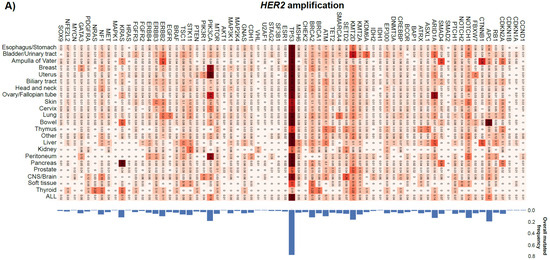
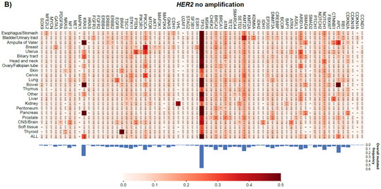
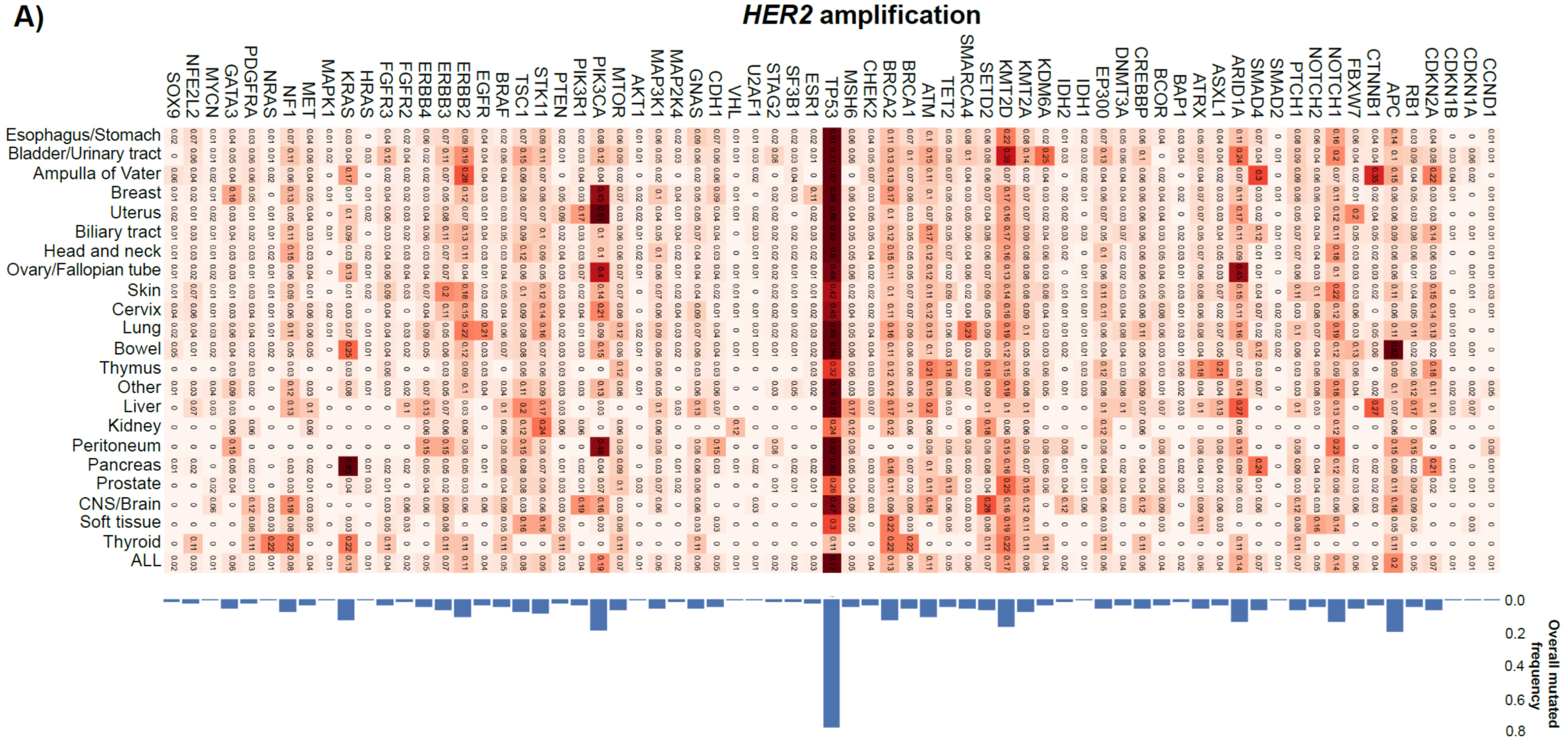
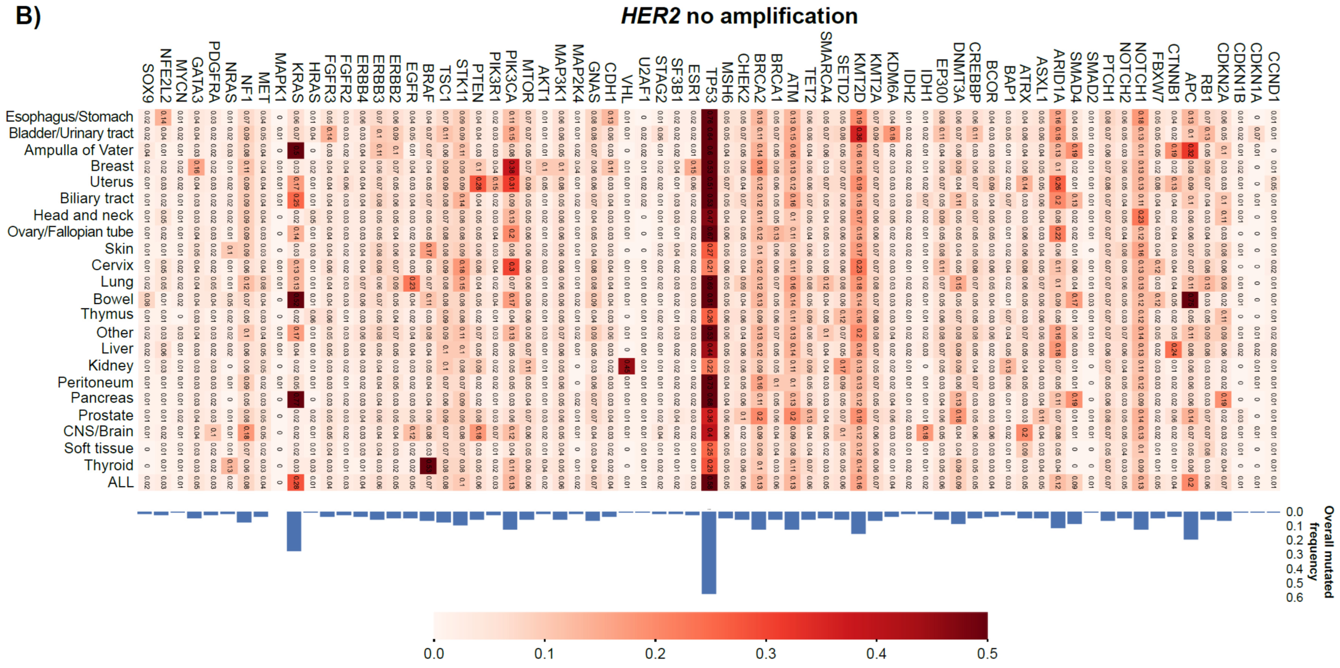
There were more numbers and types of co-occurring gene mutations in the HER2 amplification group than in the HER2 no-amplification group. KRAS mutations were more common in the HER2 no-amplification group (28%) than in the HER2 amplification group (13%). TP53 mutations were common across tumor types in both the HER2 amplification and HER2 no-amplification groups. The frequency of TP53 mutations was also similar across tumor types in both the HER2 amplification (77%) and HER2 no-amplification (58%) groups (Figure 4). Amplified genes in both the HER2 amplification and HER2 no-amplification groups were identified. In the HER2 amplification group, co-amplification occurred in 83% of cases. The most frequent co-amplified genes were CDK12 (33%) and MYC (16%). In contrast, in the HER2 no-amplification group, the most frequent genes were MYC (7%), RAD21 (5%), and CCND1 (4%). CDK12 gene showed the highest frequency ratio (HER2 amplification/no-amplification group) (Figure S1). In comparison, gene fusions were infrequent (2.7%) in the HER2 amplification group. The main driver genes were ERBB2 (n = 15), FGFR3 (n = 12), FGFR2 (n = 6), BRAF (n = 6), RET (n = 6), ALK (n = 5), and ERG (n = 2). The fusion partners of ERBB2 were heterogeneous, while those of other genes were largely restricted to specific partners: TACC3 for FGFR3, TACC2 for FGFR2, DENND2A for BRAF, CCDC6 for RET, EML4 and HMBOX1 for ALK, and TMPRSS2 for ERG (Table S6).
Figure 4.
Heat map of gene mutation frequency by CGP. (A) HER2 amplification group; (B) HER2 no-amplification group. CGP: comprehensive genomic profiling; CNS: central nervous system; HER2: human epidermal growth factor receptor 2.
3.7. Status of Treatment After EP Evaluation
In the HER2 amplification group, a genomically matched new therapeutic drug treatment was recommended by EP to 62.2% (3183/5119) of patients (recommendation range for all tumors: 38.2–69.2%). Among them, 23.5% (748/3183) were administered the recommended drug (Table 2). Furthermore, 39.3% (294/748) of them received anti-HER2 drugs (Table S7).
Table 2.
Evaluation of expert panel-recommended genomically matched drug treatments for the HER2 amplification group and the HER2 no-amplification group.
In the HER2 no-amplification group, a genomically matched new therapeutic drug treatment was recommended by EP to 38.9% (32,798/84,225) of patients (recommendation range for all tumors: 23.9–58.3%). Among them, 17.8% (5840/32,798) were administered the recommended drug (Table 2). Furthermore, 1.1% (62/5840) of them received anti-HER2 drugs (Table S7).
4. Discussion
This large-scale, real-world study determined the prevalence of HER2 amplification across a wide spectrum of solid tumors based on CGP testing using the C-CAT database. With approximately 90,000 patients included in the analysis and over 5100 HER2 amplification cases, this study represents one of the most extensive datasets examining HER2 amplification beyond conventionally studied tumor types such as breast and gastric cancers.
The prevalence of HER2 amplification in all solid tumors assessed by CGP testing using NGS was 5.7%. In addition to its well-known high prevalence in esophagus/stomach (12.9%) and breast (9.5%) tumors, other major tumor types with high frequencies included tumors of the bladder/urinary tract (10.6%), biliary tract (8.4%), and uterus (8.4%). In smaller fractions with fewer than 1000 tested cases, very high amplification rates were observed in salivary duct carcinoma (a Level 3 subset of head and neck cancer; 40.2%), extramammary Paget’s disease (a Level 2 subset of skin cancer; 27.7%), and ampulla of Vater (Level 1; 10.2%). These findings confirm that HER2 amplification is not confined to breast and gastric cancers but extends across a broader tumor spectrum including smaller fractions. There are several reports describing HER2 amplification rates in these tumor types using NGS. Based on published NGS analyses, the reported prevalence of HER2 amplification is 12.8% (33/256) in NSCLC [17], 11.9% (12/101) in gastric cancer, 9.4% (6/64) in esophageal cancer, 5.3% (8/151) in endometrial cancer, 5.2% (6/116) in bladder cancer, 4.9% (21/426) in biliary cancer, and 2% (26/1300) to 3.6% (30/842) in colorectal cancer [18,19]. However, due to small sample sizes, direct comparison with the present study remains difficult. Similarly, in a cohort of 37,436 sequenced cases from AACR Project GENIE, the prevalence of HER2 amplification was approximately 12% in esophagogastric cancer, 10% in breast cancer, and <4% in all other cancers [9,10]. In another study that evaluated 429,666 solid tumors profiled using F1CDx, HER2 amplification was found in 10.9% of patients with bladder cancer, 9.9% with breast cancer, 15.5% with gastroesophageal cancer, 3.9% with NSCLC, and 3.7% with colorectal cancer [20].
The HER2 amplification rates observed in this study for tumors of the esophagus/stomach (12.9%) and breast (9.5%) are generally consistent with previously reported data obtained using NGS and other methods for determining amplification [1,21]. However, considering the differences in clinical practice for HER2 testing between the United States (where testing targets overexpression by IHC) and Japan (where, for the time being, testing is expected to focus only on amplification by CGP), understanding the concordance between HER2 gene amplification and protein overexpression is of critical clinical importance. In this study, it was possible to cross-reference CGP test results with those of IHC and ISH assays prior to CGP. Relying solely on amplification may lead to missing treatable patients, underscoring the need for cautious clinical application. This finding raises concerns based on the observation that the prevalence of HER2 amplification by CGP among HER2 IHC 3+ or ISH-positive cases was not necessarily high in breast and esophagus/stomach cancers. However, these comparisons have limitations, such as cases in which different specimens were used at the time of testing.
In this study, CGP testing was predominantly performed with tissue CGP (82.8%), rather than plasma CGP (17.2%), and the prevalence of HER2 amplification was 6.5% with tissue CGP compared with 1.8% with plasma CGP. In addition, the detection rates by assay platform were 7.4% for F1CDx, 2.1% for NOP, 2.3% for GMTOP, 1.9% for F1LCDx, and 1.5% for G360CDx, indicating that the prevalence of HER2 amplification in this C-CAT cohort was substantially influenced by the detection rate of F1CDx. Furthermore, to clarify the details of differences in detection rates across assay platforms, we conducted a tumor type-specific analysis for seven tumor types. In all tumor types, the prevalence of HER2 amplification with F1CDx was markedly higher than that with the other four platforms.
Given that in the HERALD trial [6], patient enrollment was performed using G360CDx and the efficacy of T-DXd was demonstrated, the cutoff for HER2 amplification in the companion diagnostic-approved G360CDx may serve as the gold standard in Japan. On the other hand, an ongoing expansion cohort of the HERALD trial is enrolling patients with HER2 amplification confirmed by any pharmaceutical-approved CGP testing, and the results from this cohort are expected to provide further insight into tissue-selected populations (jRCT2080224635). The results of the HERALD expansion cohort might warrant close attention to assess the correlation between the detection of HER2 amplification by F1CDx and the therapeutic efficacy of T-DXd.
In addition, results from the exploratory biomarker analyses of the DESTINY-PanTumor02 trial [22] showed that the overall response rate (ORR) in IHC 3+ cases and the ORR in HER2 amplification-positive cases identified using the Guardant OMNI platform, which has analytical performance comparable to G360CDx, were similar (61.3% and 60.4%, respectively) [23]. On this basis, detection by F1CDx alone may carry the risk of designating patients as HER2-positive outside the fraction corresponding to IHC 3+, for whom treatment efficacy can be expected. To avoid this, additional IHC testing may be useful in cases with low copy numbers that are identified as HER2 amplification-positive by CGP testing [24]. However, the range considered as low copy number is not sufficiently defined and may also vary depending on the panels. This range may require further validation, for example, through cross-validation or drug response assessment.
In this study, we also analyzed differences in co-occurring genetic alterations between the HER2 amplification and HER2 no-amplification groups. With respect to co-occurrence, gene mutations were more numerous and diverse in the HER2 amplification group than in the HER2 no-amplification group. Among these, a marked difference was observed in KRAS mutations, which were more frequent in the HER2 no-amplification group (28%) than in the HER2 amplification group (13%). In some tumor types, KRAS mutations and ERBB2 (HER2) amplification have been reported to exhibit a nearly mutually exclusive relationship [25], which may have contributed to this bias. Clinically important, however, is the coexistence of KRAS mutations in the HER2 amplification group, as multiple studies, including the tumor-agnostic MyPathway basket trial [22,26], have reported that KRAS mutations confer resistance to anti-HER2 therapies. Although it is unclear if KRAS mutations are related to the efficacy of T-DXd from this study, this may be relevant in tumor types less responsive to T-DXd or in HER2 amplification-positive cases with low copy numbers, where determination of the KRAS mutation status could be critical for treatment decisions. Further studies are required to confirm the correlation between KRAS mutations and efficacy of T-DXd, as well as to elucidate the detailed mechanisms involved.
Indeed, in the DESTINY-PanTumor02 and HERALD trials, almost no efficacy was observed in pancreatic cancer, where KRAS mutations are highly prevalent. Similarly, co-amplification of HER2 and CDK12 in breast cancer has been reported to be associated with poor prognosis and resistance to anti-HER2 therapy (NGS enables assessment not only of the primary HER2 gene amplification status but also of the coexistence of genomic alterations related to treatment resistance, and may provide novel therapeutic options, particularly in the presence of activating mutations) [20,27]. It also holds promise for advancing future diagnostic and therapeutic strategies.
Our analysis further suggests that CGP is often performed late in the treatment continuum, particularly in common cancers, where it is usually initiated after third-line therapy. This practice may limit timely access to targeted therapies and highlights the need to integrate CGP earlier in the diagnostic and treatment pathway [28,29]. Generally, treatment achievement ratio after EP evaluation is expected to be around 10% [30,31,32,33], which was lower than that reported in this study (HER2 amplification group, 23.5%, and HER2 no-amplification group, 17.8%) and could be attributed to the differences in the denominator.
The strengths of this study include the fact that this is a large study with approximately 90,000 cases, provides information on the prevalence of HER2 amplification in a cross-section of Japanese patients from the C-CAT database, and provides details of the characteristics of the patients with HER2 amplification. With the availability of limited population-based data on HER2 expression across different cancer types, the findings of this study contribute valuable information about HER2 positivity, especially with an increasing number of HER2 targeted therapies in the treatment landscape.
As a limitation, this is a retrospective study conducted using the C-CAT database and was restricted to patients who underwent CGP testing; the frequency of HER2 amplification, clinical background, and mutational status cannot be readily generalized to the Japanese population. Another point of caution is that, in this study, HER2 amplification was defined as the presence or absence of records on the C-CAT database and not up to copy number. Additionally, as evident from the detection rates of HER2 amplification across assay platforms, the cutoff settings for gene amplification were not uniform (F1CDx [≥6 copies], NOP [≥8 copies], GMTOP [≥6 copies]). In particular, F1CDx is set to capture HER2 amplification positivity more broadly. Given that approximately 70% of the nearly 90,000 cases in this study were tested with F1CDx, it should be noted that this has a certain influence on the estimated prevalence. In addition, there may be an option to not enter data into the C-CAT database, which may have resulted in missing data. However, these missing entries occur randomly in patients with HER2 amplification and in those with HER2 no-amplification; therefore, the effects of biases may be small. The specific copy number of HER2 amplification could not be determined because it was not recorded in this database. Therefore, according to the copy number for each CGP platform, prospective validation of the usefulness of approved anti-HER2 drugs (particularly T-DXd) in patients with HER2 amplification is needed. Further, it is assumed that fresh pathological specimens were used for IHC and FISH testing, whereas CGP testing may have been performed on specimens obtained at different time points. These specimens may include long-term stored specimens, specimens re-biopsied after chemotherapy, or blood-based CGP samples. HER2 gene and protein expression may vary under these conditions. Lastly, it was not possible to assess institutional differences in test or platform selection within this population.
5. Conclusions
In conclusion, in this nationwide study of 89,374 Japanese patients with solid tumors, HER2 amplification was identified in 5.7%, with the highest prevalence in esophagus/stomach and urothelial cancers. Detection rates differed markedly by assay modality (tissue CGP 6.5% vs. plasma CGP 1.8%) and across assay platforms. Notably, F1CDx, which accounted for most tests, showed a substantially higher detection rate (7.4%) than the other four platforms (1.5–2.3%); accordingly, interpretation of the observed prevalence may need to take this distribution into account. As a clinical consideration, when using CGP-based HER2 amplification results to guide patient selection for anti-HER2 therapy, cautious interpretation by an EP may be warranted.
Supplementary Materials
The following supporting information can be downloaded at: https://www.mdpi.com/article/10.3390/curroncol33040195/s1, Figure S1: Heat map of copy number alteration A) with and B) without HER2 amplification; Table S1: Patient characteristics by tumor type; Table S2: Prevalence of HER2 amplification according to the OncoTree hierarchical classification system; Table S3: Percentage of HER2 amplification detected by CGP according to sample type; Table S4: Comparison of the HER2 status between prior companion diagnostic testing and CGP testing in selected tumor types; Table S5: Relationship between the HER2 status from CGP testing and predictive biomarker statuses from prior companion diagnostic testing in selected tumor types; Table S6: Detected driver gene fusions/rearrangements in cases with and without HER2 amplification; Table S7: Evaluation of expert panel-recommended genomically matched drug treatments (treatment policy and applied drug groups) for the HER2 amplification group and the HER2 no-amplification group.
Author Contributions
Conceptualization, Y.H., J.S. and O.T.; formal analysis, O.T.; investigation, Y.H., J.S. and O.T.; data curation, Y.H., J.S. and O.T.; writing—review and editing, Y.H., J.S. and O.T. All authors have read and agreed to the published version of the manuscript.
Funding
This study is sponsored by Daiichi Sankyo Co., Ltd. In March 2019, AstraZeneca entered into a global development and commercialization collaboration agreement with Daiichi Sankyo for trastuzumab deruxtecan (T-DXd; DS-8201).
Institutional Review Board Statement
This study was approved by the Ethical Research Practice Committee for Research on Human Tissue, Information, and Other Human Material (Approval No. 000790; 18 March 2025) and reviewed by the C-CAT Data Utilization Review Board (Approval ID: CDU2024-026N; 16 October 2024). This study was conducted in compliance with the ethical principles stipulated in the Declaration of Helsinki, "Ethical Guidelines for Medical and Health Research Involving Human Subjects (Ministry of Education, Culture, Sports, Science and Technology, Ministry of Health, Labour and Welfare, Ministry of Economy, Trade and Industry Notification No. 1 of 2021)," the Act on the Protection of Personal Information (Act No. 57 of 2003)," and related notices, as well as the study protocol (Registry and the Registration No. of the study: UMIN000057382).
Informed Consent Statement
Informed consent was obtained from all subjects involved in the study.
Data Availability Statement
The raw data supporting the conclusions of this article will be made available by the corresponding author and the funder, Daiichi Sankyo Co., Ltd., on request.
Acknowledgments
We thank the C-CAT Data Utilization Review Board for allowing access to the C-CAT Research-Use Portal site and their approval of our study protocol. Medical writing support was provided by Annirudha Chillar, of Cactus Life Sciences (part of Cactus Communications), and was funded by Daiichi Sankyo Co., Ltd.
Conflicts of Interest
Yutaka Hatanaka received honoraria from Daiichi Sankyo Co., Ltd., AstraZeneca, Eli Lilly, Chugai Pharmaceutical, MSD, and Novartis; received research funding from Daiichi Sankyo Co., Ltd., Chugai Pharmaceutical, Eli Lilly, Konica Minolta REALM, Sysmex, DNA Chip Research, and BostonGene; and also received support for the present manuscript from Daiichi Sankyo Co., Ltd. Jun Sasano and Osamu Takizawa are employees of Daiichi Sankyo Co., Ltd. The funders were involved in the study design and data analysis.
References
- Iqbal, N.; Iqbal, N. Human Epidermal Growth Factor Receptor 2 (HER2) in Cancers: Overexpression and Therapeutic Implications. Mol. Biol. Int. 2014, 2014, 852748. [Google Scholar] [CrossRef] [Scilit]
- Wolff, A.C.; Somerfield, M.R.; Dowsett, M.; Hammond, M.E.H.; Hayes, D.F.; McShane, L.M.; Saphner, T.J.; Spears, P.A.; Allison, K.H. Human Epidermal Growth Factor Receptor 2 Testing in Breast Cancer: ASCO-College of American Pathologists Guideline Update. J. Clin. Oncol. 2023, 41, 3867–3872. [Google Scholar] [CrossRef] [Scilit]
- Burstein, H.J. The Distinctive Nature of HER2-Positive Breast Cancers. N. Engl. J. Med. 2005, 353, 1652–1654. [Google Scholar] [CrossRef] [Scilit] [PubMed]
- Oh, D.Y.; Bang, Y.J. HER2-Targeted Therapies—A Role Beyond Breast Cancer. Nat. Rev. Clin. Oncol. 2020, 17, 33–48. [Google Scholar] [CrossRef] [Scilit] [PubMed]
- Yan, M.; Schwaederle, M.; Arguello, D.; Millis, S.Z.; Gatalica, Z.; Kurzrock, R. HER2 Expression Status in Diverse Cancers: Review of Results From 37,992 Patients. Cancer Metastasis Rev. 2015, 34, 157–164. [Google Scholar] [CrossRef] [Scilit] [PubMed]
- Yagisawa, M.; Taniguchi, H.; Satoh, T.; Kadowaki, S.; Sunakawa, Y.; Nishina, T.; Komatsu, Y.; Esaki, T.; Sakai, D.; Doi, A.; et al. Trastuzumab Deruxtecan in Advanced Solid Tumors With Human Epidermal Growth Factor Receptor 2 Amplification Identified by Plasma Cell-Free DNA Testing: A Multicenter, Single-Arm, Phase II Basket Trial. J. Clin. Oncol. 2024, 42, 3817–3825. [Google Scholar] [CrossRef] [Scilit]
- Kohno, T.; Kato, M.; Kohsaka, S.; Sudo, T.; Tamai, I.; Shiraishi, Y.; Okuma, Y.; Ogasawara, D.; Suzuki, T.; Yoshida, T.; et al. C-CAT: The National Datacenter for Cancer Genomic Medicine in Japan. Cancer Discov. 2022, 12, 2509–2515. [Google Scholar] [CrossRef] [Scilit]
- Horie, S.; Saito, Y.; Kogure, Y.; Mizuno, K.; Ito, Y.; Tabata, M.; Kanai, T.; Murakami, K.; Koya, J.; Kataoka, K. Pan-Cancer Comparative and Integrative Analyses of Driver Alterations Using Japanese and International Genomic Databases. Cancer Discov. 2024, 14, 786–803. [Google Scholar] [CrossRef] [Scilit]
- Meric-Bernstam, F.; Johnson, A.M.; Dumbrava, E.E.I.; Raghav, K.; Balaji, K.; Bhatt, M.; Murthy, R.K.; Rodon, J.; Piha-Paul, S.A. Advances in HER2-Targeted Therapy: Novel Agents and Opportunities Beyond Breast and Gastric Cancer. Clin. Cancer Res. 2019, 25, 2033–2041. [Google Scholar] [CrossRef] [Scilit]
- André, F.; Arnedos, M.; Baras, A.S.; Baselga, J.; Bedard, P.L.; Berger, M.F.; Bierkens, M.; Calvo, F.; Cerami, E.; Chakravarty, D.; et al. AACR Project GENIE: Powering Precision Medicine Through an International Consortium. Cancer Discov. 2017, 7, 818–831. [Google Scholar] [CrossRef] [Scilit]
- Cancer Genomic Medicine and Cancer Gene Panel Testing. C-CAT Registration Status. Available online: https://for-patients.c-cat.ncc.go.jp/registration_status/ (accessed on 29 September 2025).
- Nakata, E.; Osone, T.; Ogawa, T.; Taguchi, T.; Hattori, K.; Kohsaka, S. Prevalence of Neurotrophic Tropomyosin Receptor Kinase (NTRK) Fusion Gene Positivity in Patients With Solid Tumors in Japan. Cancer Med. 2024, 13, e7351. [Google Scholar] [CrossRef] [Scilit]
- National Cancer Center Japan. List of Information Providers and Issues. Available online: https://www.ncc.go.jp/jp/c_cat/use/release/index.html (accessed on 30 August 2025).
- Kundra, R.; Zhang, H.; Sheridan, R.; Sirintrapun, S.J.; Wang, A.; Ochoa, A.; Wilson, M.; Gross, B.; Sun, Y.; Madupuri, R.; et al. OncoTree: A Cancer Classification System for Precision Oncology. JCO Clin. Cancer Inform. 2021, 5, 221–230. [Google Scholar] [CrossRef] [Scilit]
- von Elm, E.; Altman, D.G.; Egger, M.; Pocock, S.J.; Gøtzsche, P.C.; Vandenbroucke, J.P. Strengthening the Reporting of Observational Studies in Epidemiology (STROBE) Statement: Guidelines for Reporting Observational Studies. BMJ 2007, 335, 806–808, Erratum in BMJ 2007, 335, 0–a. https://doi.org/10.1136/bmj.39386.490150.94. [Google Scholar] [CrossRef] [Scilit] [PubMed]
- Schulz, K.F.; Altman, D.G.; Moher, D. CONSORT 2010 Statement: Updated Guidelines for Reporting Parallel Group Randomised Trials. BMJ 2010, 340, c332. [Google Scholar] [CrossRef] [Scilit] [PubMed]
- Kıvrak, H.; Özakıncı, H.; Karasoy, D.; Sak, S.D. HER2 Amplification by Next-Generation Sequencing in Lung Carcinoma: A Comparison of NGS Amplified and Non-Amplified Cases by Immunohistochemistry and In Situ Hybridization. Balkan Med. J. 2022, 39, 21–29. [Google Scholar] [CrossRef] [Scilit]
- Dumbrava, E.E.I.; Balaji, K.; Raghav, K.; Hess, K.; Javle, M.; Blum-Murphy, M.; Ajani, J.; Kopetz, S.; Broddus, R.; Routbort, M.; et al. Targeting ERBB2 (HER2) Amplification Identified by Next-Generation Sequencing in Patients with Advanced or Metastatic Solid Tumors Beyond Conventional Indications. JCO Precis. Oncol. 2019, 3, 1–12. [Google Scholar] [CrossRef] [Scilit]
- Cenaj, O.; Ligon, A.H.; Hornick, J.L.; Sholl, L.M. Detection of ERBB2 Amplification by Next-Generation Sequencing Predicts HER2 Expression in Colorectal Carcinoma. Am. J. Clin. Pathol. 2019, 152, 97–108. [Google Scholar] [CrossRef] [Scilit] [PubMed]
- Liu, D.; Jee, J.; Drilon, A.; Heilmann, A.M.; Allen, J.M.; Schrock, A.B.; Keller-Evans, R.B.; Li, B.T. Diverse ERBB2/ERBB3 Activating Alterations and Coalterations Have Implications for HER2/3-Targeted Therapies Across Solid Tumors. Cancer Res. Commun. 2025, 5, 680–693. [Google Scholar] [CrossRef] [Scilit]
- Bonomi, M.; Spada, D.; Baiocchi, G.L.; Celotti, A.; Brighenti, M.; Grizzi, G. Targeting HER2 in Gastroesophageal Adenocarcinoma: Molecular Features and Updates in Clinical Practice. Int. J. Mol. Sci. 2024, 25, 3876. [Google Scholar] [CrossRef] [Scilit]
- Meric-Bernstam, F.; Hurwitz, H.; Raghav, K.P.S.; McWilliams, R.R.; Fakih, M.; VanderWalde, A.; Swanton, C.; Kurzrock, R.; Burris, H.; Sweeney, C.; et al. Pertuzumab Plus Trastuzumab for HER2-Amplified Metastatic Colorectal Cancer (MyPathway): An Updated Report From a Multicentre, Open-Label, Phase 2a, Multiple Basket Study. Lancet Oncol. 2019, 20, 518–530. [Google Scholar] [CrossRef] [Scilit]
- Makker, V.; Lee, J.Y.; Oh, D.Y.; Oaknin, A.; Puvvada, S.D.; Cecchi, F.; Mcewen, R.; Michelini, F.; Meric-Bernstam, F. 148P DESTINY-PanTumor02 study of Trastuzumab Deruxtecan (T-DXd) in Patients (pts) With HER2-Expressing Solid Tumors: Exploratory Biomarker Analyses of HER2 Expression and Gene Amplification in Tissue and Plasma. Ann. Oncol. 2023, 34, S239–S240. [Google Scholar] [CrossRef] [Scilit]
- FoundationOne®CDx. Technical Information. Available online: https://www.accessdata.fda.gov/cdrh_docs/pdf17/P170019S006C.pdf (accessed on 27 October 2025).
- Hechtman, J.F.; Liu, W.; Sadowska, J.; Zhen, L.; Borsu, L.; Arcila1, M.E.; Won, H.H.; Shah, R.H.; Berger, M.F.; Vakianiet, E.; et al. Sequencing of 279 Cancer Genes in Ampullary Carcinoma Reveals Trends Relating to Histologic Subtypes and Frequent Amplification and Overexpression of ERBB2 (HER2). Mod. Pathol. 2015, 28, 1123–1129. [Google Scholar] [CrossRef] [Scilit]
- Sweeney, C.J.; Hainsworth, J.D.; Bose, R.; Burris, H.A.; Kurzrock, R.; Swanton, C.; Friedman, C.F.; Spigel, D.R.; Szado, T.; Schulze, K.; et al. MyPathway Human Epidermal Growth Factor Receptor 2 Basket Study: Pertuzumab + Trastuzumab Treatment of a Tissue-Agnostic Cohort of Patients With Human Epidermal Growth Factor Receptor 2-Altered Advanced Solid Tumors. J. Clin. Oncol. 2024, 42, 258–265. [Google Scholar] [CrossRef] [Scilit] [PubMed]
- Rüschoff, J.; Nagelmeier, I.; Jasani, B.; Stoss, O. ISH-Based HER2 Diagnostics. Pathologe 2021, 42, 62–68. [Google Scholar] [CrossRef] [Scilit] [PubMed]
- Matsubara, J.; Mukai, K.; Kondo, T.; Yoshioka, M.; Kage, H.; Oda, K.; Ikeda, S.; Ebi, H.; Muro, K.; Kajiura, S.; et al. First-Line Genomic Profiling in Previously Untreated Advanced Solid Tumors: 1-Year Follow-Up of the FIRST-Dx Study. Cancer Sci. 2025, 116, 1908–1919. [Google Scholar] [CrossRef] [Scilit] [PubMed]
- Mizuno, T.; Yoshida, T.; Sunami, K.; Koyama, T.; Okita, N.; Kubo, T.; Sudo, K.; Shimoi, T.; Ueno, H.; Saito, E.; et al. Study Protocol for NCCH1908 (UPFRONT-Trial): A Prospective Clinical Trial to Evaluate the Feasibility and Utility of Comprehensive Genomic Profiling Prior to the Initial Systemic Treatment in Advanced Solid Tumour Patients. Jpn. J. Clin. Oncol. 2021, 51, 1757–1760. [Google Scholar] [CrossRef] [Scilit]
- Supplement to the 2nd Edition of the HER2 Pathological Diagnosis Guidance for Solid Tumors [Internet]. Japanese Society of Pathology. Available online: https://www.pathology.or.jp/news/20231218_guidance20221228.pdf (accessed on 13 September 2025).
- Sunami, K.; Naito, Y.; Komine, K.; Amano, T.; Ennishi, D.; Imai, M.; Kage, H.; Kanai, M.; Kenmotsu, H.; Koyama, T.; et al. Chronological Improvement in Precision Oncology Implementation in Japan. Cancer Sci. 2022, 113, 3995–4000. [Google Scholar] [CrossRef] [Scilit]
- Ida, H.; Koyama, T.; Mizuno, T.; Sunami, K.; Kubo, T.; Sudo, K.; Tao, K.; Hirata, M.; Yonemori, K.; Kato, K.; et al. Clinical Utility of Comprehensive Genomic Profiling Tests for Advanced or Metastatic Solid Tumor in Clinical Practice. Cancer Sci. 2022, 113, 4300–4310. [Google Scholar] [CrossRef] [Scilit]
- Fukada, I.; Mori, S.; Hayashi, N.; Hosonaga, M.; Xiaofei, W.; Yamazaki, M.; Ueki, A.; Kiyotani, K.; Tonooka, A.; Takeuchi, K.; et al. Prognostic Impact of Cancer Genomic Profile Testing for Advanced or Metastatic Solid Tumors in Clinical Practice. Cancer Sci. 2023, 114, 4632–4642. [Google Scholar] [CrossRef] [Scilit]

Disclaimer/Publisher’s Note: The statements, opinions and data contained in all publications are solely those of the individual author(s) and contributor(s) and not of MDPI and/or the editor(s). MDPI and/or the editor(s) disclaim responsibility for any injury to people or property resulting from any ideas, methods, instructions or products referred to in the content. |
© 2026 by the authors. Licensee MDPI, Basel, Switzerland. This article is an open access article distributed under the terms and conditions of the Creative Commons Attribution (CC BY) license.