


Simple Summary
Thymoma is a rare tumor that usually grows slowly and stays confined to the chest and only rarely spreads to distant organs such as the liver. This report describes an unusual patient whose first sign of an occult type B1 thymoma was a large hepatic mass. Although type B1 thymoma is generally considered to have a favorable prognosis, the patient later developed repeated tumors involving several sites, including a second hepatic recurrence eight years after the initial presentation. Complete surgical removal of the liver tumors, including a second liver operation, achieved long-term disease control, and the patient remained free of recurrence two years after the most recent surgery. This case shows that even apparently low-risk thymoma can behave aggressively and recur many years later. Long-term imaging of both the chest and abdomen, careful assessment of unusual hepatic tumors, and multidisciplinary treatment may help improve the management of patients with rare metastatic thymoma.
Abstract
Thymoma is a rare tumor of thymic epithelial cells, accounting for 20–30% of mediastinal tumors. It typically grows slowly and remains confined to the anterior mediastinum, with distant metastasis, such as to the liver, being extremely rare. Liver metastasis and recurrence are particularly rare in type B1 thymoma, making clinical diagnosis more challenging. We report the case of a 60-year-old female who presented with right upper quadrant pain, and imaging revealed a large liver mass. She underwent hepatic bisegmentectomy of S4b and S5 and cholecystectomy, with postoperative pathology confirming hepatic type B1 thymoma. Further evaluation identified a soft tissue mass in the anterior-superior mediastinum, leading to right anterior mediastinal tumor resection and right middle lobectomy, with pathology again confirming type B1 thymoma. The patient received adjuvant chemotherapy. Four years later, tumor recurrence prompted diaphragmatic, paraspinal, and pulmonary nodule resections. Another recurrence occurred after 4 years, necessitating a left hemihepatectomy, which revealed recurrent type B1 thymoma in the liver. She remains recurrence-free during postoperative follow-up. Type B1 thymoma presenting with hepatic metastasis as the initial manifestation is exceedingly rare. Despite its conventionally favorable prognosis, aggressive biological behavior including hematogenous dissemination and multifocal recurrence may occur. Aggressive surgical resection, including repeat hepatectomy in selected patients, may prolong survival. Long-term abdominal surveillance and multidisciplinary management are strongly recommended for such patients.
1. Introduction
Thymoma is a tumor arising from thymic epithelial cells, presenting with diverse clinical manifestations and biological behaviors [1]. Thymoma is rare, accounting for 20–30% of mediastinal tumors, and is most commonly seen in middle-aged and elderly individuals, with no significant sex difference in incidence [2]. Thymoma typically grows slowly and is primarily confined to the anterior mediastinum. It tends to be locally invasive, affecting nearby structures like the pericardium and large blood vessels, while distant metastasis to organs such as the liver, lungs, and bones is exceedingly rare [3].
The WHO classifies thymoma into types A, AB, B1, B2, B3, and C (thymic carcinoma) based on the morphology and biological behavior of tumor cells. Type B1 thymoma typically exhibits relatively benign biological behavior and often resembles the normal cortical tissue of the thymus [4,5]. Hepatic metastases from thymoma are extremely rare in clinical practice, typically occurring in advanced cases, making clinical diagnosis challenging. This article reports a rare case of hepatic metastasis from occult Type B1 thymoma with multifocal spread and multiple recurrences. The clinical features, differential diagnosis, and treatment strategies are reviewed in light of the available literature, aiming to offer guidance for clinicians managing similar cases.
2. Case Presentation
The patient, a 60-year-old woman, presented with right upper quadrant pain for 10 days. Abdominal CT at the local hospital revealed a large mass occupying the liver. The patient reported no associated symptoms such as abdominal distension, nausea, vomiting, or significant weight change. Laboratory tests revealed normal platelet counts and liver function, with negative results for viral infection markers and tumor markers. The patient had no history of hepatitis and had undergone a total hysterectomy for uterine fibroids over 20 years ago.
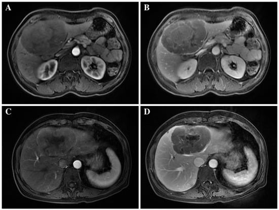
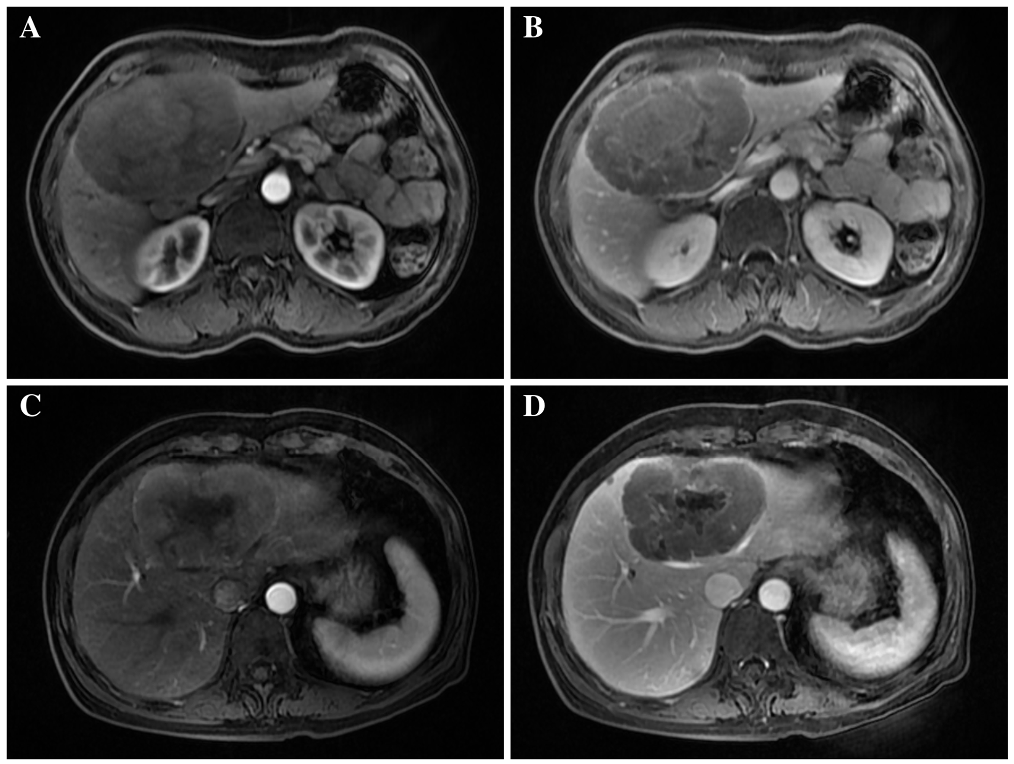
The patient was admitted and underwent enhanced MRI, revealing an irregular, shallow lobulated mass in the S4-S5 segments of the liver, with clear borders, measuring approximately 11.0 × 7.7 × 9.4 cm (Figure 1A,B). The lesion showed hypointensity on T1-weighted imaging and heterogeneous hyperintensity on T2-weighted imaging, with internal areas of markedly increased T2 signal. It also demonstrated marked hyperintensity on diffusion-weighted imaging (DWI) and mild enhancement following contrast-enhanced imaging. After comprehensive preoperative evaluation, no contraindications to surgery were identified, and the patient subsequently underwent surgical treatment. Intraoperatively, the mass was located in segments S4b and S5. It had a soft texture with clear borders, and no metastatic nodules were found in the rest of the liver. Thus, resection of hepatic segments S4b and S5, together with cholecystectomy, was performed. Rapid frozen pathology from the intraoperative hilar lymph node biopsy revealed a proliferative lymphoid lesion with predominantly T-cells. The operation proceeded smoothly, achieving complete resection of the mass. Suprahepatic and subhepatic abdominal drains were placed for postoperative monitoring of potential postoperative complications.
Figure 1.
Preoperative imaging findings of the initial and recurrent hepatic lesions. (A,B) Preoperative contrast-enhanced MR images at the initial presentation demonstrate a large, mildly lobulated and well-defined mass involving hepatic segments S4–S5, measuring approximately 11.0 × 7.7 × 9.4 cm. (C,D) Imaging obtained at the time of hepatic recurrence demonstrates a large recurrent lesion in the left liver, predominantly involving segment S4 and compressing the adjacent hepatic veins and bile duct branches.
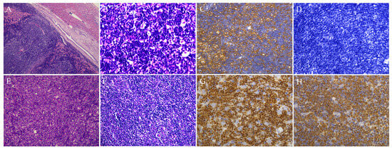
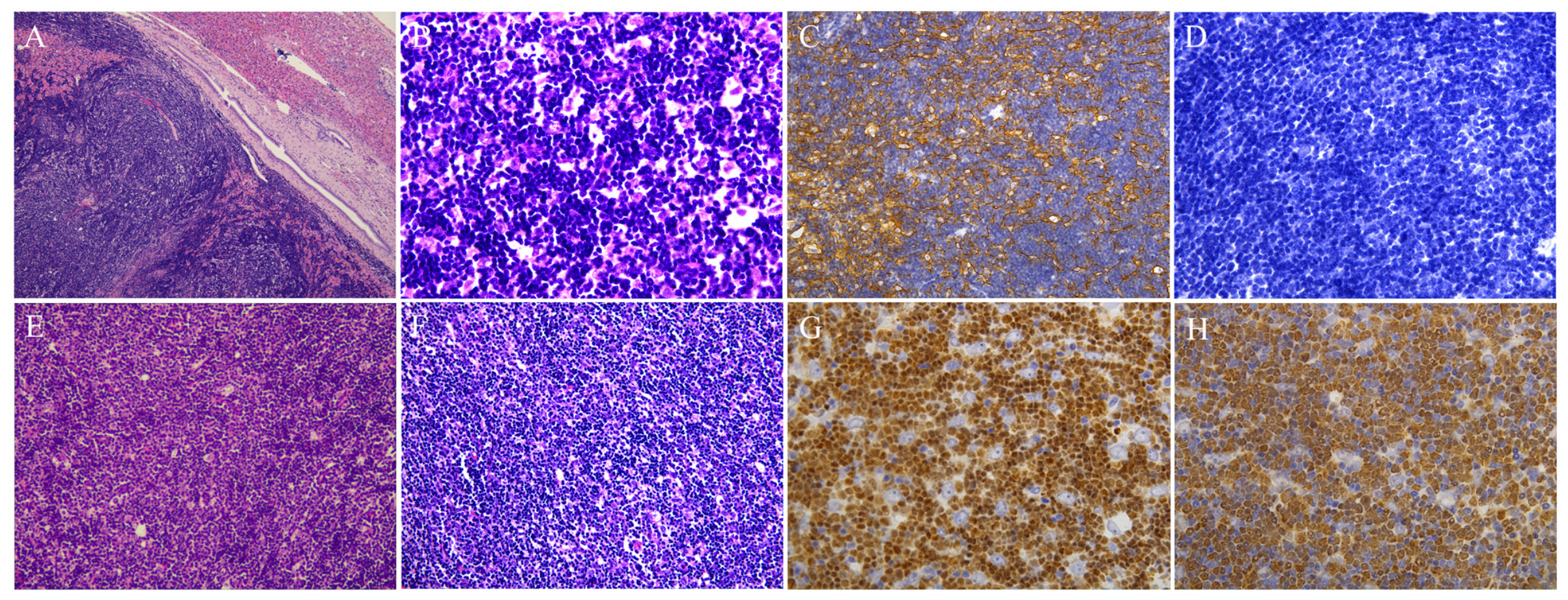
The postoperative pathology revealed a large, lobulated tumor within the liver tissue, composed of scattered ovoid epithelioid cells in a background of small lymphocytes, accompanied by lymphocyte hyperplasia, lamellar necrosis, interstitial edema, and focal lymphocyte infiltration in the surrounding liver tissue (Figure 2A,B). Immunohistochemistry showed positivity for CD3, CD5, CD2, CD7, CD4, CD8, CD43, CD1a, TdT, and CD99 (lymphocytes); CK (Figure 2C), CK5/6, p63, and p40 (ovoid cells); CD10; and Ki-67 with 80% positivity. Negative markers included EMA, CD20, PAX-5, CD23, Cyclin D1, CD56, CD30, CD15, CD34, TIA-1, Desmin, SMA, GramB, CD38, CEA, and hepatocytes (Figure 2D). In situ hybridization for EBER was negative. Lymph node specimens showed normal architecture with lymphoid tissue hyperplasia and small-to-medium cell sizes. Immunohistochemistry showed negativity for CK, CK5/6, and p63; positivity for CD3, TdT, and CD99; and Ki-67 positivity at 5%, consistent with proliferative lymphoid lesions with a predominance of T cells.
Figure 2.
Histopathological and immunohistochemical findings of the initial and recurrent hepatic lesions. (A) Low-power hematoxylin and eosin (H,E) staining of the initial hepatic tumor showing a lymphocyte-rich lobulated lesion adjacent to liver tissue (×50). (B) Higher-power H&E staining demonstrates numerous small lymphocytes with scattered ovoid epithelial cells (×400). (C) Immunohistochemical staining for cytokeratin highlights the scattered epithelial component (×400). (D) Negative immunohistochemical staining for the hepatocyte marker in the initial hepatic lesion (×400). (E) H&E staining of the primary mediastinal thymoma showing characteristic histological features of type B1 thymoma (×200). (F) H&E staining of the recurrent hepatic lesion demonstrates morphology similar to that of the initial lesion, with abundant small lymphocytes and interspersed epithelial cells (×200). (G) TdT immunostaining highlights the abundant immature lymphoid population (×400). (H) Ki-67 staining demonstrates a high overall proliferative index in the tumor area of the recurrent hepatic lesion, involving both the epithelial and lymphoid components (×400).
Based on the pathological and immunohistochemical findings, a hepatic type B1 thymoma was diagnosed. A postoperative chest CT revealed an irregularly shaped soft tissue mass in the anterior superior mediastinum, measuring approximately 4.8 × 2.4 cm, with irregular borders, and associated with multiple enlarged mediastinal and right internal mammary lymph nodes. After consultation with thoracic surgery, thymoma was considered likely. The patient recovered well postoperatively. The suprahepatic and subhepatic abdominal drains were removed on the 6th and 10th days, respectively, and the patient was discharged on the 12th postoperative day.
The patient was rehospitalized in the Department of Thoracic Surgery after a 1-month postoperative review following hepatectomy, which revealed no abnormalities. Following preoperative evaluation and exclusion of surgical contraindications, the decision was made to proceed with surgery. Intraoperatively, a space-occupying lesion measuring approximately 6 × 4 cm with a hard texture was found in the right anterior mediastinum. The tumor had peripheral invasion and extended directly into the base of the right middle lobe, the pericardium, and the right phrenic nerve, with approximately 5 cm of the phrenic nerve involved. The affected lung tissue in the right middle lobe was resected, and the right lower pole of the thymus was freed. Attempts to isolate the infiltrated phrenic nerve were unsuccessful, as the tumor had completely encircled it. Therefore, the phrenic nerve was transected distal to the affected area. The pericardium was incised, revealing that the tumor had penetrated the inner wall, though the pericardial fluid remained clear. To ensure complete resection, the tumor and affected pericardium were removed, and the pericardial defect was repaired. A chest drain was placed in the 5th intercostal space at the mid-axillary line.
The postoperative pathology report revealed a lobulated tumor structure composed primarily of ovoid epithelioid cells in a background of small lymphocytes, with active lymphocyte proliferation (Figure 2E). Immunohistochemistry showed CK (+), EMA (−), CD3 (+), CD20 (+), CD5 (+), TdT (+), CD117 (−), CD34 (−), and Ki-67 positivity at 70%. Based on immunohistochemistry and histomorphology, the pathological diagnosis confirmed a predominantly B1 thymoma, with some areas showing B2 type, and invasion into surrounding adipose and fibrous connective tissues. The patient had an uneventful postoperative recovery and was discharged on the 3rd day after chest drain removal. The patient subsequently underwent 6 cycles of adjuvant chemotherapy with cyclophosphamide, adriamycin, and cisplatin at an outside hospital. The patient was followed up regularly postoperatively.
Four years after the initial surgery, due to tumor recurrence, the patient underwent resection of diaphragmatic and paraspinal nodules, as well as basal segment resection of the right lower lobe, at an outside hospital. The postoperative pathology report revealed that the diaphragmatic nodule was a thymoma, predominantly type B1 and B2 with a localized area of type B3, measuring 4 × 3 × 1.5 cm, and accompanied by periosteal invasion. The fibrous tissue from the paraspinal nodule specimen showed sheets of proliferating lymphocytes of small to medium size, interspersed with scattered epithelial cells, consistent with thymoma pathology. Focal lymphocytic infiltrates were observed in some lung resection tissues. Additionally, lymph nodes from groups 2, 4, 7, 8, 9, and 10 showed reactive hyperplasia. The patient continued regular postoperative follow-up to monitor her condition.
Four years after the second surgery (and eight years after the initial presentation), the patient underwent re-examination at an outside hospital, where PET-CT revealed a new space-occupying lesion in the left lobe of the liver. To further clarify the diagnosis, the patient was admitted to our hospital and underwent MRI, which revealed recurrent hepatic lesions with an overall extent of approximately 9.2 × 6.9 × 8.7 cm, predominantly involving segment S4 and compressing the left and middle hepatic veins and adjacent bile duct branches (Figure 1C,D). The lesion exhibited hypointensity on T1-weighted imaging and hyperintensity on T2-weighted imaging, with a centrally located area of markedly increased T2 signal. Following contrast-enhanced imaging, peripheral enhancement was observed during the arterial phase, whereas mild enhancement of the lesion as a whole was noted during the venous phase. Based on the patient’s previous history, recurrent hepatic metastasis of thymoma was considered. After ruling out surgical contraindications, another surgery was planned. Intraoperatively, the tumor was located in the S4 and S2–3 segments of the liver, measuring approximately 5 cm in the S4 segment and 2 cm in the S2–3 segment, with a hard texture and well-defined borders. The left half of the liver was completely resected, along with thorough lymph node dissection. Suprahepatic and subhepatic drains were placed postoperatively.
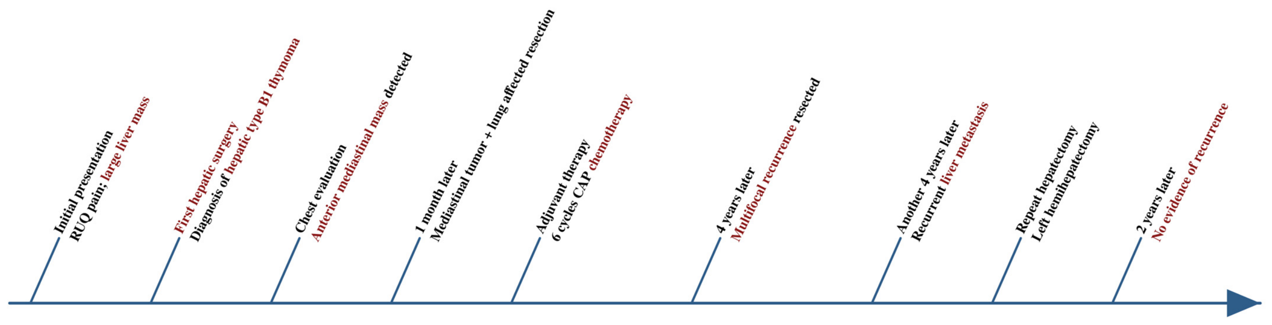
Postoperative pathology revealed tumor infiltration in the liver tissue, predominantly composed of small lymphocytes with an epithelioid cell component (Figure 2F). Immunohistochemistry showed CK (+), CD3 (+), CD20 (focal +), CD5 (+), PAX5 (individual +), Ki-67 (75% +), TdT (+) (Figure 2G,H), CD23 (−), CD117 (−), Cyclin D1 (−), and SOX11 (−), consistent with a diagnosis of liver metastasis from type B1 thymoma. The patient’s postoperative recovery was uneventful, with suprahepatic and infrahepatic drains removed on days 6 and 7, respectively. The patient was discharged on day 8 postoperatively. The patient has had regular postoperative follow-ups and is now 2 years post-surgery with no signs of recurrence. The patient’s clinical course is summarized in Figure 3.
Figure 3.
Timeline of the patient’s medical history.
3. Discussion
Thymoma is a rare tumor that originates from thymic epithelial cells, with an incidence rate of approximately 0.13 per 100,000 person-years [6]. Although distant metastases are observed in roughly 7% of thymoma patients, liver involvement is exceedingly rare [7,8]. A literature review identified 12 previously reported cases of surgically resected hepatic metastases from thymoma (Table 1). The patients ranged in age from 48 to 73 years, with a median age of 58 years. Histological subtypes were reported in 10 cases and included types A, AB, B1, B2, and B3, suggesting that hepatic metastasis may not be confined to specific WHO histological subtype. Synchronous hepatic metastases were observed in only two cases. Among the 10 patients with metachronous metastases, the median interval from primary thymoma to hepatic metastasis was 78 months (13–252 months). All 12 patients underwent hepatic resection, occasionally combined with chemotherapy or transarterial embolization. This case represents one of the few reported instances of type B1 thymoma where occult liver metastasis was the initial manifestation. The disease course spanned eight years, during which the patient underwent two liver surgeries, and the multiple lesions recurred repeatedly.
Table 1.
Reported Cases of Thymoma with Hepatic Metastasis, Including the Present Case.
The patient presented with right upper quadrant abdominal pain and a large hepatic mass measuring 11.0 × 7.7 × 9.4 cm on enhanced MRI, without any identifiable primary lesion on preoperative workup. All standard tumor markers, including AFP, CEA, CA19–9, and CA125 were negative, and the patient had no history of viral hepatitis or liver cirrhosis. These clinical and imaging features did not strongly support the diagnosis of hepatocellular carcinoma and intrahepatic cholangiocarcinoma, although neither malignancy could be definitively excluded. In this situation, the preoperative differential diagnosis of a liver mass generally encompasses focal nodular hyperplasia of the liver, hepatic adenoma, hepatic angiomyolipoma, epithelioid hemangioendothelioma, primary hepatic lymphoma, and metastatic disease from an occult primary tumor. Nevertheless, given that hepatic metastasis from thymoma is extremely rare, it is scarcely ever incorporated into the preoperative differential diagnosis, and the subtype, B1-type thymoma, which was involved here is generally associated with relatively indolent biological behavior and a favorable prognosis [19,20]. The definitive diagnosis is contingent upon the final paraffin section and immunohistochemical detection. The histopathological differential diagnosis included primary hepatic lymphoma, particularly T-lymphoblastic lymphoma, given the abundant immature lymphoid population. However, the presence of scattered ovoid epithelial cells expressing CK, CK5/6, p63, and p40, together with immature T lymphocytes positive for CD3, CD5, TdT, and CD99, supported the diagnosis of thymoma. The subsequent pathological confirmation of a predominantly B1 thymoma in the anterior mediastinum further supported the diagnosis of hepatic metastasis. Furthermore, once the liver mass is confirmed or suspected to be of thymic origin, contrast-enhanced chest CT or whole-body PET-CT should be conducted to identify the concealed mediastinal primary lesion [21]. In this particular case, the primary anterior mediastinal thymoma was identified only after the initial hepatectomy, when postoperative chest CT revealed an anterior mediastinal mass. In retrospect, the initial preoperative evaluation was incomplete because it primarily focused on the hepatic lesion without comprehensive systemic staging. This diagnostic limitation highlights the possibility that an indeterminate hepatic mass may represent the initial manifestation of an occult extrahepatic malignancy. Although the surgery ultimately established a definitive pathological diagnosis and achieved macroscopic tumor clearance, the lack of a preoperative tissue diagnosis and complete staging limited the opportunity to determine the underlying disease and formulate an optimal oncological treatment strategy before surgery. Therefore, comprehensive systemic staging, including evaluation for a potential extra-hepatic primary tumor, should be considered before major hepatic resection. In addition, a preoperative core-needle biopsy may provide useful diagnostic clues in indeterminate hepatic masses and help guide further staging and treatment decisions.
The first hepatic operation in this case consisted of resection of hepatic segments S4b and S5 with cholecystectomy, achieving macroscopically complete resection of an 11.0 × 7.7 × 9.4 cm tumor. For hepatic metastases from non-colorectal, non-neuroendocrine primaries, complete resection (R0) remains a key prerequisite for achieving favorable long-term outcomes in appropriately selected patients [22]. In the rare literature on thymoma hepatic metastases, complete surgical resection has been associated with favorable long-term outcomes in selected patients [21,23]. Intraoperative frozen-section biopsy of the hilar lymph nodes was performed in this case, revealing reactive T-cell predominant lymphoproliferation. Although routine lymph node dissection is not generally indicated for all hepatic metastatic lesions, intraoperative lymph node sampling may provide additional diagnostic information in selected cases with uncertain hepatic pathology. In this patient, the examination of hilar lymph nodes revealed T-cell-predominant lymphoproliferative tissue, providing a nonspecific pathological clue that prompted further diagnostic consideration.
Eight years subsequent to the initial presentation and four years after the first recurrence treated elsewhere, the patient underwent left hepatectomy along with lymph node dissection due to the recurrence of thymoma in the left lobe of the liver, which was verified by MRI and PET—CT. The oncological significance of repeat hepatectomy in the context of recurrent metastatic thymoma warrants further exploration [11]. A substantial number of studies in the domain of colorectal liver metastases (CRLM) have firmly established that carefully selected patients who undergo repeat hepatectomy for CRLM achieve survival outcomes comparable to those after first hepatectomy, with acceptable morbidity [22,24]. In this patient, there was an eight-year disease course with multiple surgical interventions, all achieving macroscopic clearance, and no recurrence was detected during the follow-up subsequent to the second hepatectomy. Although this outcome suggests the feasibility of repeat surgical resection in carefully selected patients with recurrent hepatic metastases from thymoma, the relatively short follow-up period precludes conclusions regarding long-term disease control or cure. Furthermore, given the patient’s previous recurrence approximately four years after the initial surgery, continued long-term surveillance remains essential. The potential oncological benefit of repeat hepatectomy in this setting warrants further investigation. In addition, lymph node dissection was performed concurrently with the second hepatectomy in this patient. However, the oncologic significance of hepatic hilar and regional lymph node involvement in metastatic thymoma is not well characterized. Regional lymph node assessment may provide additional pathological and staging information in selected patients, particularly when nodal involvement is suspected. Nevertheless, whether lymph node dissection confers an independent oncological benefit in this setting remains uncertain and requires further investigation.
This case challenges the traditionally favorable prognosis associated with type B1 thymoma. According to the WHO classification, type B1 thymoma morphologically resembles the normal thymic cortex and generally exhibits relatively indolent biological behavior compared with type B2 and B3 thymomas. It is more frequently associated with early stage disease and is generally characterized by excellent long-term survival. In the WHO classification of thymic tumors, B1-type thymoma is generally regarded as the least aggressive subtype within the type B-subtypes, morphologically resembling normal thymic cortex and typically associated with Masaoka-Koga Stage I or II disease and five-year survival rates exceeding 90% [5,25]. However, the literature documents occasional cases of B1 thymoma with pleural dissemination, extrathoracic spread, and late recurrence [8,17], suggesting that it is arduous to precisely predict biological behavior solely based on histological subtypes. Several features in this case may help explain its unusually aggressive clinical course. Firstly, the pathology of thymectomy specimens demonstrated predominantly B1 morphology with focal B2 areas, and the recurrent diaphragmatic specimen showed B1/B2 with focal B3 components, indicating a trend of histological evolution toward a more aggressive phenotype over time. This clonal evolution phenomenon has been reported in the literature on thymoma, which may reflect the heterogeneous evolution of tumors under selective therapeutic pressure [26,27]. Secondly, the recurrence pattern of this case, where liver metastasis was detected prior to the primary mediastinal tumor, followed by multiple recurrences in the diaphragm, paravertebral area, lung, and liver, indicates hematogenous and contiguous spread rather than the pleural seeding more typical of thymoma recurrence. The aforementioned recurrence characteristics hold significant implications for the subsequent surveillance strategy. Standard follow-up protocols for resected thymoma focus primarily on thoracic imaging to detect mediastinal recurrence or pleural implants. This case strongly indicates that contrast-enhanced abdominal CT or MRI should be routinely incorporated into the long-term follow-up protocol, particularly for patients with known extrathoracic disease. PET-CT demonstrated particular utility in this patient, identifying the second hepatic recurrence at a resectable stage when it might otherwise have been missed on standard thoracic surveillance alone.
In this case, the patient received six cycles of adjuvant chemotherapy with the CAP regimen following mediastinal resection, which is the recognized first-line chemotherapy regimen for advanced thymoma. Nevertheless, the patient experienced multifocal recurrence 4 years later, indicating that the efficacy of systemic therapy in tumor control was limited [28,29,30]. In this particular case, the initial intervention, commencing with liver surgery, was not founded on a planned oncologic strategy but was instead driven by clinical manifestations (hepatic symptoms, large abdominal mass). In retrospect, if systematic staging (chest, abdomen, and pelvis enhanced CT or PET—CT) can be completed prior to any intervention and deliberated by the multidisciplinary oncology committee, a more optimized surgical sequential strategy may be formulated. Additionally, preoperative neoadjuvant chemotherapy may be considered as a potential strategy for controlling micrometastases in selected patients with thymoma. Emerging systemic therapies may alter the management for patients with recurrent thymoma in the future. Targeted agents including sunitinib, everolimus, and octreotide analogues have demonstrated certain activity in refractory thymoma [30,31]. Immune checkpoint inhibitors, while demonstrating initial efficacy in thymic carcinoma, carry significant risk of severe immune-related adverse events, including autoimmune hepatitis, myocarditis, and neuromuscular syndromes, in thymoma patients due to the underlying immunological dysregulation intrinsic to the disease [30,31]. Careful patient selection and close hepatic function monitoring would be essential if immunotherapy were considered in such cases. Consequently, the treatment of recurrent and metastatic thymoma necessitates multidisciplinary collaboration.
4. Conclusions
Type B1 thymoma presenting with hepatic metastasis as the initial manifestation is exceedingly rare and traditionally associated with a favorable prognosis. However, this case demonstrates that even a relatively low-risk subtype of thymoma, according to the WHO classification, may exhibit highly aggressive biological behavior, including hematogenous dissemination and multifocal recurrence. Therefore, when evaluating an unexplained hepatic mass, a thorough understanding of the diagnostic approach to thymoma liver metastasis is essential to improve diagnostic accuracy and reduce the risk of missed diagnoses. An aggressive surgical strategy, including repeated hepatectomy in carefully selected patients, may prolong survival and improve outcomes. In addition, long-term follow-up, optimization of systemic adjuvant therapies, and the development of multidisciplinary management represent significant directions for improving the management of such rare tumors, which will require further evidence from accumulated case reports and multicenter studies.
Author Contributions
Conceptualization, Y.L., P.W. and Z.L.; methodology, Y.L. and P.W.; investigation, L.Q., Y.P., Z.K., A.A., J.G. and Z.L.; data curation, Y.L., P.W., L.Q., Y.P. and Z.K.; writing—original draft preparation, Y.L., P.W., L.Q., Y.P., A.A. and Z.K.; writing—review and editing, Y.L., P.W., A.A. and Z.L.; supervision, J.G. and Z.L.; funding acquisition, Z.L. All authors have read and agreed to the published version of the manuscript.
Funding
This research was funded by the Noncommunicable Chronic Diseases-National Science and Technology Major Project (Project No. 2023ZD0502003), the National Natural Science Foundation of China (Grant No. 82472662), and the Beijing Municipal Natural Science Foundation (Grant No. L258067).
Institutional Review Board Statement
The study was conducted in accordance with the Declaration of Helsinki, and approved by the Ethics Committee of Peking University People’s Hospital (protocol code 2026PHB557-001, approval date 28 July 2026).
Informed Consent Statement
Written informed consent has been obtained from the patient to publish this paper.
Data Availability Statement
The original contributions presented in this study are included in the article. Further inquiries can be directed to the corresponding author.
Acknowledgments
The authors thank the patient for allowing us to publish this case.
Conflicts of Interest
The authors declare no conflicts of interest.
Abbreviations
The following abbreviations are used in this manuscript:
| WHO | World Health Organization |
| AFP | alpha-fetoprotein |
| CA125 | cancer antigen 125 |
| CA19-9 | carbohydrate antigen 19-9 |
| CEA | carcinoembryonic antigen |
| CAP | cyclophosphamide, adriamycin, and cisplatin |
| CRLM | colorectal liver metastases |
| CD | cluster of differentiation |
| CK | cytokeratin |
| EBER | Epstein–Barr virus-encoded RNA |
| EMA | epithelial membrane antigen |
| H&E | hematoxylin and eosin |
| PAX5 | paired box 5 |
| SMA | smooth muscle actin |
| SOX11 | SRY-box transcription factor 11 |
| TdT | terminal deoxynucleotidyl transferase |
| TIA-1 | T-cell intracellular antigen 1 |
| MRI | magnetic resonance imaging |
| CT | computed tomography |
| PET-CT | positron emission tomography-computed tomography |
References
- Scorsetti, M.; Leo, F.; Trama, A.; D’Angelillo, R.; Serpico, D.; Macerelli, M.; Zucali, P.; Gatta, G.; Garassino, M.C. Thymoma and thymic carcinomas. Crit. Rev. Oncol. Hematol. 2016, 99, 332–350. [Google Scholar] [CrossRef] [Scilit] [PubMed]
- von der Thüsen, J. Thymic epithelial tumours: Histopathological classification and differential diagnosis. Histopathology 2024, 84, 196–215. [Google Scholar] [CrossRef] [Scilit] [PubMed]
- Dhahir Ali, F.; Kuebler, S.; Lakenberg, N.; Hermann, L.; Mall, J.; Fangmann, J. A rare case of hepatic metastasis 20 years after surgical resection of a thymoma: A case report. Int. J. Surg. Case Rep. 2021, 87, 106406. [Google Scholar] [CrossRef] [Scilit] [PubMed]
- Marx, A.; Chan, J.K.; Coindre, J.M.; Detterbeck, F.; Girard, N.; Harris, N.L.; Jaffe, E.S.; Kurrer, M.O.; Marom, E.M.; Moreira, A.L.; et al. The 2015 World Health Organization Classification of Tumors of the Thymus: Continuity and Changes. J. Thorac. Oncol. 2015, 10, 1383–1395. [Google Scholar] [CrossRef] [Scilit] [PubMed]
- Marx, A.; Chan, J.K.C.; Chalabreysse, L.; Dacic, S.; Detterbeck, F.; French, C.A.; Hornick, J.L.; Inagaki, H.; Jain, D.; Lazar, A.J.; et al. The 2021 WHO Classification of Tumors of the Thymus and Mediastinum: What Is New in Thymic Epithelial, Germ Cell, and Mesenchymal Tumors? J. Thorac. Oncol. 2022, 17, 200–213. [Google Scholar] [CrossRef] [Scilit] [PubMed]
- Engels, E.A. Epidemiology of thymoma and associated malignancies. J. Thorac. Oncol. 2010, 5, S260–S265. [Google Scholar] [CrossRef] [Scilit] [PubMed]
- Vladislav, T.; Jain, R.K.; Alvarez, R.; Mehta, R.J.; Gökmen-Polar, Y.; Kesler, K.A.; Henley, J.D.; Loehrer, P.J., Sr.; Badve, S. Extrathoracic metastases of thymic origin: A review of 35 cases. Mod. Pathol. 2012, 25, 370–377. [Google Scholar] [CrossRef] [Scilit] [PubMed]
- Speisky, D.; de Davila, M.T.; Vigovich, F.; Mendez, J.; Maurette, R.; Ejarque, M.G.; Spina, J.C.; Iotti, A.; Dezanzo, P. Hepatic metastasis of thymoma: Case report and immunohistochemical study. Ecancermedicalscience 2016, 10, 693. [Google Scholar] [CrossRef] [Scilit] [PubMed]
- Moretti, R.; Nasuelli, D.; Torre, P.; Antonello, R.M.; Cazzato, G.; Stanta, G. Hepatic metastasis of thymoma. Eur. J. Neurol. 2000, 7, 127–128. [Google Scholar] [CrossRef] [Scilit] [PubMed]
- Kawata, T.; Tsukagoshi, H.; Mashimo, T.; Kakizaki, S.; Kasahara, T.; Iwasaki, J.; Shimizu, Y.; Mori, M. KL-6-producing invasive thymoma. Intern. Med. 2002, 41, 979–982. [Google Scholar] [CrossRef] [Scilit] [PubMed]
- Lococo, F.; Cesario, A.; Margaritora, S.; Granone, P. Twenty-one-year survival in an invasive thymoma successfully treated with seven-fold iterative surgery. Interact. Cardiovasc. Thorac. Surg. 2010, 11, 322–324. [Google Scholar] [CrossRef] [Scilit] [PubMed]
- Wang, Z.; Li, H.; Cao, H.; Zheng, J. Clinicopathological features of type AB thymoma with liver metastases. Int. J. Clin. Exp. Pathol. 2014, 7, 8700–8705. [Google Scholar] [PubMed]
- Kim, H.J.; Park, Y.E.; Ki, M.S.; Lee, S.J.; Beom, S.H.; Han, D.H.; Park, Y.N.; Park, J.Y. Spontaneous rupture of hepatic metastasis from a thymoma: A case report. World J. Gastroenterol. 2016, 22, 9860–9864. [Google Scholar] [CrossRef] [Scilit] [PubMed]
- Passuello, N.; Pozza, G.; Blandamura, S.; Valmasoni, M.; Sperti, C. Thymoma metastatic to liver and pancreas: Case report and review of the literature. J. Int. Med. Res. 2017, 45, 868–874. [Google Scholar] [CrossRef] [Scilit] [PubMed]
- Ishibashi, H.; Hirose, M.; Ohta, S. Successful treatment of combined thymic epithelial tumor with hepatic metastasis: A long disease-free survival case. Gen. Thorac. Cardiovasc. Surg. 2020, 68, 1551–1554. [Google Scholar] [CrossRef] [Scilit] [PubMed]
- Utsunomiya, T.; Sakamoto, K.; Tsukamoto, D.; Shine, M.; Nishi, Y.; Matsui, T.; Tamura, K.; Inoue, H.; Takai, A.; Ogawa, K.; et al. Ruptured metastatic liver tumor secondary to a thymoma: A case report. J. Surg. Case Rep. 2021, 2021, rjab341. [Google Scholar] [CrossRef] [Scilit] [PubMed]
- Mallick, J.; Peterson, J.M.; Pina-Oviedo, S.; Patel, N.; Venkatesan, R.; Stevenson, H.L.; Qiu, S.; Shen, Y.; Lyapichev, K.A. Liver Metastasis of Thymoma: Case Report and Review of the Literature. Int. J. Surg. Pathol. 2023, 31, 755–760. [Google Scholar] [CrossRef] [Scilit] [PubMed]
- Sakai, H.; Tominaga, Y.; Yoshida, K.; Fukudome, I.; Mizukami, Y.; Watanabe, T.; Sano, K.; Hiraguri, M. Solitary Hepatic Metastasis of Thymoma: A Case Report. Surg. Case Rep. 2026, 12, 25–0400. [Google Scholar] [CrossRef] [Scilit] [PubMed]
- Kim, H.K.; Choi, Y.S.; Kim, J.; Shim, Y.M.; Han, J.; Kim, K. Type B thymoma: Is prognosis predicted only by World Health Organization classification? J. Thorac. Cardiovasc. Surg. 2010, 139, 1431–1435.e1431. [Google Scholar] [CrossRef] [Scilit] [PubMed]
- Weis, C.A.; Yao, X.; Deng, Y.; Detterbeck, F.C.; Marino, M.; Nicholson, A.G.; Huang, J.; Ströbel, P.; Antonicelli, A.; Marx, A. The impact of thymoma histotype on prognosis in a worldwide database. J. Thorac. Oncol. 2015, 10, 367–372. [Google Scholar] [CrossRef] [Scilit] [PubMed]
- Abusuliman, M.; Aboeldahb, M.; Olimy, A.; Abbas, O.; Abusuliman, A.; Jamali, T.; Gavidia Rosario, A.; Yuan, L.; Pompa, R. Rare Thymoma With Solitary Liver Metastasis. ACG Case Rep. J. 2024, 11, e01562. [Google Scholar] [CrossRef] [Scilit] [PubMed]
- Hoffmann, K.; Bulut, S.; Tekbas, A.; Hinz, U.; Büchler, M.W.; Schemmer, P. Is Hepatic Resection for Non-colorectal, Non-neuroendocrine Liver Metastases Justified? Ann. Surg. Oncol. 2015, 22, S1083–S1092. [Google Scholar] [CrossRef] [Scilit] [PubMed]
- Ma, A.J.; Alexander, E.S. Management of liver metastases secondary to thymic epithelial tumors: A narrative review of current management and future directions. Mediastinum 2026, 10, 1. [Google Scholar] [CrossRef] [Scilit] [PubMed]
- Ardito, F. Possibility of repeat surgery for recurrence following two-stage hepatectomy for colorectal liver metastases: Impact on patient outcome. Hepatobiliary Surg. Nutr. 2020, 9, 83–85. [Google Scholar] [CrossRef] [Scilit] [PubMed]
- Markowiak, T.; Hofmann, H.S.; Ried, M. Classification and staging of thymoma. J. Thorac. Dis. 2020, 12, 7607–7612. [Google Scholar] [CrossRef] [Scilit] [PubMed]
- Nabel, C.S.; Hung, Y.P.; Kurilovich, A.; Lopareva, A.; Dias-Santagata, D.; Batashkov, N.; Tabakov, D.; Sorokina, M.; Makarov, A.; Sagaradze, G.; et al. Longitudinal Molecular Analysis of Tumor Exome and Transcriptome to Evaluate Clonal Evolution and Identify Novel Therapeutic Targets in Thymoma. JCO Precis. Oncol. 2023, 7, e2300107. [Google Scholar] [CrossRef] [Scilit] [PubMed]
- He, Y.; Waltman, P.; Kane, T.; Sigouros, M.; Chandra, P.; Gorski, K.; Zhang, C.; Proietti, A.; Singh, A.; Mittal, V.; et al. Longitudinal Genomic Analysis of Five Cases of Recurrent Thymomas. J. Thorac. Oncol. 2026, 21, 103521. [Google Scholar] [CrossRef] [Scilit] [PubMed]
- Conforti, F.; Marino, M.; Vitolo, V.; Spaggiari, L.; Mantegazza, R.; Zucali, P.; Ruffini, E.; di Tommaso, L.; Pelosi, G.; Barberis, M.; et al. Clinical management of patients with thymic epithelial tumors: The recommendations endorsed by the Italian Association of Medical Oncology (AIOM). ESMO Open 2021, 6, 100188. [Google Scholar] [CrossRef] [Scilit] [PubMed]
- Berghmans, T.; Durieux, V.; Holbrechts, S.; Jungels, C.; Lafitte, J.J.; Meert, A.P.; Moretti, L.; Ocak, S.; Roelandts, M.; Girard, N. Systemic treatments for thymoma and thymic carcinoma: A systematic review. Lung Cancer 2018, 126, 25–31. [Google Scholar] [CrossRef] [Scilit] [PubMed]
- Yu, X.T.; Cui, J.; Gao, X.; Yang, X.G.; Wang, S.D.; Yu, L. Current drug treatment methods and potential significance of glucocorticoid for inoperable advanced thymoma. Pharmacol. Ther. 2026, 284, 109039. [Google Scholar] [CrossRef] [Scilit] [PubMed]
- Canaslan, K.; Yetginoğlu, Ö.; Tsutani, Y.; Sharma, A.; Mansila, D.E.; Emami, A.; Abolhassani, H.; Ardeshir-Larijani, F. Precision Oncology in Thymic Epithelial Tumors: Therapeutic Horizons and Implementation Barriers. Int. J. Mol. Sci. 2026, 27, 6613. [Google Scholar] [CrossRef] [Scilit] [PubMed]

Disclaimer/Publisher’s Note: The statements, opinions and data contained in all publications are solely those of the individual author(s) and contributor(s) and not of MDPI and/or the editor(s). MDPI and/or the editor(s) disclaim responsibility for any injury to people or property resulting from any ideas, methods, instructions or products referred to in the content. |
© 2026 by the authors. Licensee MDPI, Basel, Switzerland. This article is an open access article distributed under the terms and conditions of the Creative Commons Attribution (CC BY) license.