


Simple Summary
Spindle-cell and sclerosing rhabdomyosarcoma is a rare variant which accounts for 10% of RMS cases diagnosed in infants. Recent studies have shown that spindle-cell RMSs in infants are associated with recurring fusions involving NCOA2 and VGLL2 genes, representing a biologically distinct entity with a favorable prognosis. However, reports of infants with unresectable tumors harboring VGLL fusions with dismal prognosis emphasize that data about outcomes and treatment in this subset of patients are still lacking. In this paper we present the case of a patient with congenital spindle-cell rhabdomyosarcoma with TEAD1-NCOA2 fusion, who is currently alive and disease-free 4 years after the end of treatment. We also provide a literature review with the aim of analyzing treatment and outcomes of cases of spindle-cell rhabdomyosarcoma in infants reported in the literature in the last ten years.
Abstract
Background: Spindle-cell and sclerosing rhabdomyosarcomas (ssRMSs) are recently described as a rare variant of rhabdomyosarcoma (RMS), categorized by the World Health Organization classification as a entity distinct from embryonal or alveolar RMSs. Recent studies have shown ssRMSs to be a group of entities with significant morphological overlap but with a great molecular heterogeneity, with some genetic alterations potentially offering a better prognosis. Tumors harboring MYOD1 (p.L122R) mutations usually occur in older children and have a poorer prognosis, while tumors with genetic translocations resulting in fusion genes involving NCOA2 and/or VGLL2 tend to be congenital. Reports in the literature mainly suggest a favorable prognosis for this subset; however, four cases of patients who experienced local progression and distant metastases have recently been described. Outcomes and best treatment options for this subset of patients are therefore not fully established. Methods: We reviewed the current literature about treatment and outcomes of ssRMSs in infants using the PubMed CENTRAL databases, including in our literature search papers published in English in the last ten years. Results: We present a very rare case of a 15-month-old female diagnosed with a spindle-cell rhabdomyosarcoma harboring TEAD1-NCOA2 fusion of the left scapular region. The patient showed no local recurrence at the last follow-up, 4 years after being treated with chemotherapy and surgery. Our literature search identified 1323 records, of which 21 were included in the review. Conclusions: Our case report confirms the few data previously published in the literature, suggesting a good prognosis for this subset of patients. Molecularly defined ssRMSs in infants likely represent a biologically distinct entity which may not require the aggressive multimodal treatment used for other subtypes of rhabdomyosarcoma.
1. Introduction
Rhabdomyosarcoma (RMS) is the most common pediatric soft tissue sarcoma, representing approximately 40% of all pediatric soft tissue sarcomas and accounting for 5–8% of all pediatric malignancies. RMSs can occur at a very young age, with 1–2% considered congenital [1,2]. RMS is a family of sarcomas with morphological and immunophenotypic evidence of skeletal-muscle differentiation comprising distinct clinical subtypes: embryonal RMS (ERMS), alveolar RMS (ARMS), spindle-cell and sclerosing RMS (ssRMS) and pleomorphic rhabdomyosarcoma [2,3,4]. The spindle-cell/sclerosing subtype of RMSs is a rare, recently described variant of RMSs that accounts for roughly 5% to 10% of all cases of adult and pediatric RMSs. ssRMS, however, represents at least three different entities, and investigations and data on how to best identify each subtype are needed [5]. This entity groups together the spindle-cell form and the sclerosing form, as both have similar clinical presentations and overlapping histological features. Historically, ssRMS was described as paratesticular and head- and neck-site tumors with an excellent outcome. However, more recent studies have identified unique molecular subgroups of ssRMSs, including those with MYOD1 mutations or VGLL2/NCOA2 fusions, which have widely different presentations and prognosis [6,7,8,9,10,11]. Tumors harboring MYOD1 (p.L122R) mutations generally occur in older children and have a poorer prognosis, while tumors with genetic translocations resulting in fusion genes involving NCOA2 and/or VGLL2 tend to be congenital, and the majority of cases reported in the literature suggest a favorable prognosis [6,7,8,9,10,11]. The third subset, lacking gene fusions or MYOD1 mutations, often presenting intra-abdominally or in the genito-urinary area, seems to follow a favorable clinical course [7]. Differently from cases previously reported in the literature, a recent publication described four infants with unresectable lesions characterized by VGLL fusions with poor outcomes, contrasting with previous suggestions of favorable outcome [12]. This disparate prognosis reflects the greatly heterogeneous pathology and biology underlying RMSs, which groups together different histological and molecular subtypes, hence highlighting the importance of every single case.
Here we report the case of an infant with a congenital ssRMS of the left scapular region with TEAD1-NCOA2 translocation. The TEAD1::NCOA2 fusion gene drives oncogenesis by creating a hyperactive chimeric transcription factor that bypasses canonical Hippo pathway regulation. Structurally, the fusion joins the N-terminal DNA-binding domain of TEAD1 with the C-terminal transcriptional activation domains of the histone acetyltransferase coactivator NCOA2. The chimeric transcription factor bypasses normal YAP/TAZ coactivators to constitutively drive TEAD target genes via direct recruitment of CBP/p300. This bypasses normal upstream off-switches, keeping target gene promoters hyperacetylated and permanently transcribed, blocking normal terminal myogenic differentiation and drives rapid cell proliferation and survival [13].
We also offer a review of the literature regarding this rare childhood tumor variant broadening the knowledge of this clinical-pathological entity, in order to choose the best treatment option.
The literature shows that high heterogeneity exists between molecular subsets—such as favorable VGLL2/NCOA2 fusions in infants versus aggressive MYOD1 mutations in older children—yet unified, genotype-specific management algorithms remain unstandardized.
The diagnosis of RMSs is based on histopathological examination of tumor tissue, supported by immunohistochemistry demonstrating skeletal-muscle differentiation, particularly nuclear expression of myogenin and MyoD1, together with desmin. Molecular characterization is increasingly important for classification and risk stratification, including assessment of PAX3::FOXO1 or PAX7::FOXO1 fusion status for tumors with embryonal and alveolar morphology and other specific molecular alterations for RMSs with spindle-cell morphology (including MYOD1 mutations, VGLL2/NCOA2 related fusions and TCFP2 fusions).
Standard clinical staging and stratification in European countries for RMSs has been performed according to the RMS 2005 trial guidelines in the last twenty years. The patients were assigned a specific risk group according to six prognostic factors, including embryonal/alveolar pathology, Intergroup RMS Study (IRS) surgical grouping, primary site and size of the tumor, presence of nodal involvement and age [14]. Ongoing clinical trials, similar to COG trials, include in risk stratification PAX-FOXO1 fusion status in place of alveolar/non-alveolar histology. However, spindle-cell morphology and its related specific molecular alterations are not mentioned in the context of risk stratification actually adopted in clinical practice [15].
Treatment is multimodal and generally combines systemic chemotherapy, including Ifosfamide, Vincristine and Actinomycin D (IVA regimen) with surgery and/or radiotherapy for local control. In neonates and infants, however, treatment requires particular adaptation because of age-related toxicity and the potential long-term consequences of intensive local therapy. Chemotherapy doses may therefore be adjusted according to age and/or body weight, while surgery is pursued when complete resection can be achieved without major functional impairment. Radiotherapy is used more cautiously, especially in very young infants, but its omission or substantial delay when otherwise indicated may increase the risk of local recurrence. Consequently, treatment should be individualized within specialized multidisciplinary pediatric sarcoma protocols, balancing adequate oncological control against acute and long-term treatment-related morbidity [14].
Optimal intensity and duration for chemotherapy and radiotherapy are poorly defined in the context of infant ssRMSs, leading to potential over-treatment in low-risk infant ssRMSs and under-treatment in high-risk genomic variants.
2. Case Description
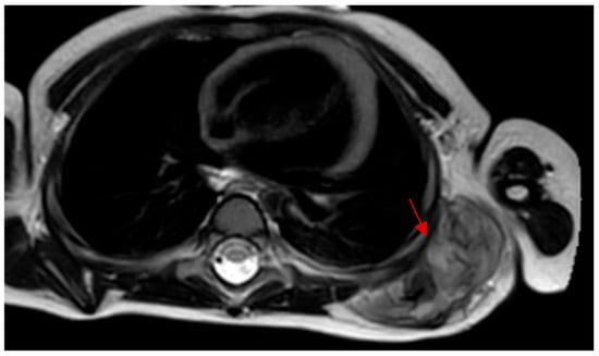
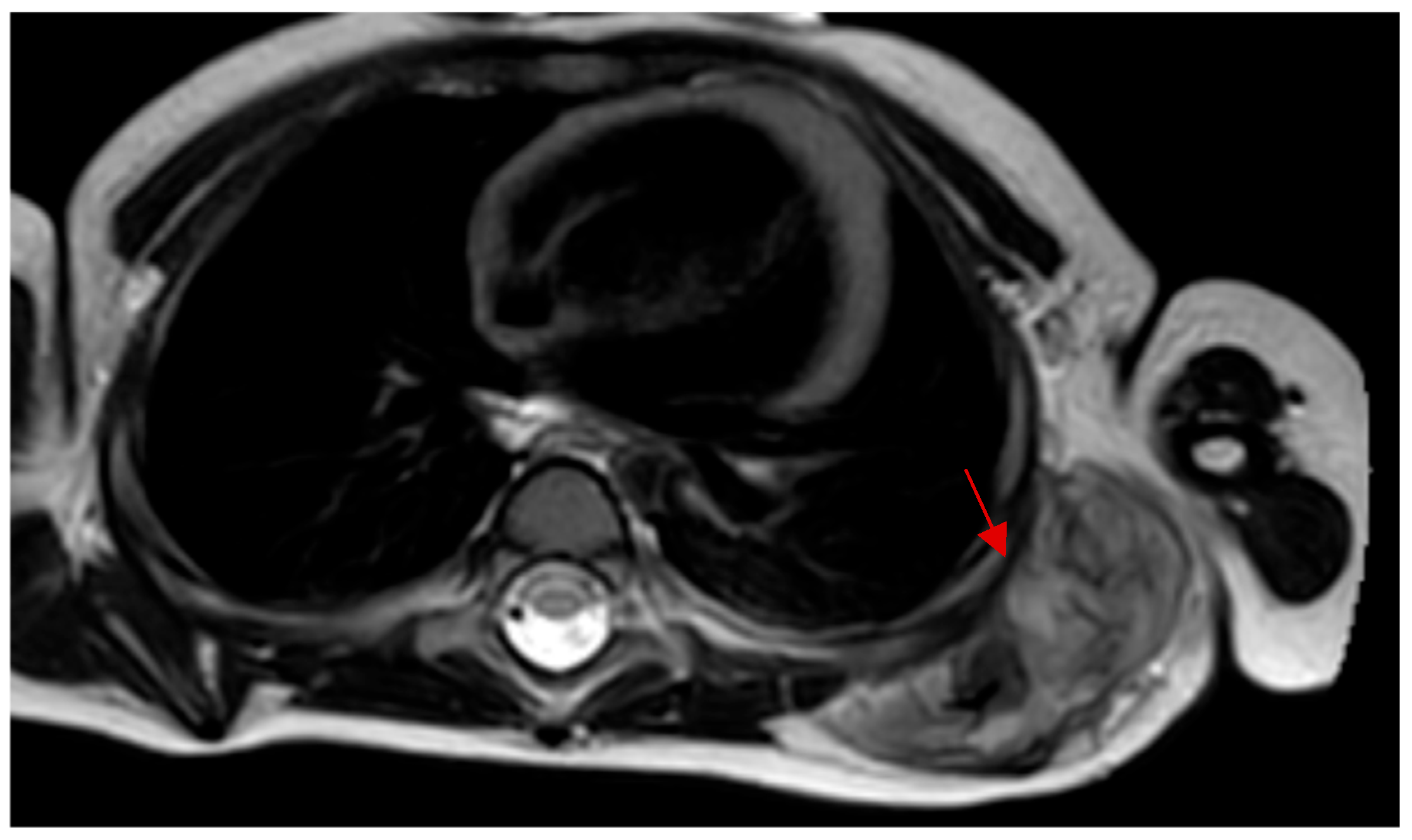
We describe the case of a female infant who presented at birth with a soft tissue mass of the left scapular area that was attached to the underlying tissues and overlapped by atrophic skin area. The soft tissue mass had grown dramatically in time, resulting in functional limitation in the abduction movement of the left shoulder. During the first months of life, she underwent numerous investigations in peripheral centers, including dermatological, rheumatological and genetic counseling in suspicion of a malformation. At the age of 15 months, after an orthopedic evaluation, she came to our attention and a magnetic resonance imaging (MRI) was performed. MRI revealed an expansive soft tissue mass approximately 56 mm × 46 mm × 57 mm in size, with lobulated and jagged margins, hyperintense in T2-weighted sequences and isointense in T1-weighted sequences; the soft tissue mass showed infiltrative aspects on the deltoid, supra- and subspinatus, and subscapularis muscles close to the inferior angle of the scapula. The soft tissue mass extensively infiltrated the subcutaneous and cutaneous plane with retractive aspects. In diffusion-weighted sequences it showed relative diffusivity restriction, with non-homogeneous enhancement due to the presence of fibrotic areas (Figure 1).
Figure 1.
MRI showing lesion of about 56 mm × 46 mm × 57 mm in the left scapular region.
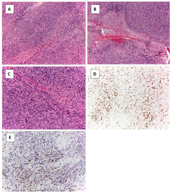
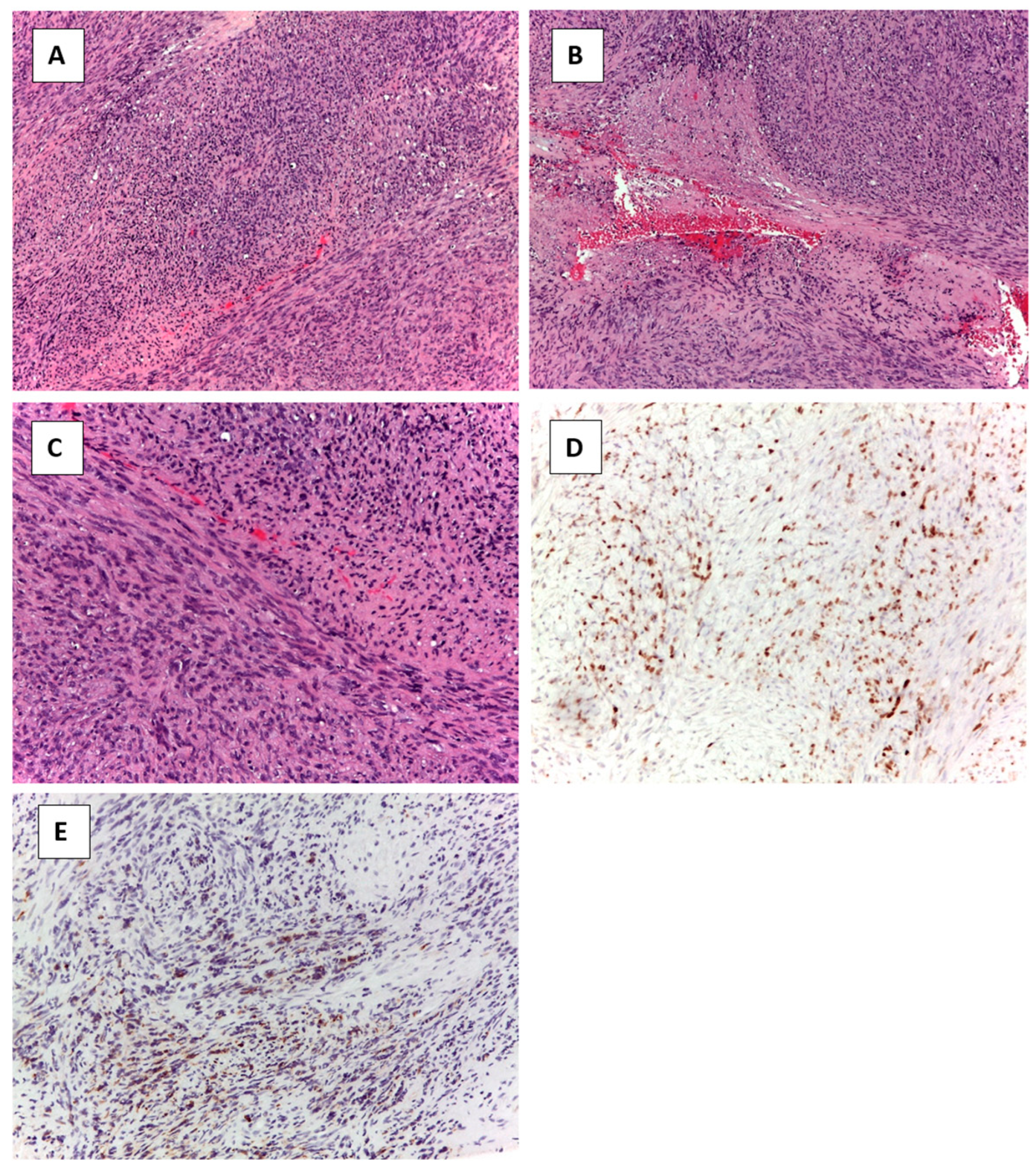
The patient underwent an incisional biopsy and the histological examination showed the presence of monomorphic spindle-cell bundles, with morphological appearance referable to a spindle-cell neoplasm with a high degree of malignancy. Immunohistochemistry showed focal nuclear staining for myogenin and cytoplasmic staining for desmin (Figure 2).
Figure 2.
(A) Spindle-cell rhabdomyosarcoma. Intersecting fascicles of spindle cells with brightly eosinophilic cytoplasm are present (hematoxylin and eosin, 50× of magnification). (B) Tumoral necrosis (hematoxylin and eosin, 50× of magnification). (C) Nuclear atypia (hematoxylin and eosin, 50× of magnification). (D) Focal nuclear staining for myogenin (100× of magnification) and (E) cytoplasmic staining for desmin (100× of magnification) by immunohistochemistry is observed.
The initial diagnosis was infantile fibrosarcoma, which was subsequently excluded by the negative translocation study for ETV6-NTRK3. Molecular testing was performed with Archer Anchored multiplex PCR and revealed a fusion breakpoint between exon 5 of TEAD1 and exon 12 of NCOA2 (Figure 3). The final diagnosis was therefore a TEAD1-NCOA2 fusion-positive ssRMSs.
Figure 3.
Identification of the TEAD1-NCOA2 fusion transcript. Analysis of anchored multiplex PCR result of the Archer FusionPlex Sarcoma Panel (Version Expanded sarcoma 16696 V.1.1) showed TEAD1-NCOA2 fusion in tumor sample.
Diagnostic work-up for staging was completed with chest–abdomen and neck Computed Tomography (CT), which revealed some enlarged lymph-nodes of uncertain significance in the homolateral axillary region, but with a short axis of under 10 mm. At the age of 15 months neoadjuvant chemotherapy was started according to protocol EpSSG RMS 2005, after consultation with the national reference center [16]. Given the onset of the disease, present at birth, the favorable pathology and the molecular testing positive for TEAD1-NCOA2 fusion, the patient was assigned to the standard risk group, subgroup C, according to the clinical grouping system adapted from the International Rhabdomyosarcoma Study Group (IRS) [17]. After three cycles of neoadjuvant chemotherapy with IVA (Ifosfamide, Vincristine, Actinomycin), a radiological reevaluation of disease did not show any substantial change in the mass. The patient underwent en bloc resection of the tumor at the age of 17 months, by performing a partial resection of the scapula. The histological examination confirmed the diagnosis of ssRMSs with residual vital areas and focal areas of necrosis without evidence of infiltration of bone tissue. Treatment was continued, according to protocol, with adjuvant chemotherapy, until completion of six postoperative IVA cycles. MRI performed after the end of treatment showed signal alteration suspicious for residual disease but in differential diagnosis with radiological signs of post-surgery inflammation. The case was discussed by the multidisciplinary board, and we decided to avoid radiotherapy given the age of the patient and to proceed with radiological follow-up every three months. Subsequent MRIs showed a progressive reduction in the signal alteration, consistent with post-surgical outcomes. At the last follow-up, the MRI showed no local recurrence, 4 years after the last chemotherapy administration.
Written informed consent for participation was obtained from the legal guardian of the participant involved in this study.
3. Materials and Methods for Literature Review
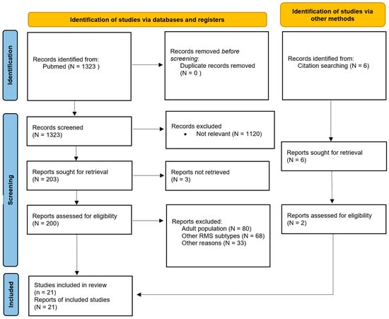
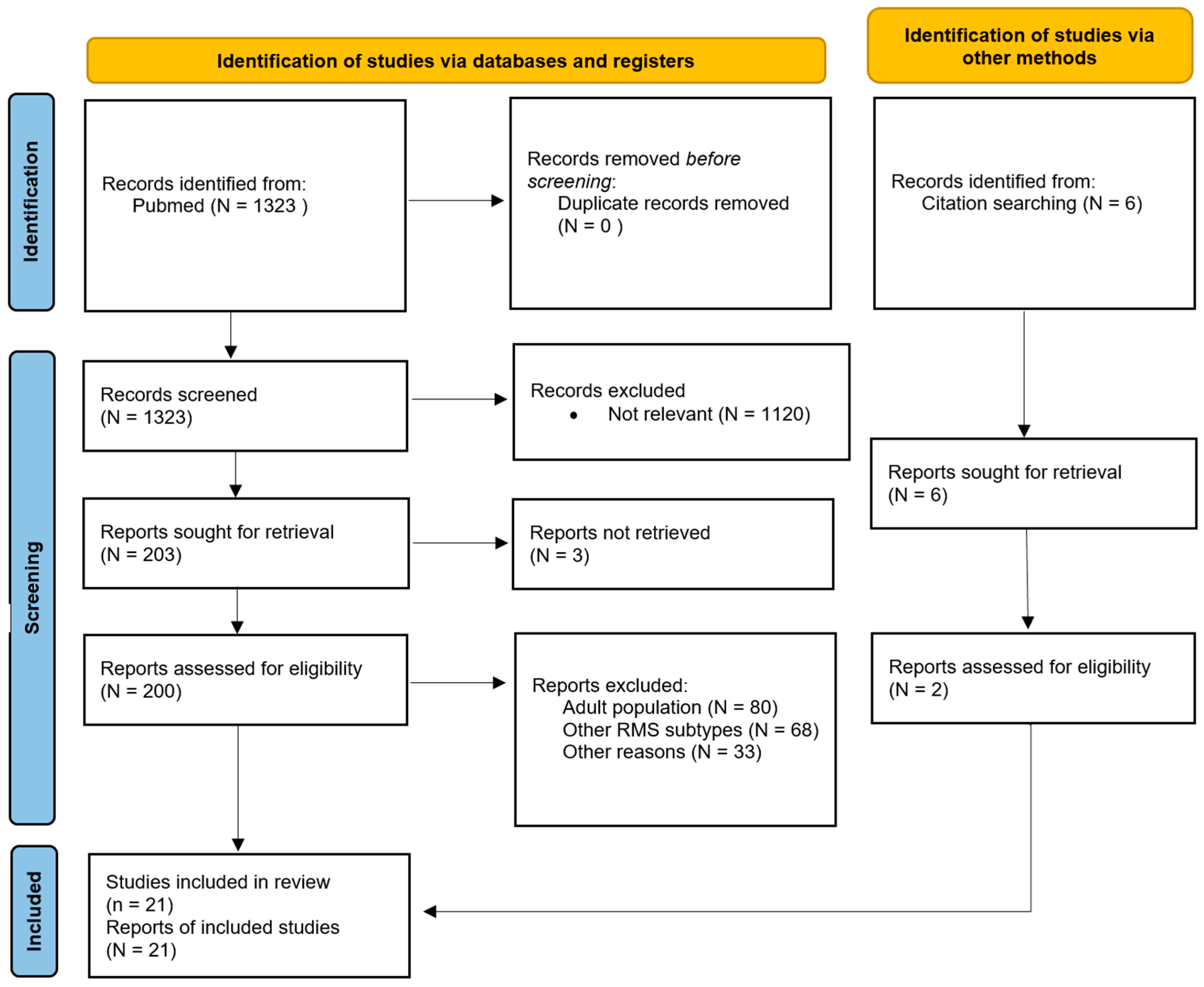
The literature search was conducted independently by two investigators (AP and ML) using the PubMed CENTRAL databases. The aim of this review is to analyze the state of the art with regard to ssRMSs in infants, children and adolescents, focusing on management and prognosis of the subset characterized by fusion genes involving NCOA2 and/or VGLL2. The following combinations of keywords were used: (“Rhabdomyosarcoma” [MeSH Terms] OR “Spindle cell rhabdomyosarcoma” OR “Congenital Rhabdomyosarcoma”) AND (“Child” [MeSH Terms] OR “Adolescent” [MeSH Terms] OR “Pediatrics” OR “Infant”). The research was limited to papers published in English in the last ten years. The reference lists of retrieved studies have also been reviewed to identify studies that may have not been spotted by the search strategy. Given the rarity of the disease, we decided to include in our analysis case reports, case series, observational studies, randomized-controlled studies, metanalysis and systematic reviews focused on outcomes and treatment of patients diagnosed with ssRMSs under the age of 18 years. In total, 1323 Pubmed CENTRAL studies were independently assessed by two investigators (AP and ML) using the PRISMA 2020 checklist and explained in the flow diagram (Figure 4). We excluded all the studies focusing on animals and in vitro models; we also excluded papers focused on different population of patients, for example adult patients, or different subgroups of the disease RMS not including in their analysis the specific subtype of ssRMS with fusion genes. We finally included in our analysis 21 studies.
Figure 4.
PRISMA flow diagram for selection of articles. Adapted from: Page, M.J. et al. [18].
4. Literature Review and Discussion
4.1. Epidemiology
RMS is the most common soft tissue sarcoma of children and about 5–8% of all malignancies in childhood, with a median age at diagnosis of about 5 years [16]. The ssRMS is a rare variant of RMS initially described in the head and neck and paratesticular region in children, which accounts for 5–10% of all RMS. In infants, ssRMSs represent 10–22% of the cases of RMS. The clinical and biological characteristics of RMSs among infants are different from those of older children or adults with RMSs [19].
4.2. Anatomopathology
Since 2013, the WHO recognizes four histological variants of RMS: alveolar (approximately 15–20% of all RMS), embryonal (about 80% of all RMS), and the rarer subtypes spindle-cell/sclerosing and pleomorphic [5,13].
ssRMS is a molecularly heterogeneous entity with some genetic alterations having a strong impact on prognosis [11]. At least three distinct clinicopathologic and molecular subclasses of ssRMSs are recognized at present [4] (Table 1).
Table 1.
Spindle-cell RMS subtypes.
MYOD1 mutated ssRMSs are typically found in older children and adolescent with a poor prognosis and most commonly arise in the head and neck, trunk and extremities [4,7,9,10,20,21]. These tumors are characterized by an L122R mutation in MyoD1 and are reported to harbor, in some cases, coexistent mutations including PI3KA, KRAS and HRAS whose clinical significance remains uncertain [9]. By immunohistochemistry, this subgroup of RMS shows weak or absent myogenin staining and variable desmin expression, with a strong positive expression of MyoD1 [4].
Infantile ssRMSs occur in children under two years of age, with 1–2% of RMSs described to be congenital, and have a predilection for the neck and trunk [1,4]. Morphologically these tumors can appear similar to infantile fibrosarcoma, with rare high-grade features described but with well-differentiated rabdhomyoblasts which help in diagnosis. Recurring fusions involving VGLL2, TEAD1 and NCOA2 are characteristics of this subset of ssRMSs and are described to be associated with a better prognosis [4].
Mosquera et al. first described recurrent NCOA2 gene rearrangements in 2013 in a small subset of ssRMSs arising in infants, involving SRF and TEAD1 as partner genes, which are involved in skeletal-muscle differentiation [8]. In 2016 Alaggio et al. described seven additional fusion-positive ssRMSs in infants. In this case series the authors identified novel VGLL2 gene rearrangements with either NCOA2 or CITED2 gene partners as the most common genetic abnormality in the group of congenital/infantile ssRMSs (64%) [7] (Table 2).
Table 2.
Recurrent fusions identified in congenital/infantile ssRMSs.
A third subset of ssRMSs has been described, characterized by fusions between TCFP2 and EWSR1/FUS or less commonly by MEIS1:NCOA2. This group of ssRMSs tend to arise in the axial skeleton or facial bones and occur more commonly in adults or older children [4,22].
4.3. Treatment Modality and Outcomes
The subset of ssRMSs in infants characterized by fusions involving VGLL2 and NCOA2 has been reported as having a favorable prognosis since the first reports available in the literature [6] (Table 3). Whittle et al. reported in 2019 four cases of infants treated in their institution for ssRMSs [6]. Among the four cases reported, three tested positive for the fusion TEAD1-NCOA2 while for one of them tissue was not available for the molecular characterization. All patients were treated with chemotherapy and surgical resection. Of note is the fact that one patient never achieved R0 resection but, due to age and size, did not receive radiation therapy as local control strategy and was reported to be alive with no recurrence signs after one year off-therapy [6]. All these patients were reported to have an excellent outcome regardless of surgical margin or lack of radiation [6].
In the same paper Whittle et al. discuss 11 other cases already published before in the literature, all characterized by fusion involving NCOA2 or VGLL2 and all treated with chemotherapy and surgical resection [6,7,8,23]. Alaggio et al. [7] reported in 2016 eleven cases of patients diagnosed at birth (congenital ssRMS) or within the first year of life (infantile ssRMS), all except one located in the trunk. Ten of the eleven described cases showed recurrent fusion genes and all of them with available long-term follow-up were alive and well at that time, after a median time of 7 years [7].
In 2019 the French MMT Committee reported 37 cases of infants aged less than 6 months treated for an RMS in France from 1989 to 2015. Eight patients had ssRMSs and among them three had VGLL2- involving fusions. All the three patients received chemotherapy (Vincristine, cyclophosphamide and vinblastine, VAC, VAC/IVA); surgery was performed in all patients achieving incomplete macroscopic or microscopic resection and none of the three patients received radiotherapy [24]. All three patients were reported to be in complete remission at the time of data collection, at a mean time of 3 years off-therapy [24].
Ze Lin Tan et al., in 2020, described a case of infantile ssRMS with the fusion gene TEAD1-NCOA2 treated with neoadjuvant chemotherapy, late surgical resection and adjuvant chemotherapy, alive with no evidence of recurrence of disease 21 months from birth at the time of the publication [11].
Bisogno et al. described in 2022 the population of infants with congenital RMSs diagnosed before 2 months of age, enrolled in the EpSSG RMS 2005 protocol. Among 24 patients, all children presented with a favorable histology except for one with alveolar histology and metastatic disease, and four tumors showed spindle-cell morphology. Three patients were reported to be positive for VGLL2-CITED2/NCOA2 among the seven patients investigated; one of them was of the spindle-cell subtype [1]. In this report from EpSSG, 19 out of 24 patients achieved a complete remission during first-line treatment with resection and chemotherapy (VA, IVA or IVADo depending on risk group); no patients received radiotherapy [1].
In 2022 Whittle et al. described a cooperative analysis of 39 children of less than 12 months at diagnosis of localized ssRMSs identified from the Children’s Oncology Group (COG), Cooperative Weichteilsarkom Studiengruppe (CWS), European pediatric Soft tissue sarcoma Study Group (EpSSG) and Italian Soft tissue Sarcoma Committee (STSC), clinical trial and registry databases, as well as the Texas Children’s Hospital (TCH) pathology archives [13]. Fusion data was not available for all cases and among 26 patients with molecular characterization available, 13 showed VGLL2/NCOA2 rearrangements. A multimodal approach of surgery combined with chemotherapy and radiotherapy was used with different combinations determined by the clinical trial of enrolment and the disease staging. The chemotherapy regimens used always included viincristine and dactinomycin, often combined with cyclophosphamide or ifosfamide (VAC/IVA) or doxorubicin (VAIA). Surgical resection was performed upfront when feasible without mutilation or delayed in 37 cases. RT was performed only in five cases [13].
In contrast, in 2021 a case series of four patients diagnosed with VGLL2-rearranged infantile RMSs who experienced local progression and, in two cases, had a fatal outcome, has been published [12]. In this paper the authors underscore two important differences in terms of clinical management compared to other published cases. In all the four cases reported the tumors were initially considered unresectable, and three out of four patients were initially treated with chemotherapy regimens different from the RMS type, due to the tumors being misdiagnosed as low-grade entities [12].
The recent cooperation analysis published by Whittle et al. in 2022 [13] describes treatment and outcomes of 39 patients aged <12 months with localized spindle-cell RMSs treated from 1997 to 2018. In the international cohort described by Whittle et al., in 2022 the five-year EFS and OS for infants with localized ssRMSss were 86% and 91% respectively [13].
Of these cases only 13 were VGLL2- and/or NCOA2-positive, and among these the four cases treated at Texas Children’s Hospital and previously described in 2019 were included [11]. As previously reported in the literature, these tumors are frequently found in axial locations and are almost always localized at presentation. In this study, the extent of resection was one prognostic factor not previously described. Very few patients in this cohort received RT and no statistically significant difference in outcomes was reported based on the use of RT; this is reassuring because, as in the case of our patient, these are children of age less than 3 years, and a radiotherapy-sparing approach could mean reduced late effects. In this cooperative analysis, the case of a patient with metastatic disease at diagnosis, with NCOA2 fusion, is described, but excluded from the overall analysis. This patient was treated with chemotherapy and underwent a delayed R2 resection without radiotherapy, achieving partial response. The patient died 3 months after completion of therapy for a Cytomegalovirus pneumonitis. These data clearly suggest the good prognosis of these tumors, and favor the potential sparing of a multimodal treatment, also considering the very young age at diagnosis for this subclass of patients.
In contrast with these data, Cyrta et al. reported four infants with unresectable VGLL2-rearranged RMSs and unfavorable outcomes [12]. The authors describe four cases with several years’ follow-up that showed metastatic progression, including death due to disease in two of the four cases. However, as the authors underline, of these cases three were initially diagnosed as fibromatosis or infantile fibrosarcoma and managed as such with a sub-optimal chemotherapy regimen, with a subsequent transformation in high-grade morphology tumors. This underlines the importance of initial expert pathologic diagnosis and suggests that RMS-type chemotherapy should be considered for unresectable rearranged tumors, as stated by Whittle et al. [13]. Our case also confirms that ssRMSs can closely mimic other more commonly described mesenchymal tumors, making the differential diagnosis challenging if not supported by molecular studies. The preliminary diagnosis based solely on morphologic appearance was that of infantile fibrosarcoma, as in the case described by Ze Li Tan and colleagues [11]. This further confirms that the diagnosis shouldn’t be performed without immunohistochemistry and molecular studies. Table 3 summarizes studies including fusion-positive ssRMSs in infants in the last ten years.
Evidence accumulated since the initial descriptions of this molecular subgroup supports an overall favorable prognosis for localized congenital/infantile VGLL2/NCOA2-rearranged ssRMSs and the possibility of radiotherapy-sparing strategies in selected patients. Overall, the available literature suggests, as the current therapeutic strategy, treatment de-escalation aimed at minimizing long-term developmental toxicity.
Management strategies favor complete initial surgical resection (R0) when feasible without functional morbidity, followed by multi-agent chemotherapy regimens (such as low-risk VA [vincristine, dactinomycin] or VAC/IVA). Radiation therapy is omitted or strictly restricted to salvage settings for gross residual or unresectable disease to spare infants severe long-term radiation-induced sequelae.
However, the aggressive course of unresectable VGLL2-rearranged tumors described by Cyrta et al. indicates that fusion status alone should not be considered sufficient to define a uniformly low-risk population. Molecular characterization should therefore always be integrated with clinical features, resectability and response to RMS-directed treatment when considering therapeutic de-escalation [12].
Table 3.
Summary table of studies including fusion-positive ssRMSs in infants in the last ten years.
5. Conclusions
This paper provides a new clinical report of congenital ssRMSs, with TEAD1-NCOA2 translocation, in which numerous features common to this subgroup are found. The patient is alive and in complete remission four years after end of treatment. The patient was treated with an RMS-type chemotherapy, R1 surgery and adjuvant chemotherapy, sparing radiotherapy. Limitations of this study are represented by the relatively short follow-up period and the literature review based only on the PUBMED central database. However, this case report underlines some important key points about this rare entity.
In particular, this case report highlights the importance of an integrated pathological diagnosis which takes into account morphological assessment, immunohistochemistry and molecular testing, not only for a diagnostic purpose but also with great implications for prognosis.
The histopathological assessment, with immunochemistry and genetic testing, is necessary in patients with congenital mesenchymal tumors, especially in spindle-cell and sclerosing rhabdomyosarcoma.
The available literature also highlights the lack of consensus guidelines regarding the treatment of these rare congenital/infantile ssRMS subtypes, highlighting the need to collect more data about treatment and outcomes of patients with molecular defined ssRMSs.
Author Contributions
Conceptualization, A.P., M.L., E.E. and I.M.; methodology, A.P. and L.I.; data acquisition and analysis, A.P., I.M., M.d.C.C.G., B.P., A.R. and C.E.; writing—original draft preparation, A.P., E.E. and M.L.; writing—review and editing, A.P., M.L., E.E., I.M., M.d.C.C.G., A.R., C.E., L.I. and G.P.; supervision, L.I. and G.P. All authors have read and agreed to the published version of the manuscript.
Funding
This research received no external funding.
Institutional Review Board Statement
Ethical approval was not required for this single-patient case report in accordance with institutional and international ethical standards. All procedures were conducted in compliance with the Declaration of Helsinki.
Informed Consent Statement
Informed written consent was obtained from parents of the subject involved to publish this paper.
Data Availability Statement
The original contributions presented in this study are included in the article. Further inquiries can be directed to the corresponding author.
Conflicts of Interest
The authors declare no conflicts of interest.
Abbreviations
The following abbreviations are used in this manuscript:
| RMS | Rhabdomyosarcoma |
| ssRMS | Spindle-Cell and Sclerosing Rhbdomyosarcoma |
| MRI | Magnetic Resonance Imaging |
| CT | Computed Tomography |
| IRS | International Rhabdomyosarcoma Study Group |
| IVA | Ifosfamide, Vincristine, Actinomycin D |
| IVADo | Ifosfamide, Vincristine, Actinomycin D, Doxorubicin |
| VAC | Vincristine, Actinomycin D, Cyclophosphamide |
| VAIA | Vincristine, Actinomycin D, Ifosfamide |
References
- Bisogno, G.; Minard-Colin, V.; Arush, M.B.; Daragjati, J.; Coppadoro, B.; Gallego, S.; Alaggio, R.; Smeulders, N.; Mudry, P.; Zin, A.; et al. Congenital Rhabdomyosarcoma: A Report from the European Paediatric Soft Tissue Sarcoma Study Group. Pediatr. Blood Cancer 2022, 69, e29376. [Google Scholar] [CrossRef] [Scilit]
- Skapek, S.X.; Ferrari, A.; Gupta, A.A.; Lupo, P.J.; Butler, E.; Shipley, J.; Barr, F.G.; Hawkins, D.S. Rhabdomyosarcoma. Nat. Rev. Dis. Primers 2019, 5, 1. [Google Scholar] [CrossRef] [Scilit] [PubMed]
- Chen, C.; Dorado Garcia, H.; Scheer, M.; Henssen, A.G. Current and Future Treatment Strategies for Rhabdomyosarcoma. Front. Oncol. 2019, 9, 1458. [Google Scholar] [CrossRef] [Scilit] [PubMed]
- Dehner, C.A.; Rudzinski, E.R.; Davis, J.L. Rhabdomyosarcoma: Updates on Classification and the Necessity of Molecular Testing beyond Immunohistochemistry. Hum. Pathol. 2024, 147, 72–81. [Google Scholar] [CrossRef] [Scilit] [PubMed]
- Rudzinski, E.R.; Kelsey, A.; Vokuhl, C.; Linardic, C.M.; Shipley, J.; Hettmer, S.; Koscielniak, E.; Hawkins, D.S.; Bisogno, G. Pathology of Childhood Rhabdomyosarcoma: A Consensus Opinion Document from the Children’s Oncology Group, European Paediatric Soft Tissue Sarcoma Study Group, and the Cooperative Weichteilsarkom Studiengruppe. Pediatr. Blood Cancer 2021, 68, e28798. [Google Scholar] [CrossRef] [Scilit] [PubMed]
- Whittle, S.B.; Hicks, M.J.; Roy, A.; Vasudevan, S.A.; Reddy, K.; Venkatramani, R. Congenital Spindle Cell Rhabdomyosarcoma. Pediatr. Blood Cancer 2019, 66, e27935. [Google Scholar] [CrossRef] [Scilit] [PubMed]
- Alaggio, R.; Zhang, L.; Sung, Y.-S.; Huang, S.-C.; Chen, C.-L.; Bisogno, G.; Zin, A.; Agaram, N.P.; LaQuaglia, M.P.; Wexler, L.H.; et al. A Molecular Study of Pediatric Spindle and Sclerosing Rhabdomyosarcoma: Identification of Novel and Recurrent VGLL2-Related Fusions in Infantile Cases. Am. J. Surg. Pathol. 2016, 40, 224–235. [Google Scholar] [CrossRef] [Scilit] [PubMed]
- Mosquera, J.M.; Sboner, A.; Zhang, L.; Kitabayashi, N.; Chen, C.; Sung, Y.S.; Wexler, L.H.; LaQuaglia, M.P.; Edelman, M.; Sreekantaiah, C.; et al. Recurrent NCOA2 Gene Rearrangements in Congenital/Infantile Spindle Cell Rhabdomyosarcoma. Genes Chromosom. Cancer 2013, 52, 538–550. [Google Scholar] [CrossRef] [Scilit] [PubMed]
- Agaram, N.P.; LaQuaglia, M.P.; Alaggio, R.; Zhang, L.; Fujisawa, Y.; Ladanyi, M.; Wexler, L.H.; Antonescu, C.R. MYOD1-Mutant Spindle Cell and Sclerosing Rhabdomyosarcoma: An Aggressive Subtype Irrespective of Age. A Reappraisal for Molecular Classification and Risk Stratification. Mod. Pathol. 2019, 32, 27–36. [Google Scholar] [CrossRef] [Scilit] [PubMed]
- Rekhi, B.; Upadhyay, P.; Ramteke, M.P.; Dutt, A. MYOD1 (L122R) Mutations Are Associated with Spindle Cell and Sclerosing Rhabdomyosarcomas with Aggressive Clinical Outcomes. Mod. Pathol. 2016, 29, 1532–1540. [Google Scholar] [CrossRef] [Scilit] [PubMed]
- Tan, G.Z.L.; Saminathan, S.N.; Chang, K.T.E.; Odoño, E.G.; Kuick, C.H.; Chen, H.; Lee, V.K.M. A Rare Case of Congenital Spindle Cell Rhabdomyosarcoma with TEAD1-NCOA2 Fusion: A Subset of Spindle Cell Rhabdomyosarcoma with Indolent Behavior. Pathol. Int. 2020, 70, 234–236. [Google Scholar] [CrossRef] [Scilit] [PubMed]
- Cyrta, J.; Gauthier, A.; Karanian, M.; Vieira, A.F.; Cardoen, L.; Jehanno, N.; Bouvet, M.; Bouvier, C.; Komuta, M.; Le Loarer, F.; et al. Infantile Rhabdomyosarcomas With VGLL2 Rearrangement Are Not Always an Indolent Disease: A Study of 4 Aggressive Cases With Clinical, Pathologic, Molecular, and Radiologic Findings. Am. J. Surg. Pathol. 2021, 45, 854–867. [Google Scholar] [CrossRef] [Scilit] [PubMed]
- Whittle, S.; Venkatramani, R.; Schönstein, A.; Pack, S.D.; Alaggio, R.; Vokuhl, C.; Rudzinski, E.R.; Wulf, A.-L.; Zin, A.; Gruver, J.R.; et al. Congenital Spindle Cell Rhabdomyosarcoma: An International Cooperative Analysis. Eur. J. Cancer 2022, 168, 56–64. [Google Scholar] [CrossRef] [Scilit] [PubMed]
- Slater, O.; Gains, J.E.; Kelsey, A.M.; De Corti, F.; Zanetti, I.; Coppadoro, B.; Jorgensen, M.; Gallego, S.; Orbach, D.H.; Glosli, H.; et al. Localised Rhabdomyosarcoma in Infants (<12 Months) and Young Children (12–36 Months of Age) Treated on the EpSSG RMS 2005 Study. Eur. J. Cancer 2022, 160, 206–214. [Google Scholar] [CrossRef] [Scilit] [PubMed]
- Chisholm, J.; Mandeville, H.; Adams, M.; Minard-Collin, V.; Rogers, T.; Kelsey, A.; Shipley, J.; van Rijn, R.R.; de Vries, I.; van Ewijk, R.; et al. Frontline and Relapsed Rhabdomyosarcoma (FaR-RMS) Clinical Trial: A Report from the European Paediatric Soft Tissue Sarcoma Study Group (EpSSG). Cancers 2024, 16, 998. [Google Scholar] [CrossRef] [Scilit] [PubMed]
- Merks, J.H.M.; Brack, E.; Ebinger, M.; Minard-Colin, V.; Defachelles, A.-S.; Hladun, R.; Rogers, T.; Fuchs, J.; Godzinski, J.; Van Scheltinga, S.T.; et al. European Standard Clinical Practice Recommendations for Children and Adolescents with Rhabdomyosarcoma a Joint EpSSG, CWS and ERN PaedCan Project. EJC Paediatr. Oncol. 2025, 5, 100229. [Google Scholar] [CrossRef] [Scilit]
- De Salvo, G.L.; Del Bianco, P.; Minard-Colin, V.; Chisholm, J.; Jenney, M.; Guillen, G.; Devalck, C.; Van Rijn, R.; Shipley, J.; Orbach, D.; et al. Reappraisal of Prognostic Factors Used in the European Pediatric Soft Tissue Sarcoma Study Group RMS 2005 Study for Localized Rhabdomyosarcoma to Optimize Risk Stratification and Generate a Prognostic Nomogram. Cancer 2024, 130, 2351–2360. [Google Scholar] [CrossRef] [Scilit] [PubMed]
- Page, M.J.; McKenzie, J.E.; Bossuyt, P.M.; Boutron, I.; Hoffmann, T.C.; Mulrow, C.D.; Shamseer, L.; Tetzlaff, J.M.; Akl, E.A.; Brennan, S.E.; et al. The PRISMA 2020 statement: An updated guideline for reporting systematic reviews. BMJ 2021, 372, n71. [Google Scholar] [CrossRef] [Scilit] [PubMed]
- Yan, A.P.; Venkatramani, R.; Bradley, J.A.; Lautz, T.B.; Urla, C.I.; Merks, J.H.M.; Oberoi, S. Clinical Characteristics, Treatment Considerations, and Outcomes of Infants with Rhabdomyosarcoma. Cancers 2023, 15, 2296. [Google Scholar] [CrossRef] [Scilit] [PubMed]
- Rekhi, B.; Dodd, L.; Dharavath, B.; Dutt, A. Cytomorphology of Spindle Cell/Sclerosing Rhabdomyosarcoma, Including MYOD1 (LI22R) Mutation Result. Diagn. Cytopathol. 2022, 50, E367–E372. [Google Scholar] [CrossRef] [Scilit] [PubMed]
- Chisholm, J.C.; Selfe, J.L.; Alaggio, R.; Cheesman, E.; Zin, A.; Tombolan, L.; Parafioriti, A.; Milano, G.M.; Adams, M.; Popov, S.; et al. Clinicopathological Analysis of a European Cohort of MYOD1 Mutant Rhabdomyosarcomas in Children and Young Adults. Pediatr. Blood Cancer 2025, 72, e31428. [Google Scholar] [CrossRef] [Scilit] [PubMed]
- Haug, L.; Doll, J.; Appenzeller, S.; Kunzmann, V.; Rosenwald, A.; Maurus, K.; Gerhard-Hartmann, E. Epithelioid and Spindle Cell Rhabdomyosarcoma with EWSR1::TFCP2 Fusion Mimicking Metastatic Lung Cancer: A Case Report and Literature Review. Pathol. Res. Pract. 2023, 249, 154779. [Google Scholar] [CrossRef] [Scilit] [PubMed]
- Waldman, L.E.; Williamson, A.K.; Amodio, J.B.; Collins, L. Congenital Rhabdomyosarcoma Presenting as a Neck Mass at Birth. Pediatrics 2018, 2018, 1243436. [Google Scholar] [CrossRef] [Scilit] [PubMed]
- Butel, T.; Karanian, M.; Pierron, G.; Orbach, D.; Ranchere, D.; Cozic, N.; Galmiche, L.; Coulomb, A.; Corradini, N.; Lacour, B.; et al. Integrative Clinical and Biopathology Analyses to Understand the Clinical Heterogeneity of Infantile Rhabdomyosarcoma: A Report from the French MMT Committee. Cancer Med. 2020, 9, 2698–2709. [Google Scholar] [CrossRef] [Scilit] [PubMed]



Disclaimer/Publisher’s Note: The statements, opinions and data contained in all publications are solely those of the individual author(s) and contributor(s) and not of MDPI and/or the editor(s). MDPI and/or the editor(s) disclaim responsibility for any injury to people or property resulting from any ideas, methods, instructions or products referred to in the content. |
© 2026 by the authors. Licensee MDPI, Basel, Switzerland. This article is an open access article distributed under the terms and conditions of the Creative Commons Attribution (CC BY) license.