


Abstract
Coronary artery fistulae (CAF) are uncommon congenital or acquired coronary anomalies. A CAF occurs when a coronary artery bypasses the myocardial capillary bed to directly communicate with a cardiac chamber, a great vessel, or another vascular structure. Many CAFs are found by chance. If haemodynamically significant, a CAF may cause a variety of phenomena e.g., myocardial ischaemia, arrhythmias, heart failure, pulmonary hypertension, infective endocarditis/endarteritis, aneurysm formation, and late thrombotic complication. Management is anatomy-driven and dependent on the precise definition of the CAF’s origin, course, termination, multiplicity, associated coronary remodeling, and complications, together with an assessment of physiological relevance. Invasive coronary angiography is indispensable for real-time haemodynamics and transcatheter therapy, yet the two-dimensional projection nature can incompletely characterize complex CAF anatomy. Gated computed tomography coronary angiography (CTCA) produces high-resolution volumetric imaging with robust three-dimensional (3D) reconstruction and is central to contemporary diagnosis, quantitative risk stratification, procedural planning, and follow-up. This review examines the role of CTCA for the diagnosis and management of CAF and aims to provide a comprehensive overview for physicians managing this esoteric group of patients.
1. Literature Search Strategy
This narrative review was based on a structured search of PubMed/MEDLINE, Embase, and Google Scholar for relevant studies published up to April 2026. Search terms included combinations of “coronary artery fistula”, “coronary arteriovenous fistula”, “cardiac CT”, “coronary CT angiography”, “computed tomography”, “transcatheter closure”, “aneurysm”, “thrombosis”, and “surgical management”. Reference lists of key articles were also reviewed manually to identify additional relevant publications. Priority was given to larger contemporary CT series, interventional outcome studies, society guidelines, and systematic reviews, while selected case reports were retained when they illustrated unusual anatomy, multimodality imaging findings, or management challenges.
2. Background and Classification
CAFs are rare coronary anomalies whereby the coronary artery (or branch) abnormally terminates in a cardiac chamber or a major vessel [1]. In the general population, the incidence of CAFs is circa 0.002% but is far greater in patients with congenital heart disease—circa 0.2–0.4% [2,3]. A 12 year study of 32,000 coronary angiograms identified only 55 CAF [4].
CAFs are most often attributed to persistence of embryonic coronary sinusoids or disrupted vessel morphogenesis. Acquired fistulae may also occur after cardiac surgery, percutaneous interventions, trauma, or infection [5,6].
Embryological development of CAF is not fully understood. Some early experimental work suggests that failure of compact myocardial development leads to CAF development in utero [7]. CAF represents 50% of all congenital coronary anomalies without a particular predisposition by gender or race [8].
2.1. Symptomatology
Most patients (~75%) with CAFs are asymptomatic and are incidentally found. A small number of patients present de novo with complications such as angina, arrhythmias, heart failure, pulmonary hypertension, and endarteritis/endocarditis. Such presentations are more likely when lesions are large, draining into low-pressure chambers and associated with aneurysmal remodeling [9,10]. Clinical significance is determined by fistula resistance, the pressure/compliance of the receiving structure, and the degree of coronary remodeling proximal and distal to the fistulous origin. High volume of blood flow and low-resistance terminations promote coronary steal which may cause ischemia even without any obstructive coronary disease. Chronic low resistance terminal runoff can drive progressive dilation and aneurysm formation which may become clinically relevant independent of shunt magnitude [11].
Symptomatically, most presentations of CAF occur >60 years old and about a fifth have symptoms <25 years old [12]. There is an important caveat—CAF may be seen in up to 15% of autopsies in sudden death in children, although the causal relationship between anatomical findings and sudden demise is difficult to ascertain [13].
2.2. Types and Locations
CAF can be classified by coronary origin, termination site, number, morphological complexity, and associated aneurysmal change (Table 1). Origins from all coronary arteries have been described and multiple feeders can coexist (Figure 1) [14]. Terminations typically occur into low-pressure structures such as the right ventricle, right atrium, pulmonary artery, or coronary sinus; less common sites are left-sided chambers and extracardiac vascular beds (e.g., bronchial circulation) [15]. Anecdotally, it is perceived that the RCA is the commonest coronary artery affected but in the largest review published, the most frequent CAF origin point was the left main stem or LAD coronary artery and most frequent run-off connection was the pulmonary artery [3].
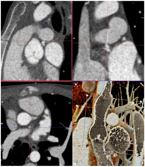
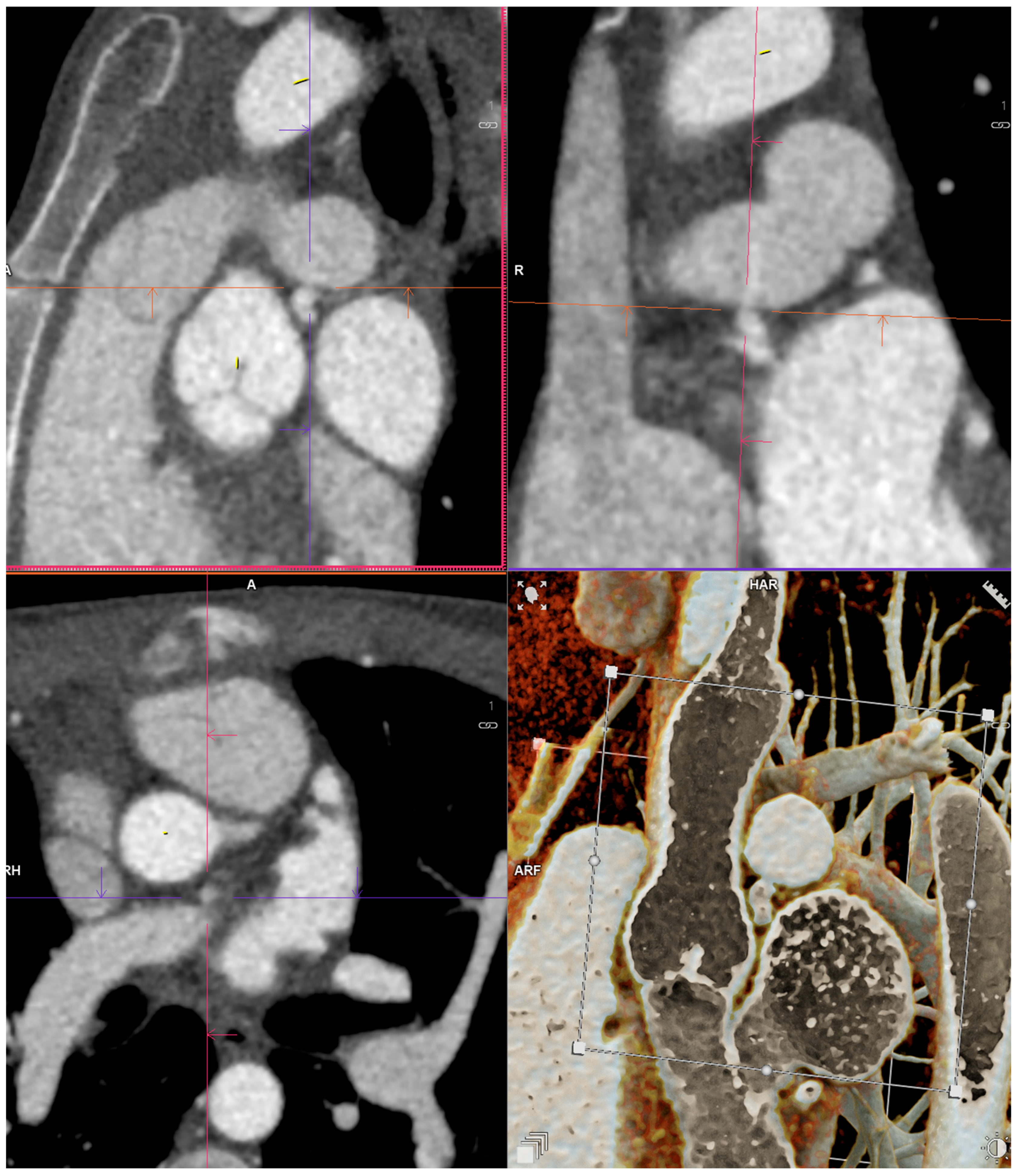
Figure 1.
Abnormal termination of RCA that runs a retro-aortic course to the left atrioventricular groove to terminate as a fistula with a contrast shift in the LPA.
Morphologically, CAFs range from focal communications with discrete narrowing and relatively straight segments—often favourable for coil or plug occlusion—to long, tortuous, aneurysmal systems with large terminal sacs or multiple distal communications [16,17,18]. Aneurysmal or “giant” CAF form a clinically important subgroup in which symptoms and complications reflect not only shunt physiology but also the hemodynamic and thrombotic consequences of massively remodeled vessels [19,20,21].
Histologically, there is significant variation in the makeup of the fistulous vessels. The fistulous portion of the vessel may have a very thick muscular medial layer with well-defined elastic lamina before a change is observed in the aneurysmal run-off vessel which may lack elastic lamina. In the aneurysmal portion of the vessel, histological changes have been seen such as an increase in mucopolysaccharides and desmin-negative spindle cells relative to the fistulous portion of the vessel [22].
2.3. The Role of Cardiac Catheterisation
Selective coronary artery angiography provides dynamic assessment and is essential for transcatheter intervention. Angiography can only give limited visualisation of the full 3D geometry of tortuous fistulous pathways, the true extent of aneurysmal segments, and the relationship to adjacent branches and extracardiac structures [23]. The only area in which invasive angiography may still be a preferred option may be in the extremely small child where very high heart rates may limit CT accuracy. With the development of transcatheter techniques, percutaneous closure is a well-established alternative to surgery for many anatomically suitable lesions, achieving high acute success while highlighting late issues such as residual shunt, persistent ectasia, and thrombosis in selected anatomies [24,25,26].
CTCA is now the first-line imaging modality to provide comprehensive 3D mapping for diagnosis, planning, and follow-up of CAFs [27].
Table 1.
Definitions of CAF Morphologies [28].
3. CT Technique for Coronary Artery Fistula
Imaging of coronary artery fistulae (CAF) may require protocol modifications to the standard CTCA with a tailored protocol to delineate complex vascular connections and drainage anatomy. We discuss the specific technical changes needed below and these are summarised in Table 2.
Table 2.
Summary of modifications to standard cardiac CT for optimal CAF imaging.
ECG-gated CTCA accurately depicts the coronary origin and fistulous tract. Prospective gating is preferred where feasible to reduce radiation dose, although retrospective gating may be required in complex or high-flow fistulae. Submillimeter slice thickness (<0.6 mm) and high iodine concentration contrast (>350 mg/mL) are recommended to maximise spatial resolution.
A wide field of view (FOV) should be routinely considered, particularly when extracardiac drainage is suspected (e.g., pulmonary artery, bronchial circulation), or for surgical planning.
Unlike standard CTCA, contrast timing in CAF aims to opacify both coronary arteries and the receiving chambers simultaneously and therefore injection techniques are changed commonly to:
- Injection rate: 5–6 mL/s
- Bolus tracking (ascending aorta) or test bolus techniques are used to optimise the timing of contrast.
Other techniques to improve the image quality include contrast injection protocols such as:
- ○
- A 60 mL main bolus, chaser around 40–50 mls of 50% contrast −50% saline (rather than 25 contrast −75%saline in standard CTCA) to ensure contrast opacification of both right and left chambers Figure 1.
- ○
- In selected cases, eliminating contrast from the chaser phase may improve detection of subtle shunt jets into low-pressure chambers.
Delayed acquisitions are not routinely required in standard CTCA but are particularly valuable in CAF for the following key reasons:
- To confirm drainage site in high-flow fistulae
- To detect thrombus within aneurysmal segments
- To differentiate slow flow from true occlusion
- Multiphase or dynamic acquisitions may also help in complex haemodynamics.
Although modern scanners reduce motion sensitivity, heart rates < 60 bpm are desirable. Sublingual nitrate (400–800 µg) helps to improve coronary visualisation, particularly when assessing the fistula origin or coexisting coronary disease.
3.1. Radiation and Emerging Technologies
Dose-reduction strategies, including prospective triggering, iterative reconstruction, dose modulations, are especially important given the younger age of many CAF patients. Photon-counting CT, whilst less freely available, offers superior spatial resolution (ultra-high) and contrast-to-noise ratio at lower radiation dose, enhancing visualisation of small calibre fistulous channels (Figure 2) and distal terminations [29].
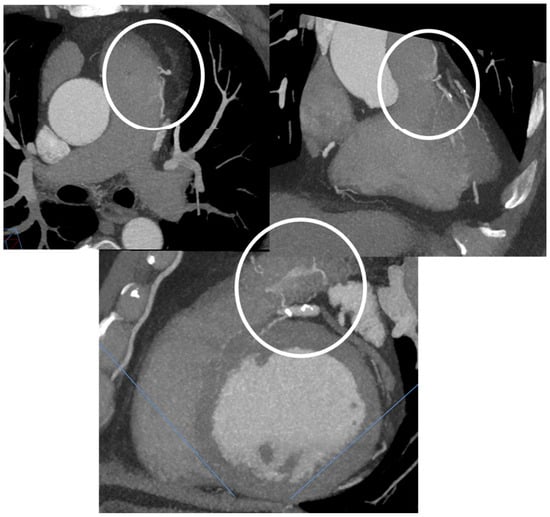
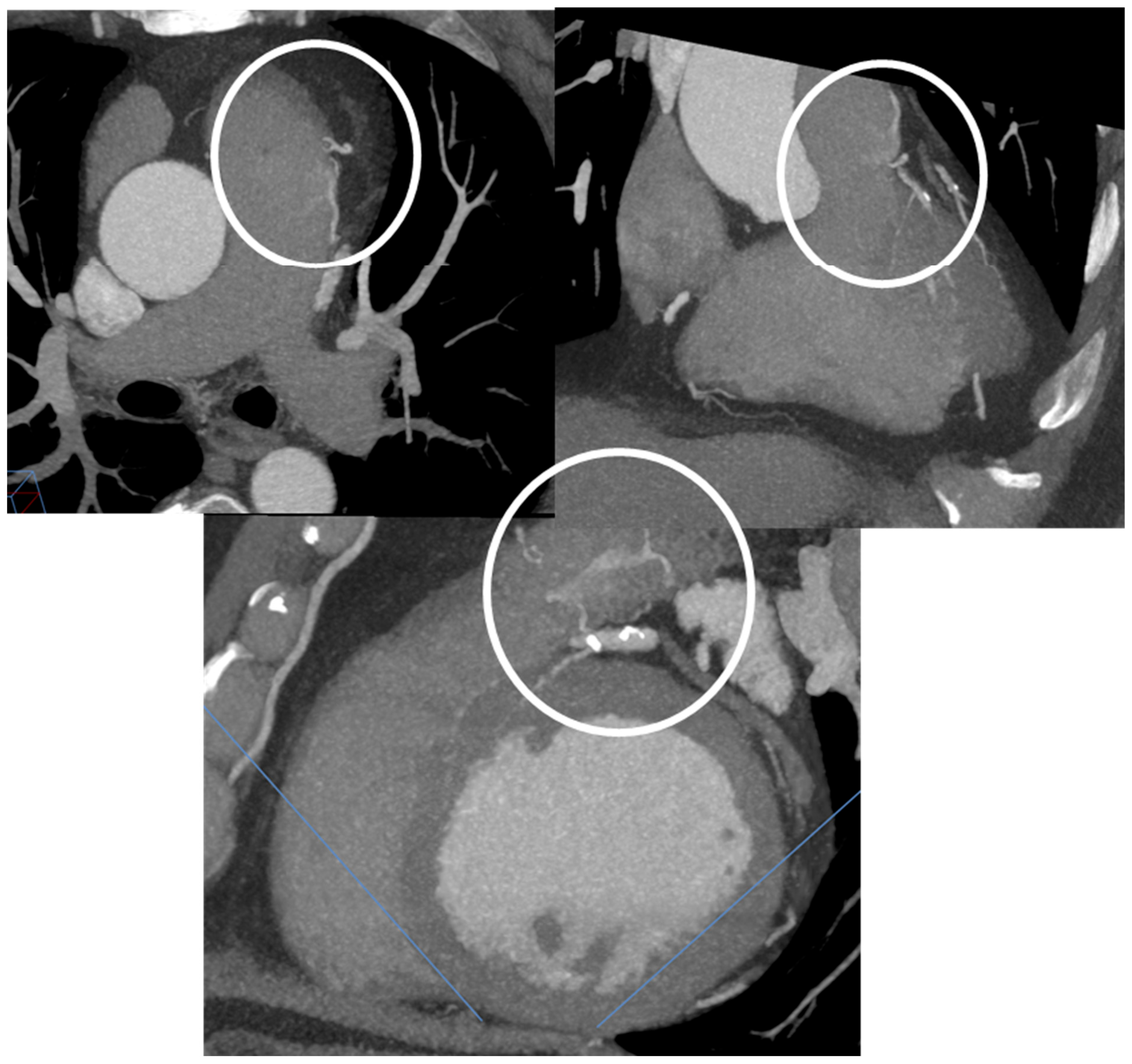
Figure 2.
MIP images showing a small fistula with MPA and contrast gradient through that small fistula.
Increasingly, there are newer techniques and technologies available for CAF assessment, and these include:
- CT-derived fractional flow reserve (CT-FFR) may be used when coincident coronary artery disease is suspected.
- Emerging dynamic/4D CT techniques in selected cases may allow visualisation of shunt flow and temporal contrast transit offering additional information [30].
- Functional CT, using wide padding with prospective ECG gating (0–100%) or retrospective gating with dose modulation, demonstrate excellent agreement with MRI in the assessment of cardiac volumes and function, and enables CT calculation of Qp:Qs through comparison of stroke volumes in the absence of significant valvular disease [31].
3.2. Multimodality Perspective
While CTCA provides superior spatial resolution for anatomical delineation and procedural planning, cardiac MRI (CMR) is complimentary. Techniques such as 2D or advanced techniques of 4D-flow CMR enable quantification of shunt flow (Qp:Qs) by direct measurements of flow at pulmonary artery and aorta offering the fistula, offering accurate assessment of its significance. CTCA and CMR are used in unison for integrating anatomical and functional information in the evaluation of CAF.
4. Role of Cardiac CT in Diagnosis
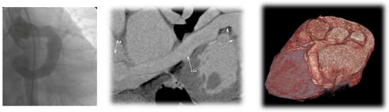
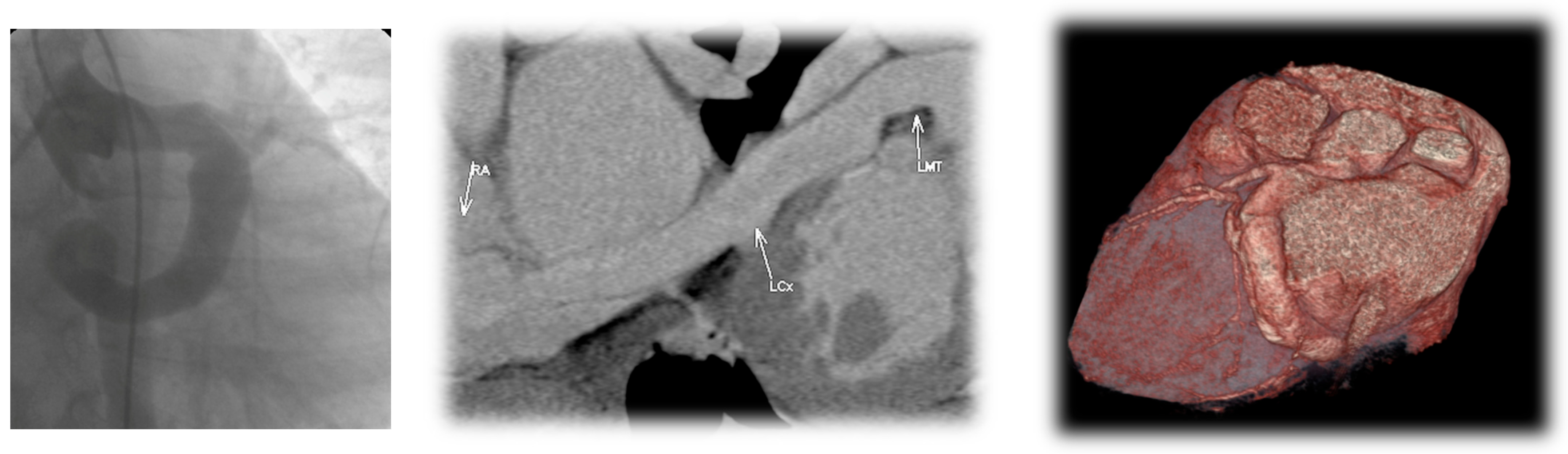
The main diagnostic advantage of CTCA over invasive angiography is the creation of a complete volumetric dataset to “unfold” tortuous anatomy and visualise the entire fistulous system from origin to terminus (Figure 3). This is particularly valuable in serpiginous or multi-communicating fistulae where angiography may demonstrate a dilated coronary with runoff but leave uncertainty regarding exact termination, multiplicity of drainage points, or the extent of aneurysmal remodeling [1,23].
Figure 3.
The left panel shows an invasive angiogram where CAF of the LCX is seen but it is not possible to determine its site of termination. The middle and right panels show a curved reconstruction and a 3D volume rendered image from the CTCA showing the origin, course and termination of a CAF originating from the LCX and terminating in the right atrium.
Termination pressure and compliance influence the amount of shunt, steal magnitude, symptom patterns, and device stability. CTCA can distinguish drainage into the main pulmonary artery versus branch pulmonary arteries, right-sided chambers, or the coronary sinus, and clarify multi-origin systems converging on a shared recipient [23,32]. Bilateral coronary-to-pulmonary artery fistulae can be clinically relevant despite modest shunt physiology and have reported with ischemia and exercise-induced ventricular arrhythmias, making precise anatomic mapping essential for causal inference and planning [33,34].
Chronic high-flow lesions can cause proximal and distal dilation, elongation, and aneurysm formation [1,16,23]. CTCA provides reproducible measurements of maximal/minimal diameters and aneurysm morphology (fusiform vs. saccular), and can detect mural thrombus—key because post-closure thrombosis and adverse remodeling have been noted in intermediate- and long-term outcome studies, especially in dilated distal systems [14,15,16]. CTCA easily defines the volume of remodeled conduit at risk for stagnation after closure and identifies pre-existing thrombus that alters urgency and antithrombotic strategy [14,15,16].
Symptoms may also be secondary to obstructive coronary disease, steal physiology, or a combination. CTCA can characterise plaque and stenoses while mapping the fistula, reducing misattribution of symptoms to an incidental CAF and supporting integrated strategies when both mechanisms are possible [16,23].
Infective endocarditis/endarteritis is an established but uncommon complication [33,34]. While echocardiography remains central for vegetation assessment, CTCA can clarify aneurysm anatomy and adjacent relationships relevant to subsequent closure planning after infection control [32]. For acquired fistulae, including those after CABG, CTCA is useful for defining the relationship to grafts and native coronaries; a systematic review of case reports confirms the existence of these iatrogenic aortocoronary AV fistulae and the need for careful anatomical characterisation [17].
Benefits of CT vs. Other Modalities
CTCA is the preferred imaging of choice for diagnosing and postprocedural/surgical follow up for CAFs. Invasive selective coronary angiography has conventionally been used to diagnose CAFs and offers dynamic visualisation of the CAFs at a higher spatial and temporal resolution than a CT with the current technology [35]. However, this is an invasive procedure that poses small risks of vascular complications and mortality. Invasive coronary angiography remains indispensable for transcatheter closure but can incompletely define distal course and termination due to overlap, brisk runoff, and underestimation of aneurysm size and branch relationships with limited angles of 2D projection [1,23] CAFs may be missed [36,37].
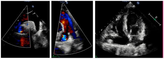
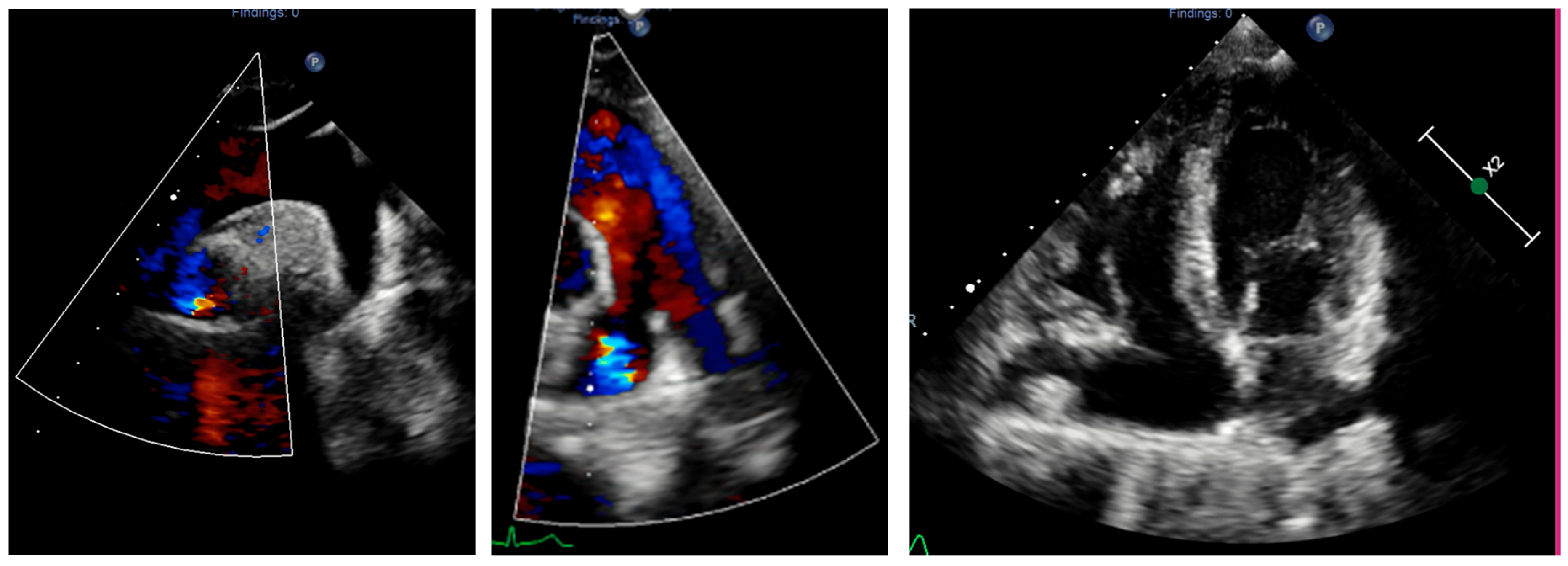
Transthoracic and transoesophageal echocardiogram are often the first modalities to detect and assess the coronary fistula by detecting a dilated blood vessel or abnormal blood flow on colour Doppler [38,39]. Transthoracic echocardiography can detect potentially fistula vessels over 3 mm in size in adult patients and the accuracy rate in patients with known CAF was reported at 19.1% [2]. Echocardiography is useful for evaluating chamber enlargement, volume loading, pulmonary pressures, and associated congenital lesions; however, it often cannot define the full coronary course in adults or in long/extracardiac pathways (Figure 4) [40].
Figure 4.
Transthoracic echocardiography, suprasternal and short-axis views showing abnormal flow in the right pulmonary artery and dilated right side of the heart in the 4-chamber view, raised the suspicion of a coronary fistula.
CMR provides functional information that is highly relevant when the haemodynamic significance of a fistula is uncertain and quantifies ventricular volumes, assesses myocardial scar and perfusion, and estimates shunt fraction without ionising radiation. Advanced techniques of 4D-flow CMR enable quantification of shunt flow (Qp:Qs) by direct measurements of flow at the pulmonary artery and aorta offering the fistula, offering an accurate assessment of its significance. Whilst assessing the significance of shunting can be difficult, CMR is useful for defining significance either as a Qp:Qs of greater than 1.5 to 1 or through the identification of dilatation of a chamber due to the CAF where no other cause is identified. CTCA nevertheless retains superior spatial resolution for defining the precise origin, distal course, aneurysm morphology, and branch relationships needed for procedural planning (34). CTCA and CMR are complementary cross-sectional modalities, can be used for integrating anatomical and functional information in the evaluation of CAF.
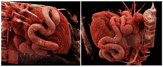
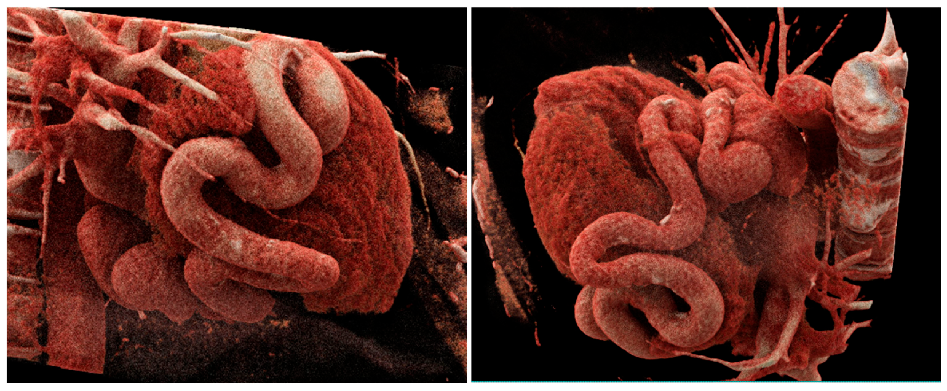
CTCA uniquely combines high spatial resolution near-isotropic volumetric acquisition, and flexible 3D reconstruction Figure 5, for comprehensive assessment of coronary origins, fistulous pathway, termination site(s), aneurysm morphology, and relations to adjacent structures, while also allowing assessment of coexistent coronary atherosclerosis in adults [27].
Figure 5.
Cinematic volume rendering 3D reconstruction of a serpiginous RCA fistula.
5. Risk Stratification and CTCA-Guided Management
Careful consideration is required as to whether closure is indicated and then the desired approach—transcatheter, surgical, or hybrid approach is most appropriate. It must be decided where occlusion should be performed to achieve durable closure with minimal risk, how device sizing and access should be planned, and how follow-up should be structured [1,9,14,28,32].
Advanced 3D processing is essential to determine the precise origin, course and termination of CAFs:
Multiplanar Reconstructions (MPR): provides cross-sectional views for accurate assessment of the vessel diameter, length and degree of tortuosity. It is key for identifying small vessels that may not be detected on axial images alone.
Maximum Intensity Projections (MIP): allows clear depiction of the entire course of the fistula from origin to the runoff (Figure 3).
3D Volume Rendering Technique: is postprocessing technique that converts the acquired CT volumetric data into an interactive 3D representation by assigning colours and opacity to voxels based on tissue density [41]. This offers 3D visualisation of the origin, course and runoff of the CAF and its anatomic relationship with other coronary arteries, cardiac chambers and large vessels [42].
A CTCA-driven pathway can classify lesions from low-risk anatomy (small calibre, no aneurysm, minimal remodeling, no thrombus) to higher-risk anatomy (large calibre, aneurysm burden, progressive ectasia, thrombus, complex architecture, bilateral feeders with plausible ischemic/arrhythmic mechanism) [23,27,32].
Low-risk: observation with structured follow-up imaging, using CTCA selectively when echocardiography is insufficient or when baseline 3D mapping is needed [23,32].
High-risk: closure considered, with CTCA guiding feasibility, access, device selection, and—critically—occlusion position to minimise residual dilated volume while preserving branches, consistent with both clinical thrombotic outcome observations and CFD evidence [14,15].
Anatomy unsuitable for percutaneous closure: surgical/hybrid strategies with CTCA providing the operative roadmap and a reproducible baseline for follow-up [20,21].
After closure, CTCA-based surveillance is targeted to expected risks: residual shunt, device position, persistence/regression of ectasia, and detection of mural thrombus in enlarged segments. “Immediate angiographic success” is not equivalent to elimination of long-term risk, supporting structured surveillance rather than discharge after initial closure [14,15,20,28,43].
5.1. Determining Whether to Close: CT-Based Risk Features
Long-term adverse outcomes correlate with larger fistula size and more extensive coronary dilation/remodeling [16]. CTCA investigates these predictors through reproducible measurements and by identifying high-risk features (giant aneurysm, mural thrombus, complex multi-channel architecture, progressive ectasia) that may support closure even when symptoms are minimal or nonspecific [19,20]. Conversely, CTCA may support observation when a fistula is small and lacks aneurysm, remodeling, or thrombus, enabling conservative management with bespoke imaging follow-up [44].
Guideline frameworks for ACHD support lesion- and risk-based decision-making rather than symptom-only strategies, particularly for lesions that are large or complicated [34]. Common consensus class 1 indications to close would include ventricular arrhythmias, ventricular dysfunction and ischaemic symptoms [45].
5.2. Selecting Closure Modality
Transcatheter closure is effective in appropriately selected anatomy, but is not free from limitations (device embolisation, residual shunt/recanalisation, ischemic complications, and thrombosis in dilated systems) [9,14,26,28,45,46]. CTCA determines feasibility by defining tortuosity, angulation, calibre variability, and the presence of a stable landing zone, while also identifying multiple distal communications or complex terminal aneurysms that may favour surgical or hybrid strategies [21,28].
Surgical series emphasize that long-term risk may persist after repair (e.g., residual fistula, late events), reinforcing the need for complete anatomic characterisation preoperatively and planned follow-up [20]. Surgery may be considered in cases requiring concomitant coronary artery bypass grafting, valve repair or replacement, and anomalies needing repair, distal CAFs, high-velocity CAFs, CAFs with tortuous courses or complex communications, with multiple drainage sites or a “fistula lake”, signs of volume overload and proximal CAFs with large aneurysms [20].
5.3. Device Selection, Sizing, and Landing-Zone Planning
CTCA provides accurate minimal/maximal diameters along candidate landing zones, available segment length for seating, and curvature that may impede deliverability or predispose to malapposition [32]. These data support rational selection between coils (often requiring narrowing/anchoring geometry) and vascular plugs (often preferable in larger, more uniform conduits) [9,26].
Large distal fistulae with substantial ectasia create a difficult dilemma: closure relieves shunt/steal but may leave a large, low-flow, “dead space” which is a risk for thrombus formation. Intermediate- and long-term outcome studies highlight thrombus formation and adverse remodeling after closure in selected anatomies [14,15,28]. CTCA helps anticipate this risk by quantifying residual dilated volume that would remain after occlusion at different positions and by defining terminal aneurysm geometry [14,15,23,32].
Patient-specific Computational Fluid Dynamics (CFD) studies derived from CTCA geometry show that occlusion position materially changes post-closure flow fields and hemodynamic parameters linked to thrombogenicity in terminal aneurysms, supporting individualised closure strategy rather than uniform approaches [43].
5.4. Preventing Closure-Related Ischemia
A critical hazard is the compromise of normal myocardial branches near the intended occlusion site. CTCA can better depict branch anatomy that may be underrepresented on angiography in high-flow fistulae due to preferential runoff, acting as a safety constraint for where occlusion can and cannot occur [8,9]. This is particularly important in proximal lesions (e.g., left main or proximal LAD involvement) where branch compromise may lead to clinically significant consequences [47].
5.5. Planning Access Route and Procedural Strategy
CTCA can predict whether an antegrade coronary approach or recipient-side retrograde approach is more feasible by defining termination angles, vessel relationships, and course complexity, potentially reducing the dose of radiation and the volume of contrast in angiography [27]. Arterial access is preferred for intervention to the proximal CAD. Distal CAF intervention is more preferably a venous approach. An arterio-venous loop with dual access is preferred in complex fistulae [45].
5.6. Covered Stents
Covered stents may be considered to block a fistulous origin but carry risks, especially side-branch compromise and thrombosis. Evidence extrapolated from coronary covered-stent use provides relevant safety context when such devices are contemplated, even though these data are not CAF-specific. CTCA informs whether a covered-stent strategy is anatomically plausible by defining branch ostia proximity and required sealing length [23].
5.7. Post Intervention/Operative Role of CT
CTCA plays an important role in the post-procedural and operative assessment of CAFs by evaluating the successful occlusion/closure, detecting complications, assessing for vessel remodeling and reoccurrence or recanalistion overtime.
During follow up: low dose CTCA techniques could be used for [27]:
- Confirming device position, stability or migration.
- Aneurysms regression.
- Presence of thrombus at the site of the device closure or within the fistula.
- Residual shunts if suspected.
- CTCA RV/LV indexed volumes in case of CMR contraindications for instances patients has pacemaker devices or claustrophobic.
- Coronary compromise or narrowing post-surgical.
- In cases of any acute extra cardiac complications for instances effusion, mediastinal hematomas, sternum or lung complications.
In previous interventional studies a degree of recanalisation in 15% over a longer period and that this occurred more than 6 months post intervention [45]. As a result of this it has been suggest that at least one follow up scan is performed up to 5 years post procedure to exclude recanalisation with successful reintervention reported following this [45].
6. Future Perspectives
CT’s role is likely to expand from anatomic depiction toward patient-specific risk prediction and strategy optimisation. CFD studies using CT-derived geometry already suggest that occlusion position can be optimised to reduce post-closure stasis in terminal aneurysms. Advances in ultra-low-dose CT and motion correction may increase feasibility of longitudinal surveillance in younger patients when clinically justified [36].
7. Conclusions
CTCA is central to contemporary management of coronary artery fistulae and provides comprehensive 3D anatomical delineation, quantitative characterisation of coronary remodeling and aneurysm burden, detection of high-risk features such as mural thrombus, and concurrent assessment of coexistent coronary disease. These capabilities influence patient selection for closure, choice of transcatheter versus surgical strategy, device/access planning, occlusion position selection with implications for late thrombosis, and structured post-closure surveillance. When integrated with echocardiography, invasive angiography, and clinical assessment, CTCA supports a reproducible, anatomy-driven management pathway aligned with outcome evidence and evolving interventional strategies.
Author Contributions
Conceptualisation, S.M.-E., R.A. and M.S.; Methodology, S.M.-E. and A.K.; Formal Analysis, S.M.-E., M.P. and A.K.; Investigation, S.M.-E., M.P. and A.K.; Resources, D.C.; Data Curation, S.M.-E., A.K., M.P. and M.S.; Writing—Original Draft Preparation, M.S. and S.M.-E.; Writing—Review & Editing, S.M.-E., DC., A.K. and R.A.; Visualization, S.M.-E.; Supervision, S.M.-E., and A.K.; Project Administration, R.A. All authors have read and agreed to the published version of the manuscript.
Funding
This research received no external funding.
Institutional Review Board Statement
Ethical review and approval were waived for this as a retrospective review paper containing historical data collected in routine care and anonymised.
Informed Consent Statement
Patient consent was waived as this is a retrospective review paper containing anonymised non-identifiable data collected as part of routine care previously.
Data Availability Statement
No new data were created or analyzed in this study.
Conflicts of Interest
The authors declare no conflict of interest.
References
- Mangukia, C.V. Coronary artery fistula. Ann. Thorac. Surg. 2012, 93, 2084–2092. [Google Scholar] [CrossRef] [Scilit]
- Goo, H.W. Imaging Findings of Coronary Artery Fistula in Children: A Pictorial Review. Korean J. Radiol. 2021, 22, 2062–2072. [Google Scholar] [CrossRef] [Scilit]
- Lim, J.J.; Jung, J.I.; Lee, B.Y.; Lee, H.G. Prevalence and types of coronary artery fistulas detected with coronary CT angiography. Am. J. Roentgenol. 2014, 203, W237–W243. [Google Scholar] [CrossRef] [Scilit]
- Torres, S.; Vasconcelos, M.; Tavares Silva, M.; Moreira, J.; Silva, J.C.; Macedo, F. Coronary artery fistulas: A 12-year single-center experience. Rev. Port. Cardiol. 2022, 41, 843–850. [Google Scholar] [CrossRef] [Scilit]
- Gowda, R.M.; Vasavada, B.C.; Khan, I.A. Coronary artery fistulas: Clinical and therapeutic considerations. Int. J. Cardiol. 2006, 107, 7–10. [Google Scholar] [CrossRef] [Scilit]
- Armsby, L.R.; Keane, J.F.; Sherwood, M.C.; Forbess, J.M.; Perry, S.B.; Lock, J.E. Management of coronary artery fistulae. Patient selection and results of transcatheter closure. J. Am. Coll. Cardiol. 2002, 39, 1026–1032. [Google Scholar] [CrossRef] [Scilit]
- Palmquist-Gomes, P.; Ruiz-Villalba, A.; Guadix, J.A.; Romero, J.P.; Bessiéres, B.; MacGrogan, D.; Conejo, L.; Ortiz, A.; Picazo, B.; Houyel, L.; et al. Origin of congenital coronary arterio-ventricular fistulae from anomalous epicardial and myocardial development. Exp. Mol. Med. 2023, 55, 228–239. [Google Scholar] [CrossRef] [Scilit]
- Reddy, G.; Davies, J.E.; Holmes, D.R.; Schaff, H.V.; Singh, S.P.; Alli, O.O. Coronary artery fistulae. Circ. Cardiovasc. Interv. 2015, 8, e003062. [Google Scholar] [CrossRef] [Scilit]
- Hobbs, R.E.; Millit, H.D.; Raghavan, P.V.; Moodie, D.S.; Sheldon, W.C. Coronary artery fistulae: A 10-year review. Clevel. Clin. Q. 1982, 49, 191–197. [Google Scholar] [CrossRef] [Scilit]
- Sunder, K.R.; Balakrishnan, K.G.; Tharakan, J.A.; Titus, T.; Pillai, V.R.; Francis, B.; Kumar, A.; Bhat, A.; Shankaran, S. Coronary artery fistula in children and adults: A review of 25 cases with long-term observations. Int. J. Cardiol. 1997, 58, 47–53. [Google Scholar] [CrossRef] [Scilit] [PubMed]
- Yun, G.; Nam, T.H.; Chun, E.J. Coronary Artery Fistulas: Pathophysiology, Imaging Findings, and Management. Radiographics 2018, 38, 688–703. [Google Scholar] [CrossRef] [Scilit]
- Kumar, R.; Kumar, J.; O’Connor, C.; Ullah, I.; Tyrell, B.; Pearson, I.; Matiullah, S.; Bainey, K. Coronary Artery Fistula: A Diagnostic Dilemma. Interv. Cardiol. 2023, 18, e25. [Google Scholar] [CrossRef] [Scilit]
- Lau, G. Sudden death arising from a congenital coronary artery fistula. Forensic Sci. Int. 1995, 73, 125–130. [Google Scholar] [CrossRef] [Scilit]
- Gowda, S.T.; Forbes, T.J.; Singh, H.; Kovach, J.A.; Prieto, L.; Latson, L.A.; Turner, D. Remodeling and thrombosis following closure of coronary artery fistula with review of management: Large distal coronary artery fistula–to close or not to close? Catheter. Cardiovasc. Interv. 2013, 82, 132–142. [Google Scholar] [CrossRef] [Scilit]
- Shah, A.H.; Osten, M.; Benson, L.; Alnasser, S.; Bach, Y.; Meier, L.; Horlick, E. Long-term outcomes of percutaneous closure of coronary artery fistulae in the adult: A single-center experience. Catheter. Cardiovasc. Interv. 2020, 95, 939–948. [Google Scholar] [CrossRef] [Scilit]
- Reidy, J.F.; Sowton, E.; Ross, D.N. Transcatheter occlusion of coronary to bronchial anastomosis by detachable balloon combined with coronary angioplasty at same procedure. Br. Heart J. 1983, 49, 284–287. [Google Scholar] [CrossRef] [Scilit]
- Chaumont, M.; Unger, P.; Debbas, N.; Vandenbossche, J.L. Unoperated giant coronary arteriovenous fistula: A thirty-year follow-up. Eur. Heart J. 2017, 38, 217. [Google Scholar] [CrossRef] [Scilit]
- Said, S.M.; Burkhart, H.M.; Schaff, H.V.; Connolly, H.M.; Phillips, S.D.; Suri, R.M.; Eidem, B.; Rihal, C.S.; Dearani, J.A. Late outcome of repair of congenital coronary artery fistulas--a word of caution. J. Thorac. Cardiovasc. Surg. 2013, 145, 455–460. [Google Scholar] [CrossRef] [Scilit]
- Sandor, B.; Bogats, G.; Toth, L.; Habon, T. Surgical treatment of heart failure due to giant coronary artery fistula: A case report. ESC Heart Fail. 2020, 7, 3203–3207. [Google Scholar] [CrossRef] [Scilit] [PubMed]
- Vijayvergiya, R.; Bhadauria, P.S.; Jeevan, H.; Mittal, B.R.; Grover, A. Myocardial ischemia secondary to dual coronary artery fistulas draining into main pulmonary artery. Int. J. Cardiol. 2010, 140, e30–e33. [Google Scholar] [CrossRef] [Scilit]
- De Roeck, F.; Franssen, C.; De Wolf, D.; Haine, S. Exercise-induced ventricular arrhythmia due to bilateral coronary to pulmonary artery fistulas. Catheter. Cardiovasc. Interv. 2019, 94, 112–116. [Google Scholar] [CrossRef] [Scilit]
- Sakata, N.; Minematsu, N.; Morishige, N.; Tashiro, T.; Imanaga, Y. Histopathologic Characteristics of a Coronary-pulmonary Artery Fistula with a Coronary Artery Aneurysm. Ann. Vasc. Dis. 2011, 4, 43–46. [Google Scholar] [CrossRef] [Scilit]
- Said, S.A. Current characteristics of congenital coronary artery fistulas in adults: A decade of global experience. World J. Cardiol. 2011, 3, 267–277. [Google Scholar] [CrossRef] [Scilit]
- Symbas, P.N.; Schlant, R.C.; Hatcher, C.R., Jr.; Lindsay, J. Congenital fistula of right coronary artery to right ventricle complicated by Actinobacillus actinomycetemcomitans endarteritis. J. Thorac. Cardiovasc. Surg. 1967, 53, 379–384. [Google Scholar] [CrossRef] [Scilit]
- Qureshi, S.A.; Tynan, M. Catheter closure of coronary artery fistulas. J. Interv. Cardiol. 2001, 14, 299–307. [Google Scholar] [CrossRef] [Scilit]
- Gowda, S.T.; Latson, L.A.; Kutty, S.; Prieto, L.R. Intermediate to long-term outcome following congenital coronary artery fistulae closure with focus on thrombus formation. Am. J. Cardiol. 2011, 107, 302–308. [Google Scholar] [CrossRef] [Scilit]
- Saboo, S.S.; Juan, Y.H.; Khandelwal, A.; George, E.; Steigner, M.L.; Landzberg, M.; Rybicki, F.J. MDCT of congenital coronary artery fistulas. Am. J. Roentgenol. 2014, 203, W244–W252. [Google Scholar] [CrossRef] [Scilit]
- Kim, H.; Beck, K.S.; Choe, Y.H.; Jung, J.I. Coronary-to-Pulmonary Artery Fistula in Adults: Natural History and Management Strategies. Korean J. Radiol. 2019, 20, 1491–1497. [Google Scholar] [CrossRef] [Scilit]
- Liu, J.; Zhang, Y. Photon-counting detector computed tomography for the diagnosis of coronary arteriovenous fistula: A new finding after repair of a coronary fistula case. Eur. Heart J. Case Rep. 2025, 9, ytaf376. [Google Scholar] [CrossRef] [Scilit]
- Han, T.H.; Kim, Y.W.; Lee, H.J.; Kim, J.-S.; Lee, S.-G.; Yang, D.H.; Oh, H.M.; Kim, D.; Shin, S.Y.; Song, S.; et al. Synthesis of coronary 4D CT Image by denoising diffusion probabilistic model. Comput. Methods Programs Biomed. 2026, 282, 109382. [Google Scholar] [CrossRef] [Scilit]
- Maffei, E.; Messalli, G.; Martini, C.; Nieman, K.; Catalano, O.; Rossi, A.; Seitun, S.; Guaricci, A.I.; Tedeschi, C.; Mollet, N.R.; et al. Left and right ventricle assessment with Cardiac CT: Validation study vs. Cardiac MR. Eur. Radiol. 2012, 22, 1041–1049. [Google Scholar] [CrossRef] [Scilit]
- Davis, J.T.; Allen, H.D.; Wheller, J.J.; Chan, D.P.; Cohen, D.M.; Teske, D.W.; Cassidy, S.C.; Craenen, J.M.; Kilman, J.W. Coronary artery fistula in the pediatric age group: A 19-year institutional experience. Ann. Thorac. Surg. 1994, 58, 760–763. [Google Scholar] [CrossRef] [Scilit]
- Said, S.A. Characteristics of Congenital Coronary Artery Fistulas Complicated with Infective Endocarditis: Analysis of 25 Reported Cases. Congenit. Heart Dis. 2016, 11, 756–765. [Google Scholar] [CrossRef] [Scilit]
- Stout, K.K.; Daniels, C.J.; Aboulhosn, J.A.; Bozkurt, B.; Broberg, C.S.; Colman, J.M.; Crumb, S.R.; Dearani, J.A.; Fuller, S.; Gurvitz, M.; et al. 2018 AHA/ACC Guideline for the Management of Adults With Congenital Heart Disease: A Report of the American College of Cardiology/American Heart Association Task Force on Clinical Practice Guidelines. J. Am. Coll. Cardiol. 2019, 73, e81–e192. [Google Scholar] [CrossRef] [Scilit]
- Zeina, A.R.; Blinder, J.; Sharif, D.; Rosenschein, U.; Barmeir, E. Congenital coronary artery anomalies in adults: Non-invasive assessment with multidetector CT. Br. J. Radiol. 2009, 82, 254–261. [Google Scholar] [CrossRef] [Scilit]
- Zhou, K.; Kong, L.; Wang, Y.; Li, S.; Song, L.; Wang, Z.; Wu, W.; Chen, J.; Jin, Z. Coronary artery fistula in adults: Evaluation with dual-source CT coronary angiography. Br. J. Radiol. 2015, 88, 20140754. [Google Scholar] [CrossRef] [Scilit]
- Erol, C.; Seker, M. Coronary artery anomalies: The prevalence of origination, course, and termination anomalies of coronary arteries detected by 64-detector computed tomography coronary angiography. J. Comput. Assist. Tomogr. 2011, 35, 618–624. [Google Scholar] [CrossRef] [Scilit]
- Kuo, C.T.; Chiang, C.W.; Fang, B.R.; Lee, C.P.; Hsu, T.S.; Lee, Y.S.; Chang, C.H. Coronary artery fistula: Diagnosis by transesophageal two-dimensional and Doppler echocardiography. Am. Heart J. 1992, 123, 218–220. [Google Scholar] [CrossRef] [Scilit]
- Calafiore, P.A.; Raymond, R.; Schiavone, W.A.; Rosenkranz, E.R. Precise evaluation of a complex coronary arteriovenous fistula: The utility of transesophageal color Doppler. J. Am. Soc. Echocardiogr. 1989, 2, 337–341. [Google Scholar] [CrossRef] [Scilit]
- Velvis, H.; Schmidt, K.G.; Silverman, N.H.; Turley, K. Diagnosis of coronary artery fistula by two-dimensional echocardiography, pulsed Doppler ultrasound and color flow imaging. J. Am. Coll. Cardiol. 1989, 14, 968–976. [Google Scholar] [CrossRef] [Scilit]
- Eid, M.; De Cecco, C.N.; Nance, J.W., Jr.; Caruso, D.; Albrecht, M.H.; Spandorfer, A.J.; De Santis, D.; Varga-Szemes, A.; Schoepf, U.J. Cinematic Rendering in CT: A Novel, Lifelike 3D Visualization Technique. Am. J. Roentgenol. 2017, 209, 370–379. [Google Scholar] [CrossRef] [Scilit]
- Detorakis, E.E.; Foukarakis, E.; Karavolias, G.; Dermitzakis, A. Cardiovascular magnetic resonance and computed tomography in the evaluation of aneurysmal coronary-cameral fistula. J. Radiol. Case Rep. 2015, 9, 10–21. [Google Scholar] [CrossRef] [Scilit]
- Cao, H.; Qiu, Y.; Yuan, D.; Yu, J.; Li, D.; Jiang, Y.; Su, L.; Peng, L.; Zheng, T. A computational fluid dynamics study pre- and post-fistula closure in a coronary artery fistula. Comput. Methods Biomech. Biomed. Eng. 2020, 23, 33–42. [Google Scholar] [CrossRef] [Scilit]
- Abbara, S.; Blanke, P.; Maroules, C.D.; Cheezum, M.; Choi, A.D.; Han, B.K.; Marwan, M.; Naoum, C.; Norgaard, B.L.; Rubinshtein, R.; et al. SCCT guidelines for the performance and acquisition of coronary computed tomographic angiography: A report of the society of Cardiovascular Computed Tomography Guidelines Committee: Endorsed by the North American Society for Cardiovascular Imaging (NASCI). J. Cardiovasc. Comput. Tomogr. 2016, 10, 435–449. [Google Scholar] [CrossRef] [Scilit]
- Al-Hijji, M.; El Sabbagh, A.; El Hajj, S.; AlKhouli, M.; El Sabawi, B.; Cabalka, A.; Miranda, W.R.; Holmes, D.R.; Rihal, C.S. Coronary Artery Fistulas: Indications, Techniques, Outcomes, and Complications of Transcatheter Fistula Closure. Cardiovasc. Interv. 2021, 14, 1393–1406. [Google Scholar] [CrossRef] [Scilit]
- Bagul, P.K.; Phadke, M.S.; Kerkar, P.G. Percutaneous Closure of Left Main Coronary Artery Fistula Using AMPLATZER Vascular Plug II and AMPLATZER Duct Occluder I. Cardiovasc. Interv. 2016, 9, e67-70. [Google Scholar] [CrossRef] [Scilit]
- Vaidya, Y.P.; Green, G.R. Coronary artery fistula. J. Card. Surg. 2019, 34, 1608–1616. [Google Scholar] [CrossRef] [Scilit]

Disclaimer/Publisher’s Note: The statements, opinions and data contained in all publications are solely those of the individual author(s) and contributor(s) and not of MDPI and/or the editor(s). MDPI and/or the editor(s) disclaim responsibility for any injury to people or property resulting from any ideas, methods, instructions or products referred to in the content. |
© 2026 by the authors. Licensee MDPI, Basel, Switzerland. This article is an open access article distributed under the terms and conditions of the Creative Commons Attribution (CC BY) license.