


Abstract
Diagnosis of vascular anomalies of the aortic arch is often delayed in adolescents and adult patients. In the presence of dyspnea that cannot be explained through a pulmonary and/or cardiac disease, and sudden dysphagia in children and younger adult patients, a cross-sectional imaging examination of the chest is mandatory to exclude or confirm malformation or malposition of the aortic arch and/or aberrant supra-aortic vessels.
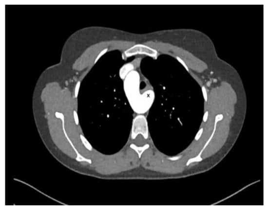
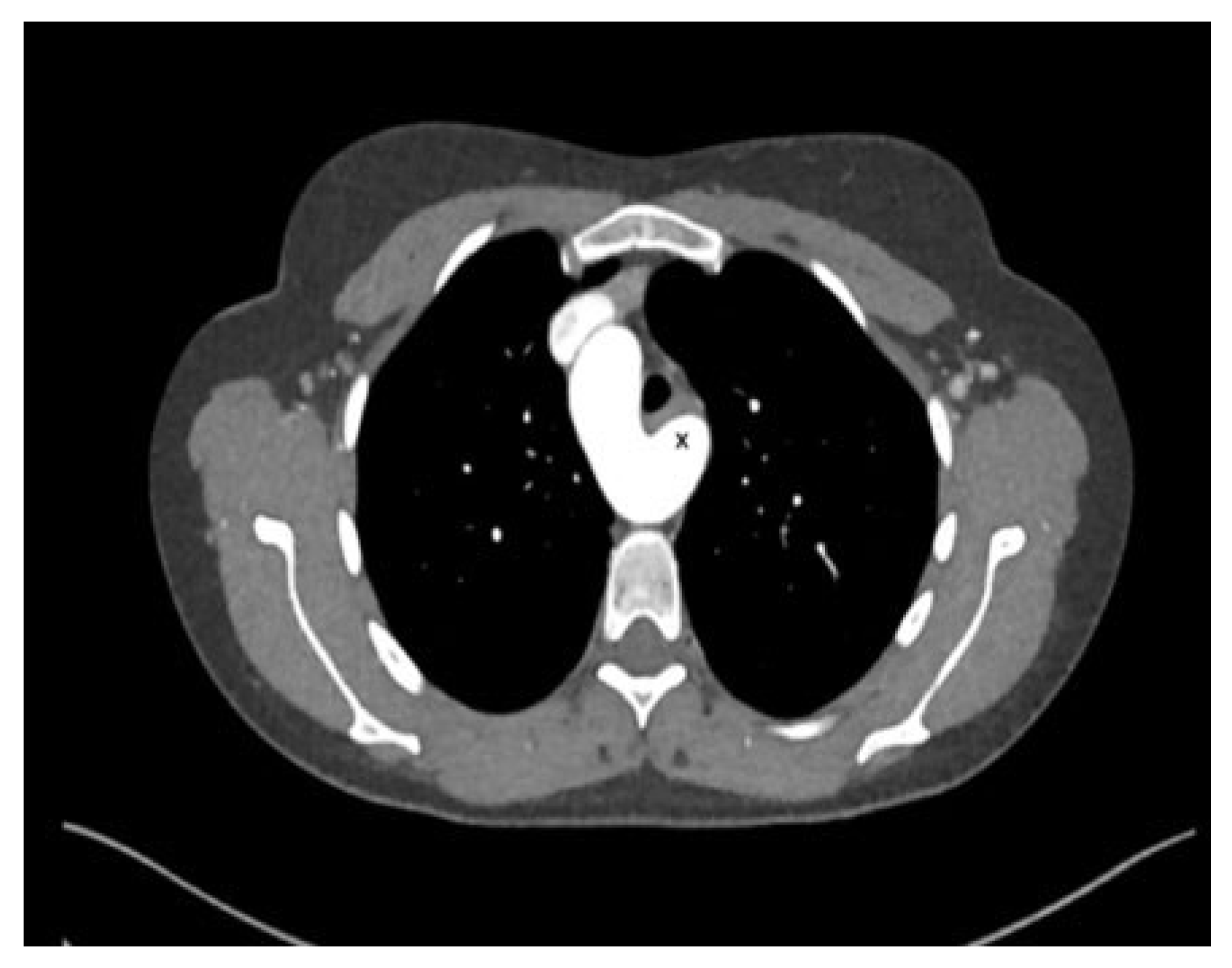
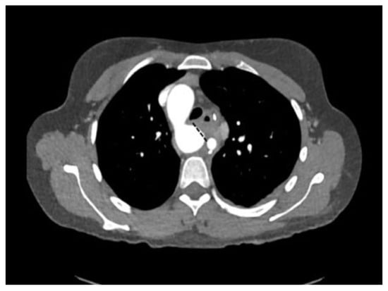
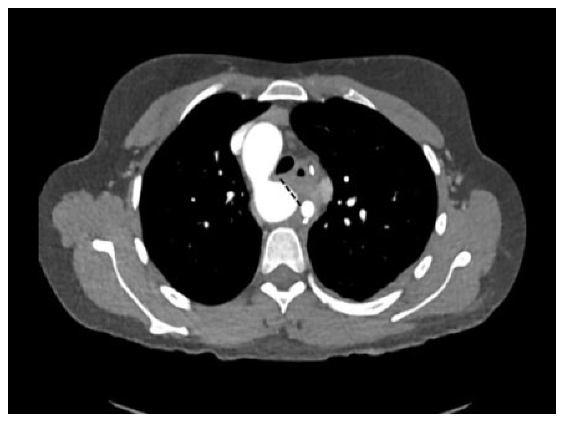
A 31-year-old woman with a history of bronchial asthma during adolescence was referred to the Cardiology Department because of recent worsening shortness of breath during minimal exercise; as she was working as an ambulatory nurse visiting patients at home, she could not work anymore. Otherwise she was in a good clinical condition and laboratory results were completely normal. A computed tomography (CT) scan surprisingly showed a right descending aortic arch with an abnormal left subclavian artery (left arteria lusoria) with a rather large Kommerell diverticulum and significant compression of the lower part of the trachea (Figure 1). A bronchoscopy confirmed this observation, revealing a pulsating mass (ascending aorta in the right upper quadrant of the trachea and a slit-like narrowing of the trachea, just cranial to the tracheo-bronchial bifurcation. Indication for surgery was discussed in the interdisciplinary team.
Figure 1.
CT scan showing the right descending arch with a large Kommerell diverticulum (x), out of which the left subclavian artery originates.
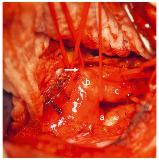
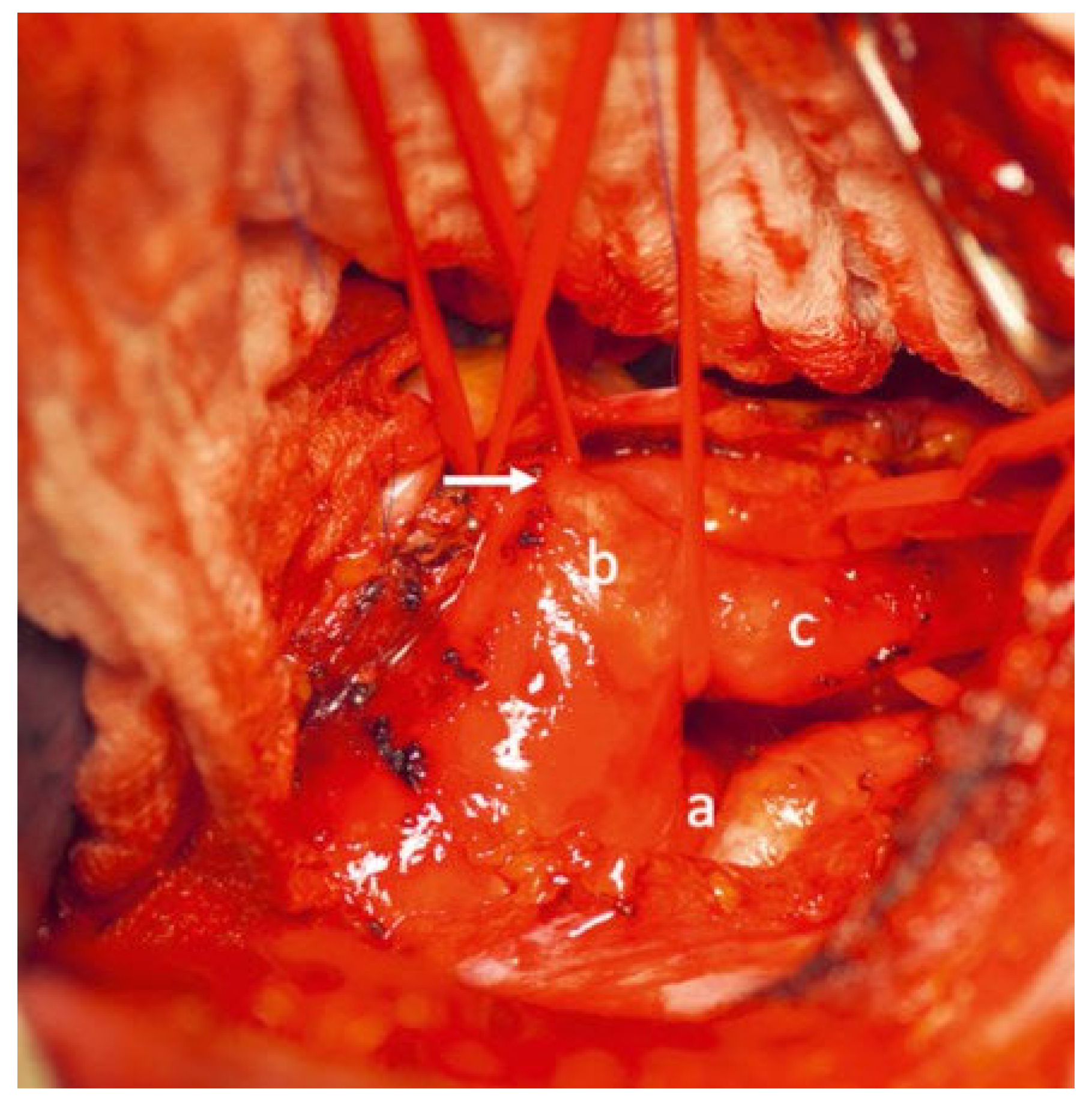
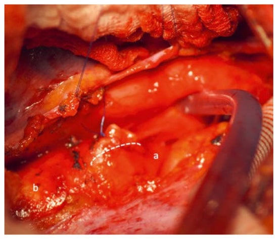
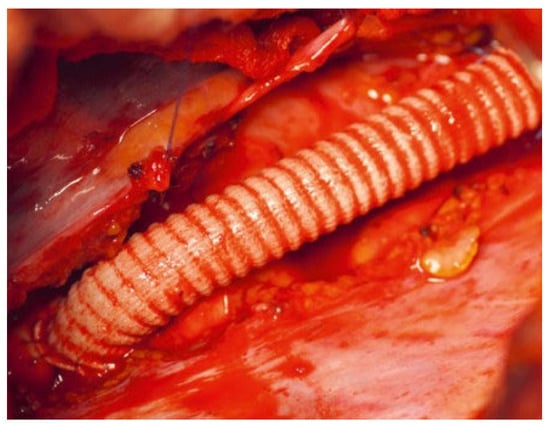
Surgical repair was performed through a postero-lateral left thoracotomy under 7500 IU intravenous heparin. The ductus ligamentum was divided, the left subclavian artery was detached from the Kommerell diverticulum and reimplanted more distally into the descending aorta using a 8 mm Dacron interposition graft and allowing thereby a more lateral position, away from esophagus and trachea. Finally, the distal aortic arch was clamped tangentially, the Kommerell diverticulum was resected and the aortic arch was closed with a running polypropylene suture (Figure 2, Figure 3, Figure 4, Figure 5 and Figure 6). The intraoperative bronchoscopy at the end of the operation showed a broad opening of the trachea. The postoperative course was uneventful except for temporary hoarseness, most probably due to the mobilisation of the recurrent nerve.
Figure 2.
Intraoperative view showing the distal aortic arch (a), the large Kommerell diverticulum (b) with the left subclavian artery (c) and the ductus ligament before division (arrow).
Figure 3.
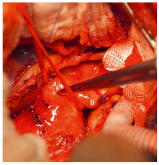
Vessel loop around the distal aortic arch. Kommerell diverticulum (a) and the proximal stump of the subclavian artery (b) before excision of the diverticle.
Figure 4.
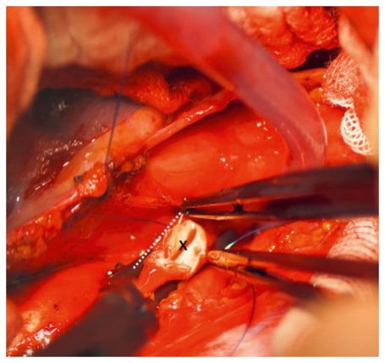
The aortic arch has been clamped tangentially (dotted line) and the Kommerell diverticulum excised (x).
Figure 5.
View after continuous suture of the excised Kommerell diverticle (dotted line) in convexity of the distal aortic arch (a). Site on the descending aorta for later subclavian artery re-implantation (b).
Figure 6.
“Distalisation and lateralisation” of the left subclavian artery in the descending aorta using a Dacron vascular graft.
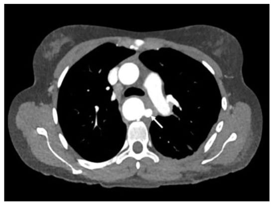
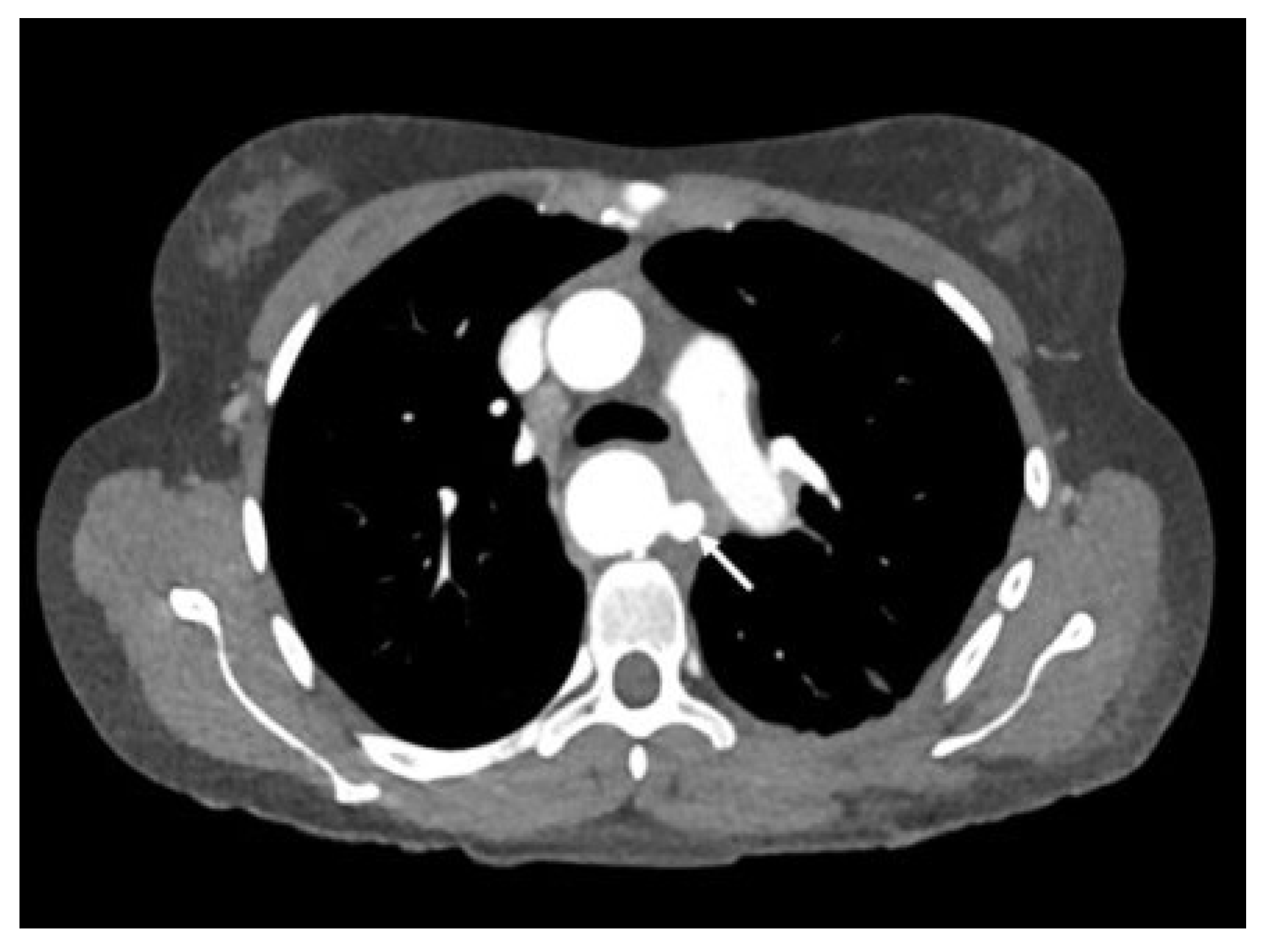
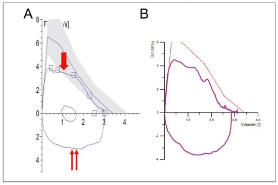
Postoperative CT scan did not show any residual tracheal compression (Figure 7). The aortic arch looked pretty normal after Kommerell resection and more distal left subclavian artery re-attachment (Figure 8). The patient received aspirin 100 mg for 6 months. At follow up 2 months postoperatively she was completely asymptomatic with normalisation of the lung function (Figure 9) and considerable improvement of her physical performance.
Figure 7.
Postoperative CT scan shows absence of left-anterior buldging of the Kommerell diverticulum (dotted line shows excision plane of the diverticle).
Figure 8.
Postoperative CT scan after resection of the diverticulum and implantation of the left subclavian artery more distally in the descending aorta (arrow). Broadly opened tracheo-bronchial bifurcation.
Figure 9.
(A) Flow-volume loop prior to relief of vascular ring, showing typical inspiratory (arrow) and expiratory (double arrow) flattening of fixed obstruction. (B) Normalisation of pattern of flow-volume loop after the operation.
Arteria lusoria, which is normally a right aberrant subclavian artery from the descending aorta, is one of the most common intrathoracic arterial anomalies, crossing behind the trachea and the oesophagus. It is often associated with other congenital cardiac malformations. A left arteria lusoria is much rarer and is usually encountered in patients with a right descending aortic arch.
This case report summarises a rare case of dyspnea in an adult patient and emphasises the fact that, in the absence of cardiac and/or pulmonary diseases, congenital vascular anomalies (vascular rings and slings) should be suspected [1,2]. To confirm the suspicion, a full cross-sectional imaging including either computed tomography and/or magnetic resonance imaging is necessary. Interestingly, the patient never presented with dysphagia or dyspnea but with a history of branchial asthma in the childhood.
Results of the surgical treatment of these malformations—division of the ductus ligament, excision of the diverticulum and/or translocation of the left subclavian artery (alternatively extra-thoracic reimplantation of the left subclavian into the left carotid artery)—are excellent and symptoms disappear usually rapidly after the intervention [3,4].
Conflicts of Interest
No financial support and no other potential conflict of interest relevant to this article was reported.
References
- Backer, C.L. Vascular rings with tracheoesophageal compression: management considerations. Semin Thorac Cardiovasc Surg Pediatr Card Surg Annu. 2020, 23, 48–52. [Google Scholar] [CrossRef] [Scilit] [PubMed]
- Saran, N.; Dearani, J.; Said, S.; Fatima, B.; Schaff, H.; Bower, T.; et al. Vascular rings in adults: outcome of surgical management. Ann Thorac Surg. 2019, 108, 1217–1227. [Google Scholar] [CrossRef] [Scilit] [PubMed]
- Vinnakota, A.; Idrees, J.J.; Rosinski, B.F.; Tucker, N.J.; Roselli, E.E.; Pettersson, G.B.; et al. Outcomes of repair of Kommerell diverticulum. Ann Thorac Surg. 2019, 108, 1745–1750. [Google Scholar] [CrossRef] [Scilit] [PubMed]
- Backer, C.L.; Russell, H.M.; Wurlitzer, K.C.; Rastatter, J.C.; Rigsby, C.K. Primary resection of Kommerell diverticulum and leh subclavian artery transfer. Ann Thorac Surg. 2012, 94, 1612–1617. [Google Scholar] [CrossRef] [Scilit] [PubMed]
© 2022 by the authors. Attribution - Non-Commercial - NoDerivatives 4.0.