


Introduction
As part of the Union Cycliste Internationale (UCI) medical monitoring programme, we perform each year a standard cardiovascular examination including a 12-lead resting electrocardiogram (ECG) in all members of a professional cycling team. Over a period of 2 years, we have analysed 45 ECGs of highly trained cyclists.
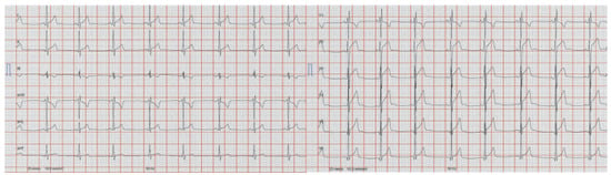
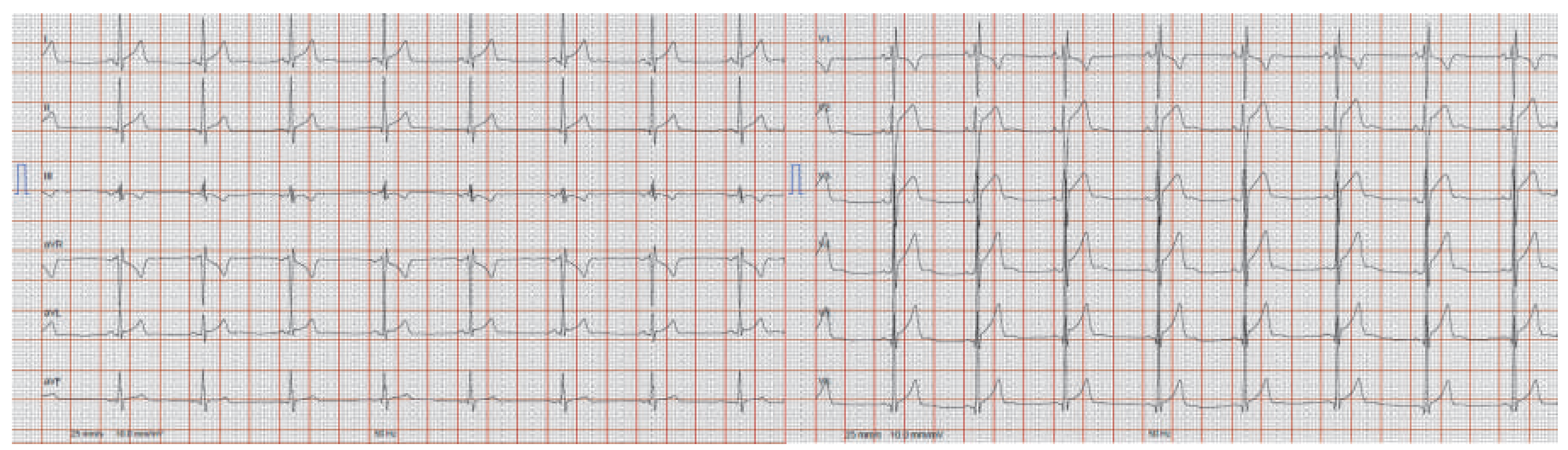
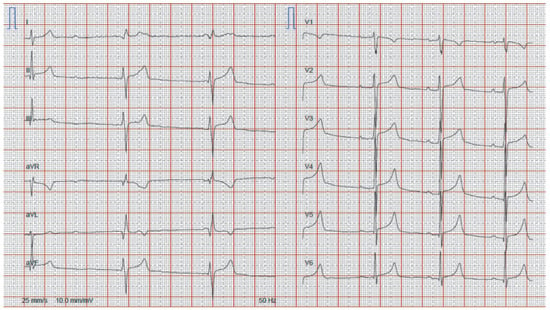
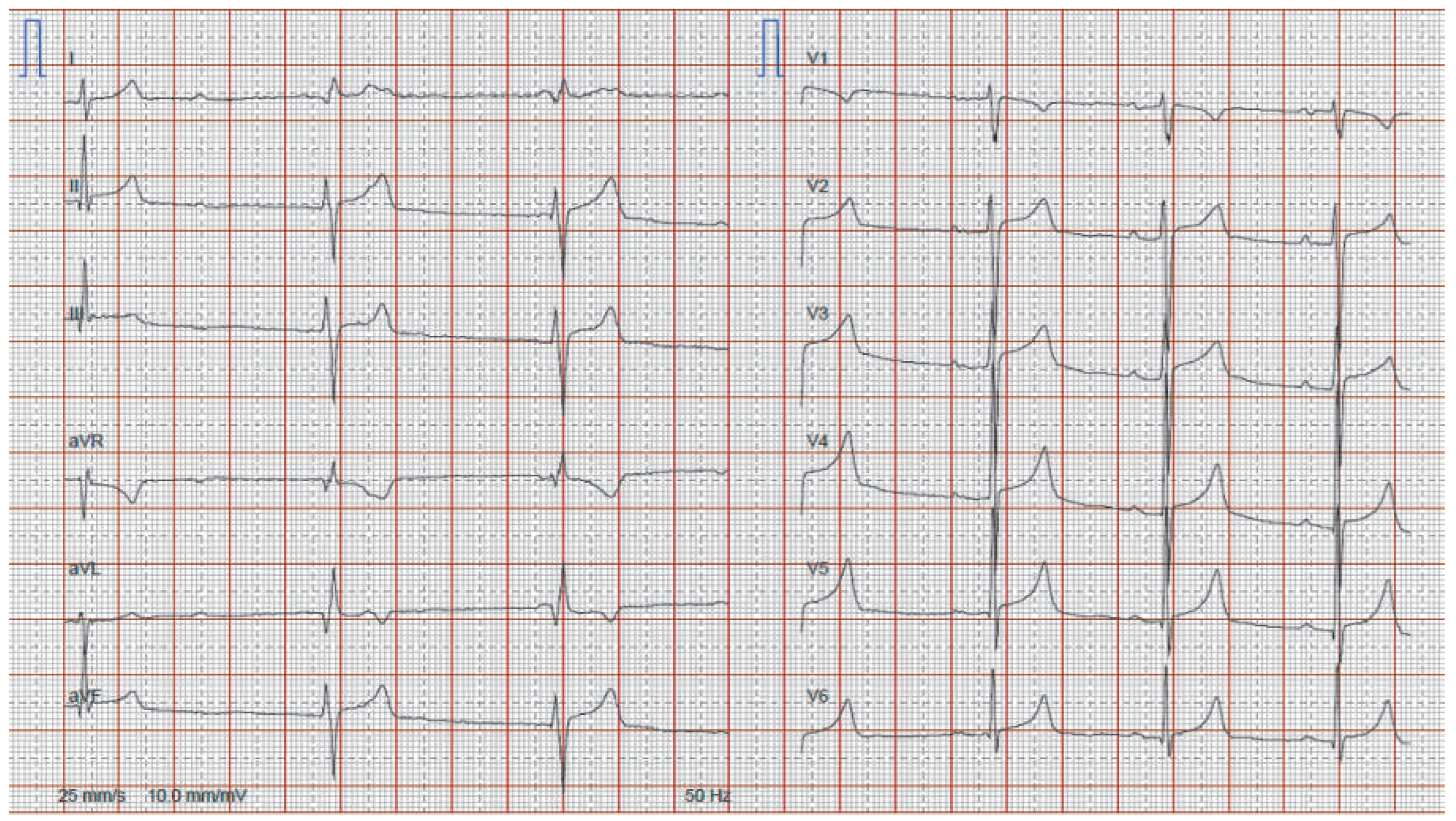
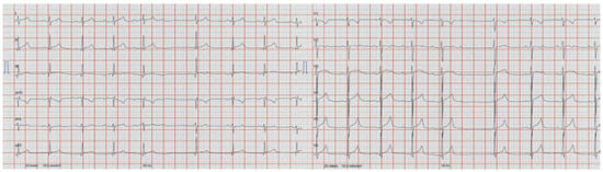
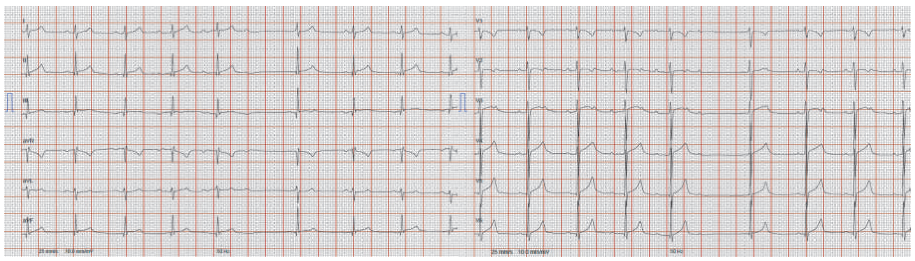
We present here the ECG recordings of two asymptomatic cyclists with characteristic changes found in this population. Neither had any personal or family history of cardiovascular disease and physical examination was unremarkable in both cases. In Figure 1 the ECG of cyclist no. 1 is displayed. The ECGs of cyclist no. 2 are presented in Figure 2 (2013 recording) and Figure 3 (2014 recording).
Figure 1.
Twelve-lead ECG of cyclist no. 1.
Figure 2.
Twelve-lead ECG of cyclist no. 2 in 2013.
Figure 3.
Twelve-lead ECG of cyclist no. 2 in 2014.
Questions
- − What ECG abnormalities should prompt further investigations in an athlete?
- − What degree of AV block is tolerable in an athlete?
Discussion
The ECG of cyclist no. 1 (Figure 1) shows sinus bradycardia, incomplete right bundle branch block (RBBB), a positive Sokolow-Lyon index (5.5 mV) for left ventricular hypertrophy (LVH), early repolarisation from V2 to V6 and in I, II and aVL, and tall precordial T waves. In Figure 2, the 2013 recording from cyclist no. 2 shows a second degree atrioventricular (AV) block, probably type I (Wenkebach), with two junctional escape beats preventing antegrade conduction of two sinus P waves. The 2014 recording (Figure 3) reveals sinus bradycardia, and clearly shows type I 2nd degree AV block, bifid P waves and early repolarisation in the precordial leads, with otherwise unremarkable findings.
High-level sports practice causes adaptive changes in the cardiovascular system, resulting in physiological alterations of the athlete’s ECG. It is important for the clinician interpreting these ECGs to know the boundaries between these frequent adaptive changes and pathological abnormalities that can signal underlying cardiac disease and/or an increased risk for arrhythmia or sudden cardiac death (SCD). To do so, specific criteria for athletes’ ECG interpretation have been established and have proven to be life-saving and costeffective. Corrado et al. wrote in 2010 a consensus statement for the European Society of Cardiology (ESC) [1], distinguishing two groups of ECG changes:
- − Common (up to 80% of athletes) and training-related changes include sinus bradycardia, first-degree AV block, incomplete RBBB, early repolarisation and isolated QRS voltage criteria for LVH. These changes, considered mild and warranting no further workup in the absence of symptoms and family history for SCD, are the result of an increased vagal tone, decreased resting sympathetic tone and left ventricular remodelling as a result of training.
- − Uncommon (fewer than 5% of athletes) and training-unrelated changes include T-wave inversion, ST-segment depression, pathological Q waves, left atrial enlargement, left-axis deviation / left anterior hemiblock, right-axis deviation / left posterior hemiblock, right ventricular hypertrophy, ventricular pre-excitation, complete left or right BBB, long or short QT interval, Brugada-like early repolarisation. These changes must be considered as potentially resulting from a pathological process and further testing is indicated.
The Seattle criteria, edited in 2012 [2], offer an equivalent tool with additional ECG criteria (sinus arrhythmia, ectopic atrial rhythm, junctional escape rhythm, ST segment elevation with T-wave inversion in V1–4 in black / African athletes considered as normal findings and intraventricular conduction delay (QRS ≥140 ms), profound sinus bradycardia (≤30 bpm or pauses ≥3s), atrial tachyarrhythmias including supraventricular tachycardia, atrial fibrillation and atrial flutter, frequent premature ventricular contractions (≥2 per 10 s), ventricular arrhythmias including couplets, triplets and nonsustained ventricular tachycardia considered as abnormal findings), and have proven epidemiologically superior to the previous ESC criteria [3]. Recently, refined criteria by Sheikh et al. added a third subgroup of ECG changes: borderline or minor abnormal findings, to be considered normal if found isolated, abnormal if found in combination, further improving specificity without any loss of sensitivity [4].
According to these recommendations, first degree and type I second degree (Wenkebach type) AV blocks are tolerated in athletes and considered to be associated with an increase in resting vagal tone. AV blocks of higher degrees are to be considered pathological. Some case reports mention asymptomatic and harmless type II second degree blocks in athletes. However, according to a literature review by Barold and Padeletti [5], these are cases of ECG misinterpretation and represent atypical type I second degree or vagally mediated AV blocks.
Returning to our cases, both cyclists met the criteria for common, training-related ECG changes. In cyclist no. 1, echocardiography was performed as an UCI obligatory examination and did not reveal LVH. Cyclist no. 2 was asymptomatic and showed a normal AV conduction and chronotropic response during the exercise stress test, therefore no additional study was performed.
Disclosure statement
No financial support and no other potential conflict of interest relevant to this article was reported.
References
- Corrado, D.; Pelliccia, A.; Heidbuchel, H.; et al. Recommendations for interpretation of 12-lead electrocardiogram in the athlete. Eur Heart J. 2010, 31, 243–59. [Google Scholar] [CrossRef] [Scilit] [PubMed]
- Drezner, J.A.; Ackerman, M.J.; Anderson, J.; et al. Electrocardiographic interpretation in athletes: the “Seattle criteria”. Br J Sports Med. 2013, 47, 122–4. [Google Scholar] [CrossRef] [Scilit] [PubMed]
- Brosnan, M.; La gerche, A.; Kalman, J.; et al. The Seattle Criteria increase the specificity of preparticipation ECG screening among elite athletes. Br J Sports Med. 2014, 48, 1144–50. [Google Scholar] [CrossRef] [Scilit] [PubMed]
- Sheikh, N.; Papadakis, M.; Ghani, S.; et al. Comparison of electrocardiographic criteria for the detection of cardiac abnormalities in elite black and white athletes. Circulation 2014, 129, 1637–49. [Google Scholar] [CrossRef] [Scilit] [PubMed]
- Barold, S.S.; Padeletti, L. Mobitz type II second-degree atrioventricular block in athletes: true or false? Br J Sports Med. 2011, 45, 687–90. [Google Scholar] [CrossRef] [Scilit] [PubMed]
© 2015 by the author. Attribution - Non-Commercial - NoDerivatives 4.0.