


Summary
Coronary vasospasm has been associated with acute myocardial infarction in patients with normal coronary arteries. A 35-year-old woman with acute myocardial infarction is described who had prolonged coronary artery spasm refractory to intracoronary vasodilators requiring primary coronary angioplasty and stenting.
Case report
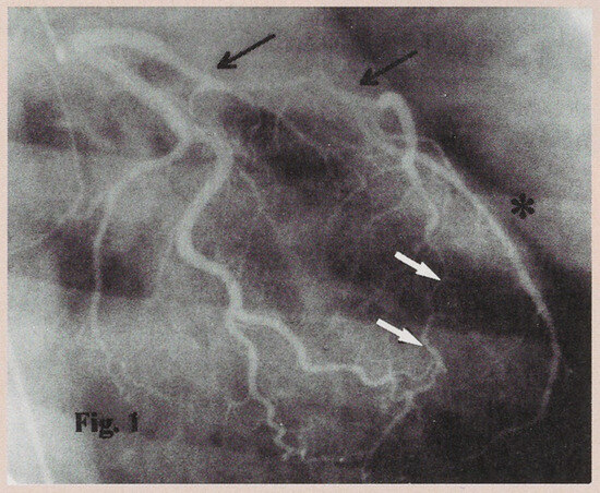
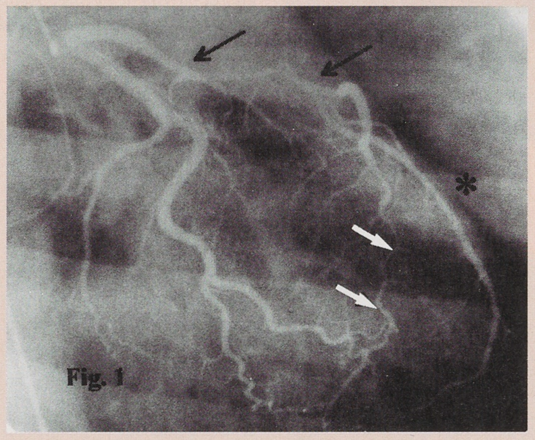
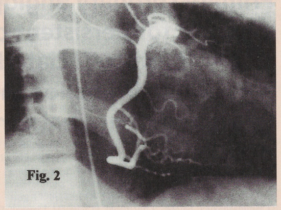
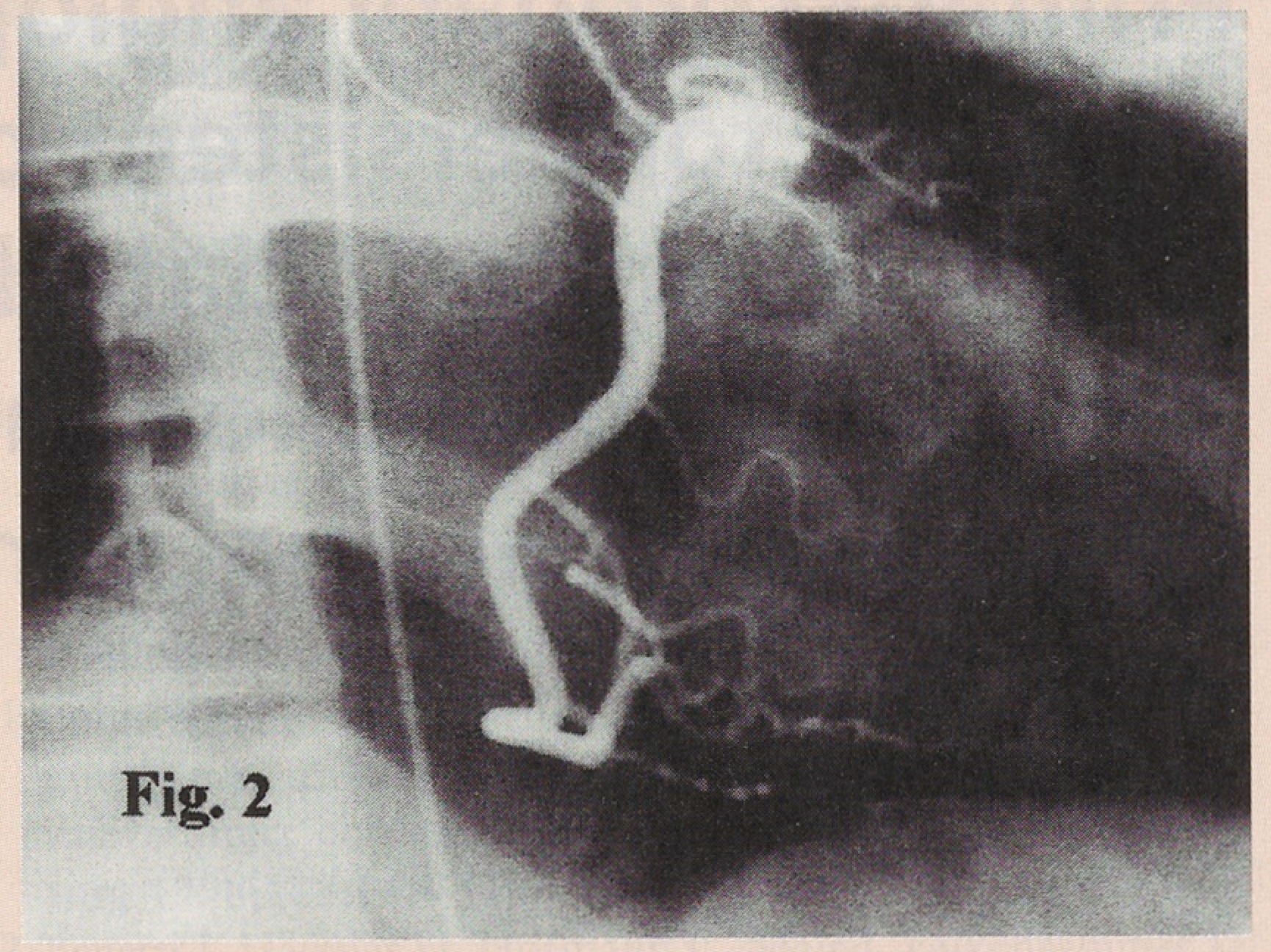
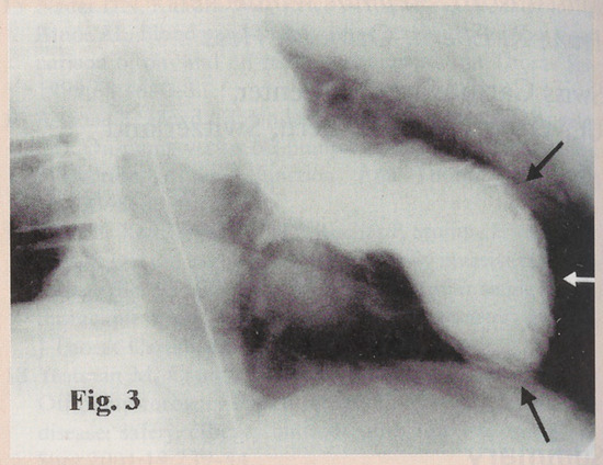
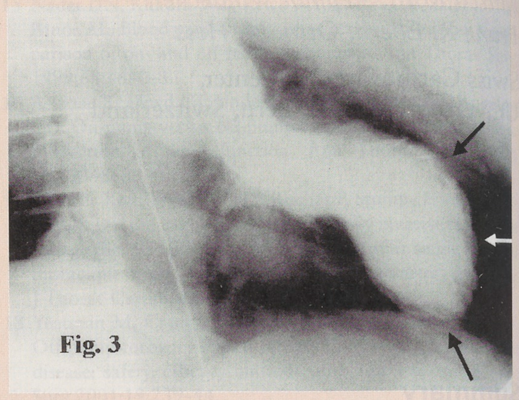
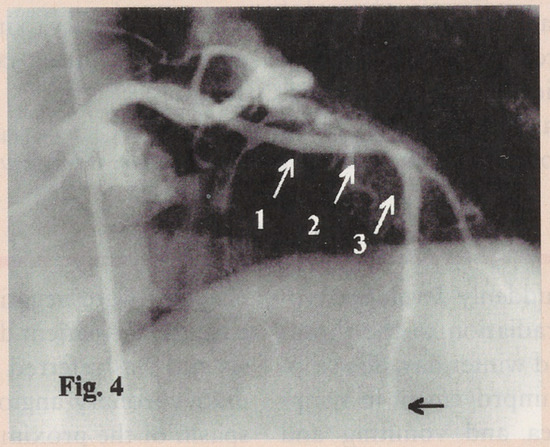
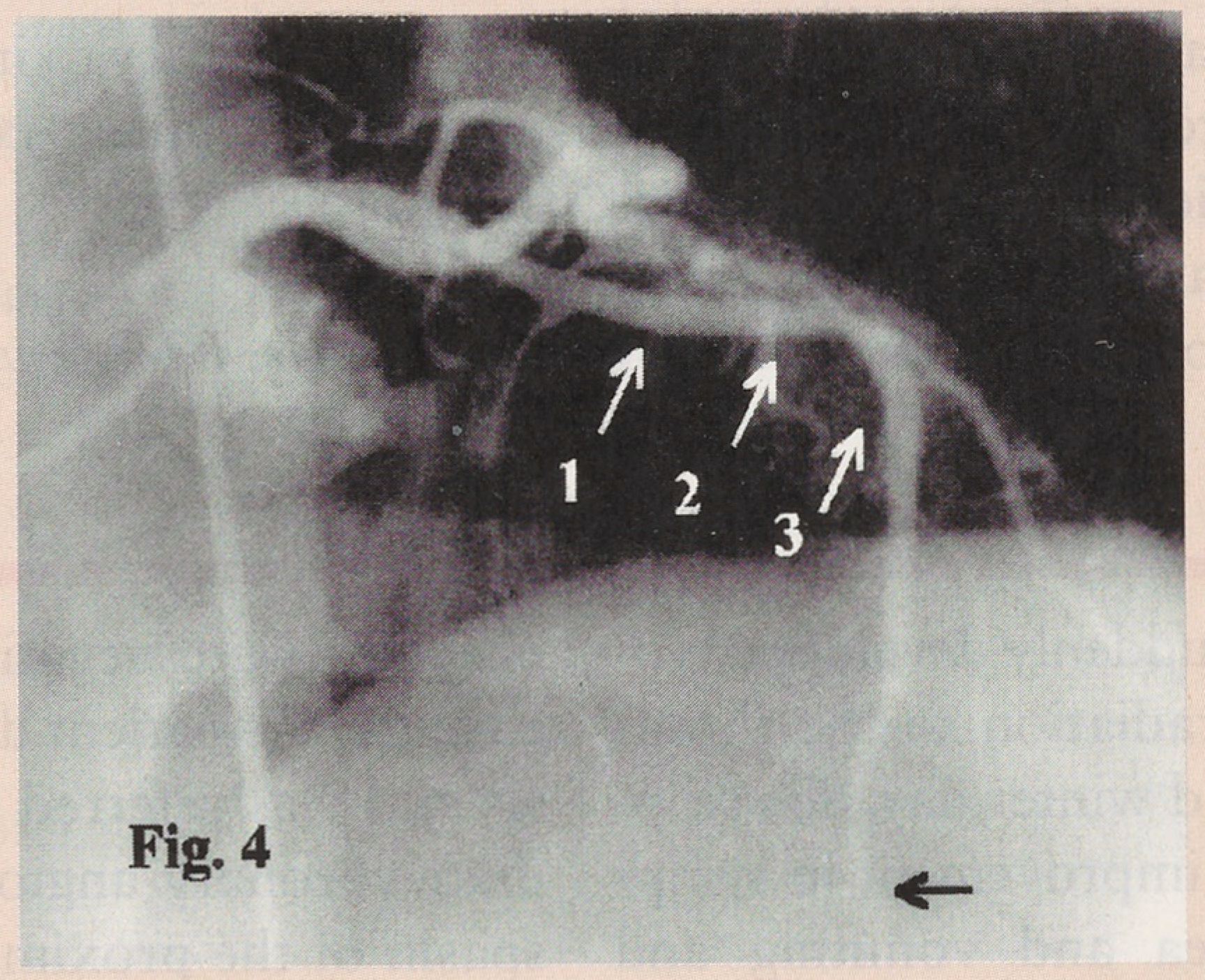
A 35-year-old woman suddenly developed typical chest pain with irradiation to the throat while bicycling on a cold winter day. She stopped exercising without improvement in symptoms, developed nausea and vomiting and suffered transient loss of consciousness. There was a similar incident the winter before. She had no risk factors for coronary artery disease and did not use cocaine or any other drugs. Her medical history was uneventful. She was admitted to a local hospital because of persisting chest pain. The ECG showed ST-segment elevation in precordial leads (V1—V5) but cardiac enzymes were negative. Assuming acute pericarditis, the patient did not receive thrombolysis and was referred to our institution. Immediate coronary angiography revealed a severe spasm of the proximal and distal left anterior descending coronary artery (LAD) (Figure 1). The left circumflex (LCX) and right coronary artery (RCA) were normal (Figure 2). Left ventricular (LV) angiography showed anterior akinesia with moderate reduction of LV systolic function (Figure 3). Despite intracoronary administration of nitroglycerin, diltiazem and adenosine, coronary artery spasm persisted. After administration of abciximab on top of heparin and aspirin, percutaneous transluminal coronary angioplasty (PTCA) of the proximal LAD was performed using a 3.0 mm balloon. The first low-pressure inflation (4 atm) led to acute vessel occlusion requiring bail-out stenting. Three 3.0 mm stents (two 13 mm long and one 8 mm long, ACS Multilink, Guidant/Advanced Cardiovascular Systems, Santa Clara, CA) were placed, two proximal to the first diagonal branch and one distal (Figure 4). The distal LAD was not approached despite a persistent, moderate to severe luminal narrowing (Figure 4). At the end of the intervention the patient was asymptomatic with a TIMI III flow in the LAD.
Figure 1.
Right anterior oblique (RAO) projection with caudal angulation of the left coronary artery showing a long spasm of the proximal (black arrows) and the distal (white arrows) LAD. The star denotes the first diagonal branch.
Figure 2.
RAO view demonstrating a normal RCA.
Figure 3.
Left ventricular systolic angiogram in 30° RAO projection during systole showing anterior akinesia (arrows).
Figure 4.
RAO cranial view demonstrating the final result of PTCA and deployment of three stents (number 1 to 3). The persistent diffuse spasm of the distal LAD (black arrow) was not approached.
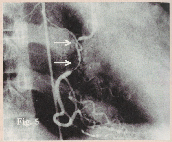
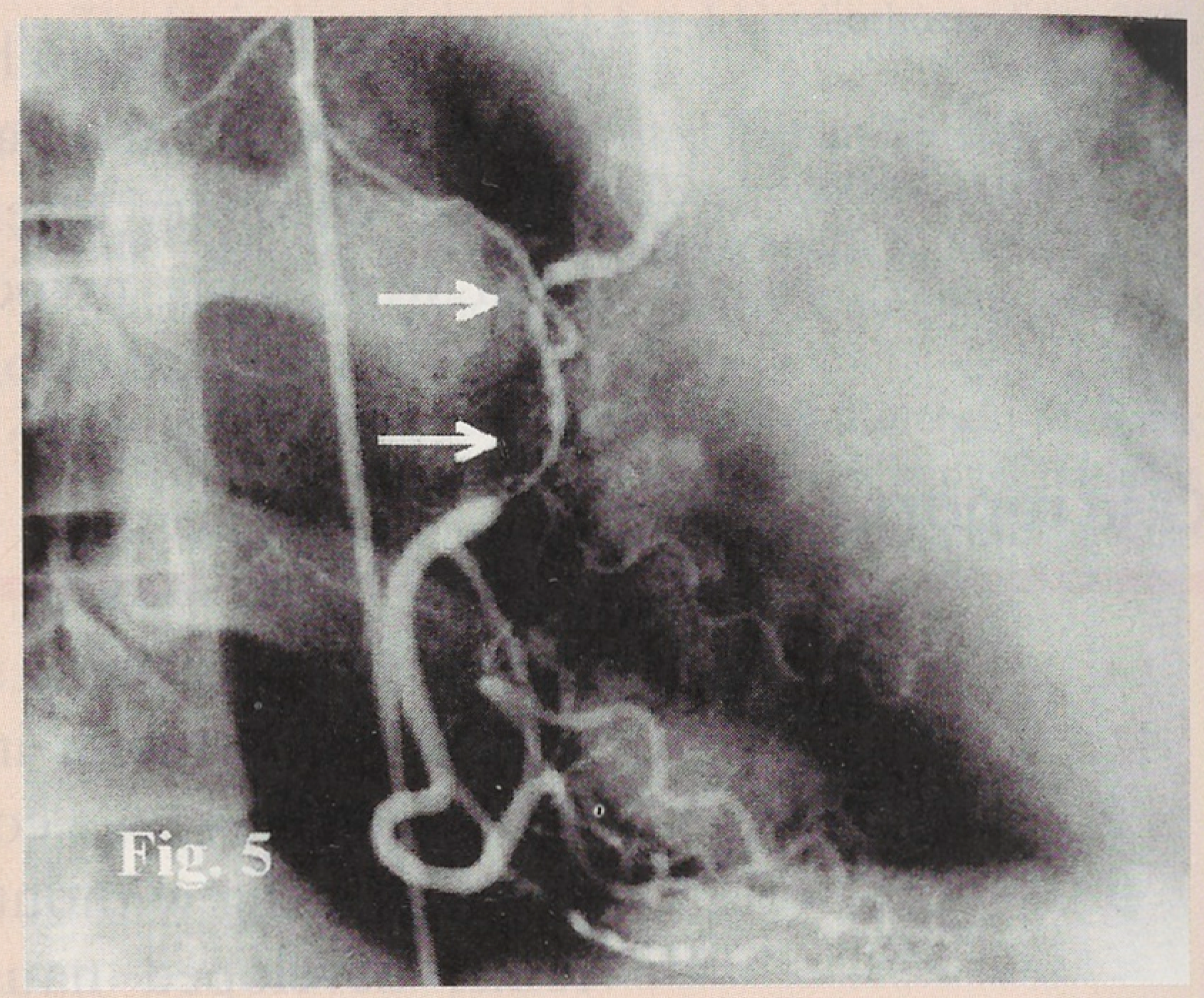
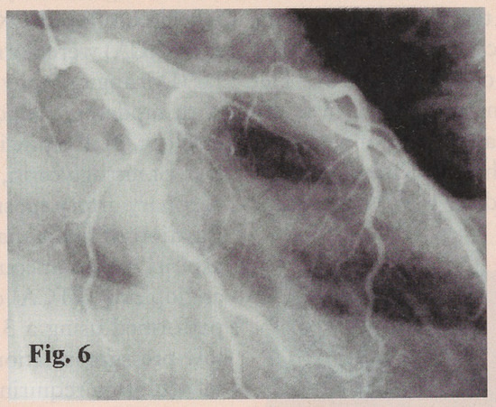
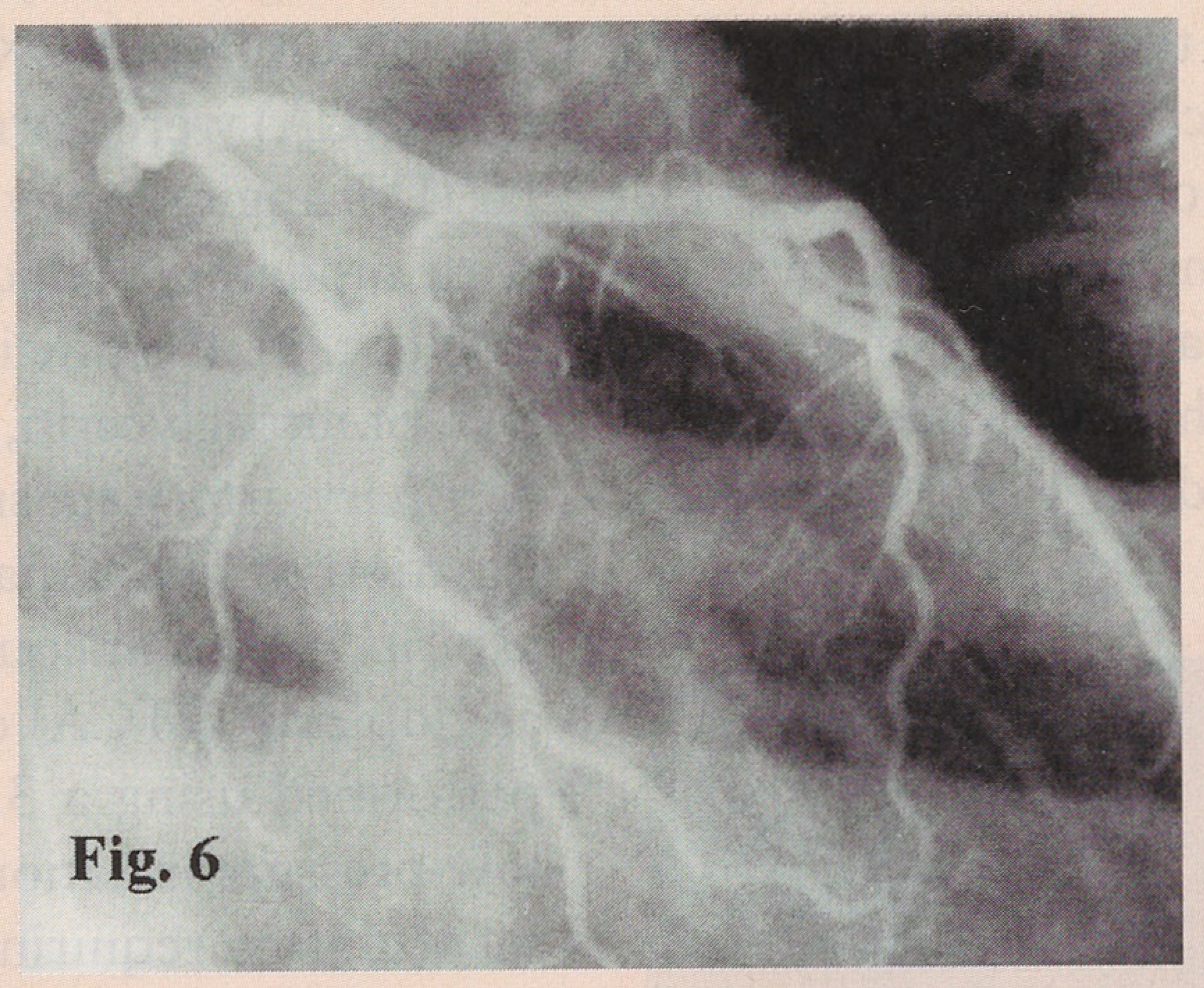
Cardiac enzymes reached already the maximum before angiography: CK 2190 U/l (normal <167 U/l), CK-MB mass 156 mmo/l (normal <4 mmo/l) and Troponin-I 260 μg/l (normal <0.6 μg/l), respectively. Follow-up ECG demonstrated loss of R-waves and symmetric T-wave inversion in the precordial leads but no Q-waves. Urine drug screening was negative for alcohol and cocaine. The patient returned to the referring hospital the following day. She was treated with a calcium-antagonist, aspirin, ticlopidine and warfarin. Due to hypotension the calcium-antagonist was later discontinued. The patient was returned to our hospital five days after discharge for recurrent chest pain. Repeat coronary angiography revealed a good result of LAD-PTCA with normal flow but persistent spasm of the distal LAD. During right coronary angiography the patient developed chest pain and hypotension. The first injection documented a long proximal subtotal spasm of the RCA (Figure 5). Administration of intracoronary nitroglycerin led to a slow resolution of the spasm. The day after the intervention, the patient was discharged on aspirin, ticlopidine, diltiazem, nitrates and warfarin and remained free of symptoms ever since. A six month follow-up angiogram was performed which showed a normal appearance of the coronary artery tree without evidence of in-stent restenosis or coronary spasm (Figure 6). The LV systolic function recovered completely, with only a slight apical akinesia.
Figure 5.
RAO view demonstrating a long subtotal spasm of the proximal RCA during repeat coronary angiogram 6 days after the intervention (arrows, compare Figure 2).
Figure 6.
Six month follow-up angiogram (RAO caudal view) showing normal appearance of the left coronary artery. No in-stent restenosis is detected. The distal LAD has normal caliber.
Discussion
A rare case of a young woman with normal coronary arteries and acute anterior myocardial infarction due to prolonged spasm of the LAD is reported. The spasm was refractory to aggressive intracoronary vasodilator therapy and required primary PTCA and coronary stenting (Figure 1, Figure 4). Another episode of chest pain was observed one year earlier under similar circumstances. There were no risk factors for coronary artery disease, and in particular no predisposing conditions for an abnormal coronary vasomotor response like hypercholesterolaemia or cigarette smoking [1]. Moreover, other triggers such as hyperventilation or cocaine abuse did not apply to this patient. The most likely pathophysiologic explanation for this event is coronary vasospasm, since the distal LAD and the RCA showed similar but reversible events. Particularly, our patient developed a long, subtotal spasm of the RCA with angina and hypotension during the second angiogram (Figure 5). Although triggered by the catheter, the length of the coronary artery spasm was unusual for a catheter-induced spasm which is typically short, just at the tip of the catheter, and usually asymptomatic. Moreover, this spasm resolved only slowly after multiple injections of intracoronary nitroglycerin. Although it has been questioned whether catheterinduced spasms are related to variant angina [2], the long and prolonged spasm seems to be suggestive for a hyperresponsiveness of the coronary tree. Variant angina is characterized by a hyperreactivity of coronary arteries to a variety of vasoconstrictor stimuli [3]. Like in our patient, cold exposure, i.e. cold pressor testing, can induce coronary artery spasm in patients with variant angina [3]. In anatomically normal coronary arteries, prolonged coronary spasm has been associated with endothelial damage, platelet adhesion, thrombus formation [4], arrhythmias [5,6], and, rarely, myocardial infarction [7]. Usually, no relation is found between physical activity and symptoms in patients with variant angina. However, exercise may induce coronary spasm as shown in patients with variant angina exercising on the cardiac catheterisation table [8]. Pharmacological treatment remains the therapy of choice in the prevention of variant angina. Recently, PTCA and stent placement have been advocated for isolated cases of recurrent variant angina resistant to medical therapy [9,10]. To our knowledge, only one case with stent placement for persistent iatrogenic coronary vasospasm has been published so far [11]. That patient had variant angina and developed refractory coronary spasm following contrast injection during coronary angiography. In our case, coronary angioplasty was mandatory due to a long subtotal LAD spasm associated with acute myocardial ischaemia persisting despite the intracoronary use of nitrates, calcium-antagonists and adenosine as well as combined antiplatelet therapy. After the first low-pressure balloon inflation, the proximal LAD occluded completely and therefore bail-out stenting with three stents was necessary. Six months after the intervention no in-stent restenosis was detected (Figure 6) and overall left ventricular function recovered except for persisting apical akinesia. Under calcium-antagonists, the patient has remained now free of symptoms for a total of 18 months. To our knowledge, this is the first report on primary PTCA and stenting for spontaneous persistent coronary spasm leading to acute myocardial infarction in a patient with prolonged coronary vasospasm in otherwise normal coronary arteries.
References
- Nobuyoshi, M.; Abe, M.; Nosaka, H.; Kimura, T.; Yokoi, H.; Hamasaki, N.; et al. Statistical analysis of clinical risk factors for coronary artery spasm: Identification of the most important determinant. Am. Heart J. 1992, 124, 32–8. [Google Scholar] [CrossRef] [Scilit] [PubMed]
- Mautner, R.K.; Cooper, M.D.; Phillips, J.H. Catheter-induced coronary artery spasm: An angiographic manifestation of vasospastic angina? Am. Heart J. 1983, 106, 659–65. [Google Scholar] [CrossRef] [Scilit] [PubMed]
- Kaski, J.C.; Crea, F.; Meran, D.; Rodriguez, L.; Araujo, L.; Chierchia, S.; Davies, G.; Maseri, A. Local coronary supersensitivity to diverse vasoconstrictive stimuli in patients with variant angina. Circulation 1986, 74, 1255–65. [Google Scholar] [CrossRef] [Scilit] [PubMed]
- Gertz, S.D.; Uretsky, G.; Wajnberg, R.S.; Navot, N.; Gotsmann, M.S. Endothelial cell damage and thrombus formation after partial arterial constriction: relevance to the role of coronary artery spasm in the pathogenesis of myocardial infarction. Circulation 1981, 63, 476–86. [Google Scholar] [CrossRef] [Scilit] [PubMed]
- Hess, O.M.; Graf, C.h.; Frey, R.; Dettli, R.; Siegenthaler, W. Coronary artery spasms with normal coronary arteries as the cause of recurrent ventricular fibrillations. Schweiz Med. Wochenschr. 1981, 111, 755–8. [Google Scholar] [PubMed]
- Miller, D.D.; Waters, D.D.; Szlachcic, J.; Théroux, P. Clinical characteristics associated with sudden death in patients with variant angina. Circulation 1982, 66, 588–92. [Google Scholar] [CrossRef] [Scilit] [PubMed]
- Vincent, G.M.; Anderson, J.L.; Marshall, H.W. Coronary spasm producing coronary thrombosis and myocardial infarction. N Engl. J. Med. 1982, 309, 220–3. [Google Scholar]
- Fuller, C.M.; Raizner, A.E.; Chahine, R.A.; Nahormek, P.; Ishimori, T.; et al. Exercise-induced coronary arterial spasm: Angiographic demonstration, documentation of ischemia by myocardial scintigraphy and results of pharmacologic intervention. Am. J. Cardiol. 1980, 46, 500–6. [Google Scholar] [CrossRef] [Scilit] [PubMed]
- Gupta, S.; Schiele, F.; Vuillemenot, A.; Bassand, J.P. Coronary stent for variant angina: Atypical presentation. Cathet Cardiovasc Diagn. 1998, 45, 439–41. [Google Scholar] [CrossRef] [Scilit] [PubMed]
- Nakamura, T.; Furukawa, K.; Uchiyama, H.; Seo, Y.; Okuda, S.; Ebizawa, T. Stent placement for recurrent vasospastic angina resistant to medical treatment. Cathet Cardiovasc Diagn. 1997, 42, 440–3. [Google Scholar] [CrossRef] [Scilit] [PubMed]
- Kultursay, H.; Can, L.; Payzin, S.; Turkoglu, C.; Alting, A.; Akin, M.; Akili, A. A rare case for stenting: Persistent coronary artery spasm. Heart Vessel 1996, 11, 165–8. [Google Scholar]
© 2001 by the author. Attribution - Non-Commercial - NoDerivatives 4.0.