


Introduction
Coronary artery bypass grafting remains the standard of care for unprotected left main trunk disease. However, in certain circumstances percutaneous coronary intervention may be appropriate. We report rotablation, ballon angioplasty and stenting of an unprotected left main trunk in a patient at high risk for bypass Surgery.
Case report
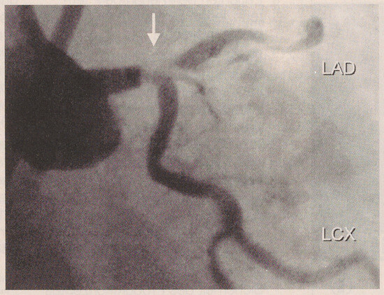
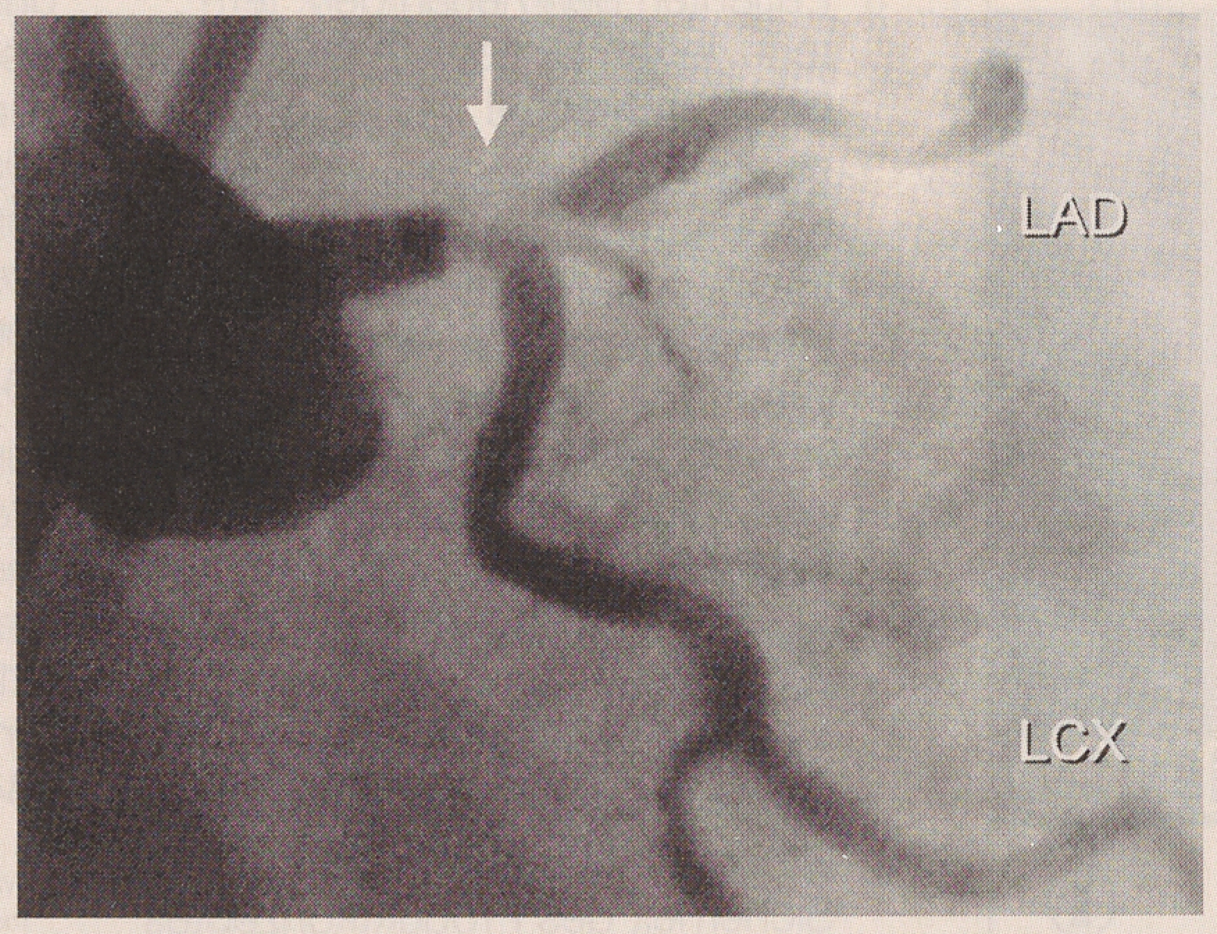
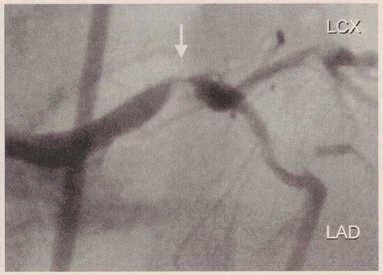
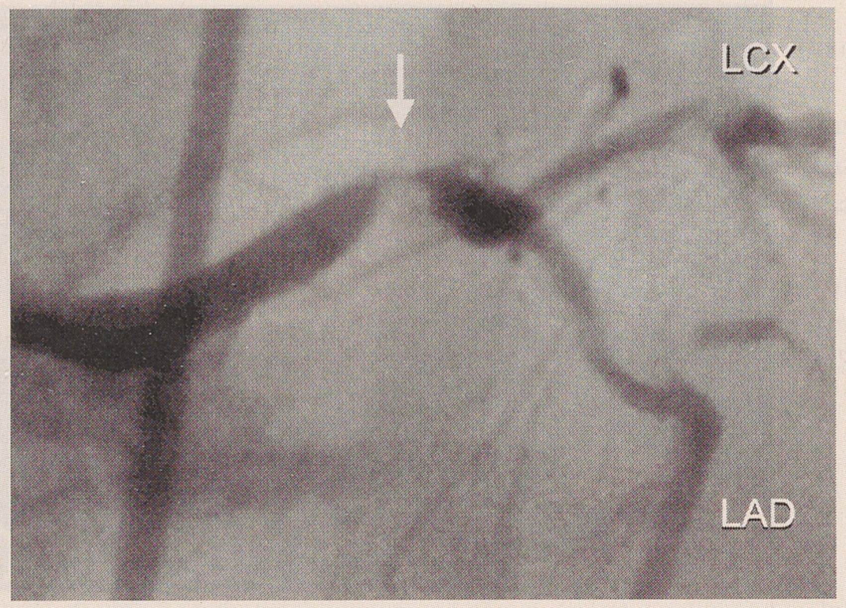
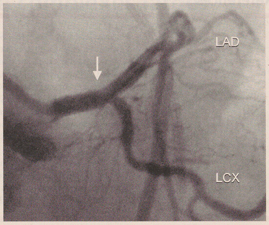
A 73-year-old man was referred for a second opinion regarding debilitating angina. At age 25, he suffered polio requiring an iron lung and causing severe restrictive lung disease. He had been on domiciliary oxygen therapy and recent pulmonary function tests revealed a FEVI of 100 ml (40% of predicted) and a total lung capacity of 1.51 (52% of predicted). Hypertension, positive family history, and previous smoking were the risk factors for coronary artery disease. He had been complaining of chest pain on mild exertion and at rest for the last 8 months. Exercices echocardiography demonstrated anterior ischemia and preserved left ventricular (LV) function. Coronary angiography revealed a critical stenosis of the distal left main trunk (LMT) involving the bifurcation of the left anterior descending coronary artery (LAD) and the left circumflex coronary artery (LCX) (Figure 1 and Figure 2). The right coronary artery (RCA) was occluded.
Figure 1.
Coronary angiogram in right anterior oblique (RAO) cranial projection showing a severe distal left main trunk stenosis (arrow) involving the proximal left anterior descending coronary artery (LAD) and the left circumflex (LCX).
Figure 2.
Same lesion (arrow) in posterior-anterior (PA) cranial view. LAD = left anterior descending coronary artery; LCX = left Figure 1 circumflex.
Due to the pulmonary comorbidity, several surgeons declined to proceed with coronary bypass surgery and the patient was treated conservatively. Despite aggressive medical treatment he continued to have severe angina with minor activities like teeth brushing.
Therefore he sought other treatment options. After consultation at our institution, the patient agreed to undergo percutaneous coronary intervention.
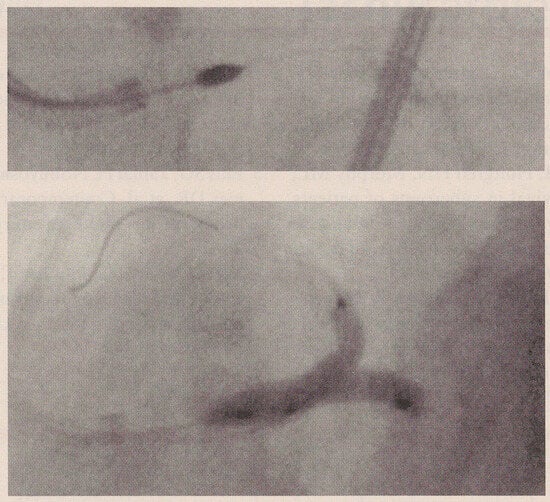
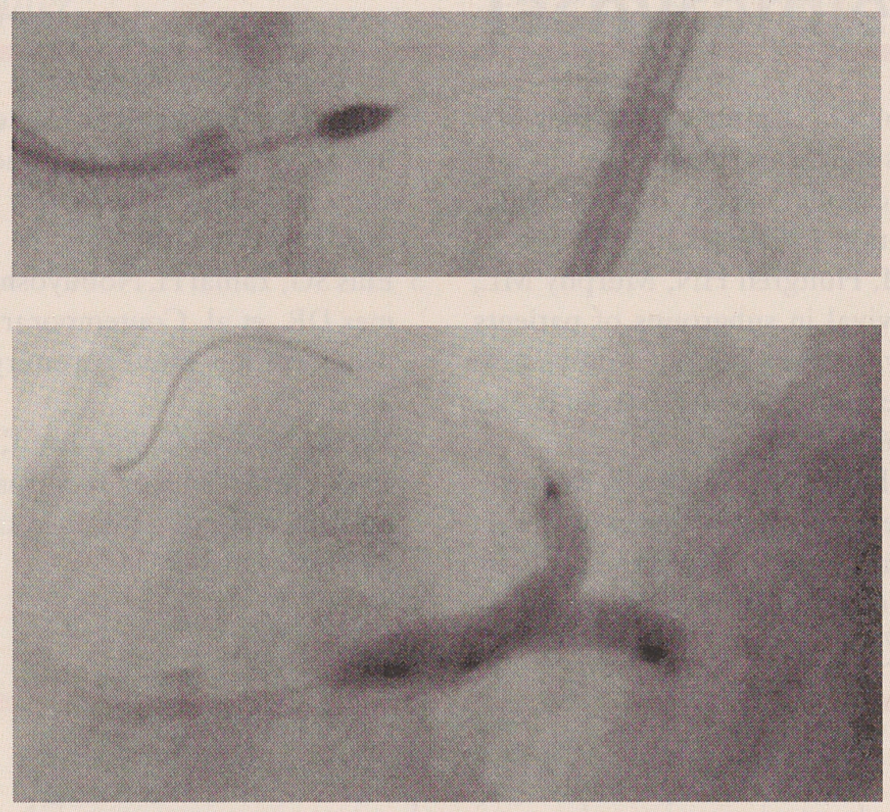
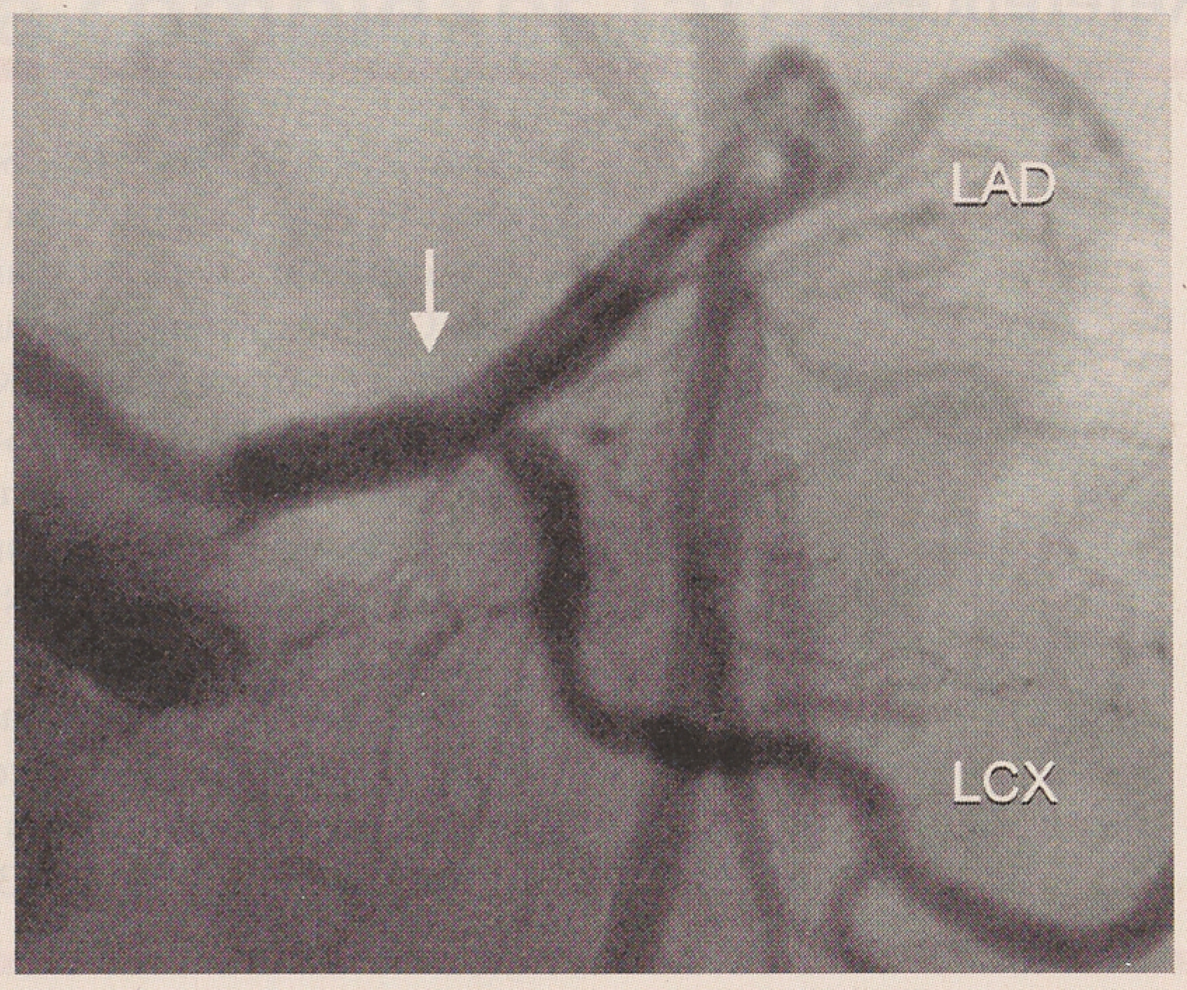
The antithrombotic/antiplatelet regimen consisted of aspirin, heparin bolus, and bolus and infusion of eptifibatide (Integrilin). A prophylactic intraaortic balloon pump and a Swan-Ganz catheter were placed before starting the procedure. High-speed rotational atherectomy (Rotablator) was performed using 1.5 and 2.0 mm burrs for the distal LMT and the proximal LAD stenoses (Figure 3). Subsequently, a second wire was placed in the LCX. Two balloons (3.0 mm of diameter) were then advanced and simultaneous inflations were performed at the LMT bifurcation ("kissing balloon", Figure 3). Following that, two stents were deployed in the distal LMT and in the proximal LAD with excellent angiographic results (Figure 4). The patient tolerated the rotational atherectomy and the balloon inflations well with no significant hypotension or increase in pulmonary artery end-diastolic pressure. The intraaortic balloon pump and the Swan-Ganz catheter were removed at the end of the procedure. The patient was discharged the next day. At 4 months he remained free of angina, but refused follow-up catheterization, even tough angiography was recommended.
Figure 3.
Rotational atherectomy with a 2.0 mm burr of the distal left main trunk and the proximal left anterior descending artery (upper panel). "Kissing balloon" inflation at the bifurcation of the left main trunk (lower panel).
Figure 4.
Final result after stenting of the proximal left anterior descending coronary artery and of the distal left main trunk (arrow), PA caudal view. LAD = left anterior descending coronary artery; LCX = left circumflex.
Discussion
Coronary artery bypass grafting (CABG) remains the standard of care for LMT disease ever since major studies established its superiority over medical therapy with regard to survival and symptom relief [1,2,3,4]. To date, no randomized clinical trials has compared percutaneous coronary intervention (PCI) with CABG in the setting of LMT disease. However, in certain circumstances PCI may be appropriate. These situations generally fall into two categories: (a) surgical high-risk or inoperable patients, and (b) the low-risk patients with good LV function who strongly desire to avoid bypass surgery. In the setting of unprotected LMT interventions, stenting appears to be superior to balloon angioplasty [5,6].
Excluding presentation with acute myocardial infarction, impaired LV function is the most important risk factor for in-hospital death after PCI in this patient population. In a multicenter registry following 81 patients undergoing elective PCI of unprotected LMT lesions, the inhospital mortality was 1.7% when LV ejection fraction was 240% and 31.8% when the LV ejection fraction was <30% [51. Patients with well preserved left ventricular function and widely patent RCA can generally tolerate relatively brief periods of left main coronary ischemia remarkably well. However, it may be prudent to place an intraaortic balloon pump to decrease LV wall stress and support systemic pressure if there is any question about the patient's capacity to tolerate ischemia. Although not essential, placement of a Swan-Ganz catheter to monitOr pulmonary pressures is often useful, providing an insight into the response of left ventricular end-diastolic pressure to ischemic stress of balloon inflation.
Patients undergoing PCI of LMT appear to be at higher risk of sudden death in the 6 months following the procedure [5]. Since severe restenosis may precipitate electrical instability even in absence of symptoms (silent ischemia), it has become our practice to perform follow-up angiography 8–12 weeks after the intervention, although the benefit of such a course of action remains unproven.
References
- Caracciolo, E.A.; Davis, K.B.; Sopko, G.; Kaiser, G.C.; Corley, S.D.; Schaff, H.; et al. Comparison of surgical and medical group surVival in patients with left main coronary artery disease. Longterm CASS experience. Circulation 1995, 91, 2325–2334. [Google Scholar] [CrossRef] [Scilit] [PubMed]
- Takaro, T.; Peduzzi, P.; Detre, K.M.; Hultgren, H.N.; Murphy, M.L.; van der Bel-Kahn, J.; et al. Survival in subgroups of patients with left main coronary artery disease. Veteran Administration Cooperative study of surgery for coronary arterial occlusive disease. Circulation 1982, 66, 14–22. [Google Scholar] [CrossRef] [Scilit] [PubMed]
- Yusuf, A.; Zucker, D.; Peduzzi, P.; Fisher, L.D.; Takaro, T.; Kennedy, J.W.; et al. Effect of coronary artery bypass graft surgery on survival; overview of 10-year results from randomized trials by the Coronary Artery Bypass Graft Surgery Trialists Collaboration. Lancet 1994, 344, 563–570. [Google Scholar] [CrossRef] [Scilit] [PubMed]
- Eagle, K.A.; Guyton, R.A.; Davidoff, R.; Ewy, G.A.; Fonger, J.; Gardner, T.J.; et al. ACC/AHA Guidelines for coronary bypass graft surgery: executive summary and recommendations. Circulation 1999, 100, 1464–1480. [Google Scholar] [CrossRef] [Scilit] [PubMed]
- Ellis, S.G.; Tamai, H.; Nobuyoshi, M.; Kosuga, K.; Colombo, A.; Holmes, D.R.; et al. Contemporary percutaneous treatment of unprotected left main coronary stenosis. Circulation 1997, 96, 3867–3872. [Google Scholar] [CrossRef] [Scilit] [PubMed]
- Marso, S.P.; Steg, G.; Plokker, T.; Holmed, D.R.; Park, S.; Kosuga, K.; et al. Catheter-based reperfusion of unprotected left main stenosis during an acute myocardial infarction. Am. J. Cardiol. 1999, 83, 1513–1517. [Google Scholar] [CrossRef] [Scilit] [PubMed]
© 2000 by the author. Attribution - Non-Commercial - NoDerivatives 4.0.