


Abstract
Heart rate variability (HRV) is a non-invasive marker of cardiac autonomic regulation that reflects the dynamic interaction between sympathetic and parasympathetic influences on the sinoatrial node. Reduced HRV is a hallmark of autonomic dysfunction and has been consistently associated with adverse outcomes across a wide range of cardiovascular conditions, particularly heart failure (HF). HRV is markedly reduced in HF, reflecting profound autonomic imbalance characterized by vagal withdrawal, impaired baroreflex sensitivity, and altered cardiorespiratory coupling. In heart failure with reduced ejection fraction (HFrEF), lower global HRV, particularly reduced SDNN, is consistently associated with mortality, sudden cardiac death, and disease progression. In heart failure with preserved ejection fraction (HFpEF), emerging observational evidence suggests that HRV alterations may accompany early autonomic dysregulation and may be associated with sex-specific patterns, although their mechanistic and prognostic significance remains incompletely defined. In acute HF, HRV assessed during hospitalization may provide additional prognostic information, although current evidence remains limited and heterogeneous. Pharmacological therapies, cardiac resynchronization therapy, exercise training, and lifestyle interventions have variably modulated HRV across studies, but the evidence is heterogeneous and changes in HRV should not be assumed to translate directly into improved clinical outcomes. Despite persistent challenges related to methodological standardization and clinical implementation, emerging technologies and multimodal approaches may refine the feasibility of integrating HRV into risk assessment frameworks. This narrative review aims to summarize the physiological basis of HRV, critically evaluate its alterations across different HF phenotypes, and discuss its prognostic associations, therapeutic modulation, and current practical limitations in clinical application.
1. Introduction
Unlike less evolutionarily advanced species, in which cardiac rhythmicity is initiated by neurons directly connected to the myocardium, the heart of mammals, especially humans, possesses an intrinsic ability to generate and sustain rhythmic contractions even when completely denervated and isolated [1]. In physiological conditions, this modulation allows the cardiovascular system to dynamically adapt to internal and external demands. While mean heart rate reflects the integrated output of sympathetic and parasympathetic activity, analysis of beat-to-beat variability and its spectral components provides a more refined characterization of autonomic modulation. Heart rate variability (HRV) analysis has become a consolidated methodology for investigating autonomic cardiovascular regulation under both physiological and pathological conditions [2]. HRV is defined as the beat-to-beat variation in RR intervals, corresponding to the temporal distance between successive systolic events [3]. Akselrod et al. first demonstrated that HRV spectral analysis enables the non-invasive assessment of sympatho-vagal interactions through the identification of discrete oscillatory components of the RR interval time series [4]. In healthy individuals, a robust high-frequency component indicates an intact and responsive vagal control, allowing rapid cardiovascular adjustments to metabolic and hemodynamic challenges [4]. However, a healthy heart does not beat with metronomic regularity; it exhibits complex, nonlinear dynamics characterized by continuous alternation between acceleration and deceleration phases, which are largely mediated by phasic vagal activity. In adaptive biological systems, higher variability and greater complexity reflect the capacity to maintain stability through change. Conversely, reduced HRV, particularly a marked depression of the high-frequency spectral component, indicates impaired parasympathetic modulation, reduced physiological flexibility, and diminished resilience to stressors [5]. In heart failure (HF), attenuation of high-frequency power is consistently observed and reflects profound vagal withdrawal, impaired baroreflex function, and disrupted cardiorespiratory coupling [6].
The prognostic relevance of HRV in cardiovascular disease has been firmly established. Both experimental and clinical studies have demonstrated that myocardial infarction and HF are associated with persistent depression of baroreflex sensitivity and vagal modulation. In preclinical models, acute ischemic injury results in a permanent reduction in baroreflex function in the majority of subjects, likely due to altered cardiac geometry, mechanical distortion, and abnormal activation of sympathetic afferent pathways [7].
Consistent with these mechanisms, time-domain measures such as the standard deviation of normal-to-normal intervals (SDNN), as well as frequency-domain indices including high-frequency power, have emerged as independent predictors of mortality in HF, even when incorporated into multivariate prognostic models [3].
In light of these considerations, the present narrative review aims to evaluate the pathophysiological and clinical relevance of HRV in HF.
2. Physiology of Heart Rate Variability
HRV reflects the physiological fluctuations in the duration of consecutive cardiac cycles and represents a non-invasive marker of autonomic nervous system (ANS) regulation of the heart [7]. Rather than indicating instability, a higher degree of beat-to-beat variability in sinus rhythm is a hallmark of a healthy cardiovascular system, reflecting the heart’s ability to dynamically adapt to internal and external stimuli [7]. For instance, reduced HRV is widely recognized as an expression of impaired autonomic modulation and diminished physiological resilience, conditions frequently observed in cardiovascular diseases and, most prominently, in HF (Figure 1).
Figure 1.
Factors influencing heart rate variability.
2.1. Autonomic Control of Heart Rate
The sinoatrial node is under continuous and dynamic modulation by the sympathetic and parasympathetic branches of the ANS. Parasympathetic activity, mediated predominantly through the vagus nerve and acetylcholine release, exerts a rapid, beat-to-beat inhibitory effect on heart rate by increasing potassium conductance and shortening the action potential duration in pacemaker cells. By contrast, sympathetic stimulation via norepinephrine acts more slowly, increasing heart rate and contractility through beta-adrenergic receptor activation and cyclic AMP-dependent pathways [8].
HRV emerges from the complex interaction between these two autonomic components, with parasympathetic influences dominating short-term variability and sympathetic influences contributing primarily to longer-term fluctuations [7].
2.2. Respiratory and Baroreflex Contributions
Respiration plays a fundamental role in shaping HRV through respiratory sinus arrhythmia (RSA), characterized by heart rate acceleration during inspiration and deceleration during expiration. This phenomenon is mediated predominantly by vagal modulation and represents one of the most prominent physiological contributors to high-frequency HRV components [9].
The arterial baroreflex constitutes another key physiological determinant of HRV. By sensing beat-to-beat changes in arterial pressure, baroreceptors modulate autonomic outflow to stabilize blood pressure. In pathological conditions such as HF, baroreflex sensitivity is often blunted, leading to a loss of autonomic flexibility and a marked reduction in HRV [9].
2.3. Domains of HRV Analysis: Physiological Meaning
HRV analysis is traditionally divided into time-domain, frequency-domain, and non-linear methods, each capturing distinct aspects of autonomic physiology [10]. Time-domain measures quantify the magnitude of variability in the RR interval series over time. They include statistical and geometrical measures derived from direct statistical calculation of MM/RR intervals over time or geometrical representation of RR interval distributions (histograms or scatter plots) respectively. Indices such as the standard deviation of normal-to-normal intervals (SDNN) reflect overall HRV and integrate both sympathetic and parasympathetic influences across the recording period [10]. Measures derived from successive beat differences, such as the root mean square of successive differences (RMSSD), predominantly capture short-term variability and are strongly linked to parasympathetic activity. The HRV triangular index (HTI) provides a geometric estimate of overall variability and may be particularly useful in longer recordings, although its interpretation depends on signal quality, preprocessing methods, and recording context rather than on a single universally applicable cutoff. From a physiological standpoint, time-domain indices provide a straightforward assessment of global and vagal-mediated autonomic modulation [10]. Table 1 provides a detailed description of the main time-domain measures. Frequency-domain analysis decomposes HRV into spectral components corresponding to specific frequency bands (Table 2). Non-linear HRV metrics aim to capture the complex, non-stationary, and fractal properties of heart rate dynamics. Poincaré plot analysis is a non-linear method for HRV assessment that evaluates beat-to-beat RR interval dynamics. It captures complex, non-linear autonomic patterns that may not be detected by time- or frequency-domain metrics. During breathing, RR intervals normally shorten and lengthen. The spectral component occurring at respiratory frequency (0.15–0.40 Hz in adults at usual breathing rates) is termed high-frequency power and predominantly reflects vagal modulation in sinus rhythm [11].
Table 1.
Heart Rate Variability (HRV) Metrics: Time-Domain measures.
Table 2.
Heart Rate Variability (HRV) Metrics: Frequency Domain measures.
Baroreflex sensitivity (BRS) quantifies the effectiveness of reflex autonomic control linking blood pressure fluctuations to heart rate modulation and has been associated with adverse outcomes, including sudden cardiac death [12]. Heart rate turbulence (HRT) comprises two derived metrics: turbulence onset (TO), typically expressed as percentage change, reflecting initial heart rate acceleration following a premature ventricular beat, and turbulence slope (TS), expressed in ms per RR interval, reflecting the subsequent deceleration of heart rate that reflects baroreflex-mediated autonomic recovery. These indices are primarily validated as post-myocardial infarction risk markers and their broader applicability in HF populations requires further investigation. Deceleration capacity (DC) and acceleration capacity (AC), derived from phase-rectified signal averaging, capture distinct components of heart rate dynamics, with DC mainly reflecting vagally mediated slowing and AC describing heart rate acceleration dynamics, often interpreted in relation to sympathetic modulation depending on context [13]. These indices have shown prognostic relevance across multiple cardiovascular conditions.
Apparently contradictory prognostic associations of low-frequency power and the low-frequency/high-frequency ratio are biologically and methodologically plausible. In earlier HF, relatively low-frequency predominance may accompany reduced vagal modulation; in advanced HF, low-frequency power may collapse despite intense sympathetic activation because of impaired baroreflex oscillation, abnormal breathing, and reduced sinus-node responsiveness. Differences in recording duration, spectral normalization, respiratory control, disease severity, rhythm selection, ectopic-beat correction, and background therapy further limit comparison across cohorts. Accordingly, no single spectral index should be interpreted as a linear measure of sympathetic drive or used as an isolated prognostic threshold. Therefore, BRS and heart-rate recovery provide related but non-interchangeable information. BRS quantifies the RR response to blood-pressure variation and depends on the provocation or spontaneous-sequence method, blood-pressure signal quality, respiration, and rhythm. Heart-rate recovery after exercise is influenced by vagal reactivation but also by exercise intensity, active versus passive recovery, chronotropic incompetence, beta-blockade, fitness, and protocol-specific thresholds. Both measures have prognostic associations, but neither is a pure measure of vagal tone, and neither should be combined with HRV without explicit methodological distinction.
3. Alterations of HRV in Heart Failure
Against this physiological and conceptual background, numerous studies have consistently documented a marked reduction in HRV in patients with HF, reflecting a profound alteration of autonomic cardiovascular regulation [14]. This reduction, generally interpreted as the combined effect of enhanced sympathetic drive, is now considered a robust and reproducible feature of the disease. Time-domain HRV indices show a well-established association with clinical severity, whereas the relationship between HF progression and frequency-domain measures remains more complex and, in some aspects, controversial. In patients with mild to moderate HF, typically corresponding to New York Heart Association (NYHA) functional classes I–II, spectral analysis frequently demonstrates a relative redistribution of power across frequency bands, characterized by an increase in low-frequency oscillations and a concomitant reduction in high-frequency power [15]. Although low-frequency power has traditionally been interpreted as a surrogate of sympathetic modulation, it is now widely recognized that this component reflects a composite signal arising from the interaction between sympathetic and parasympathetic influences, strongly modulated by baroreflex mechanisms and prevailing physiological conditions [16]. In this clinical context, the relative predominance of low-frequency oscillations likely represents an altered autonomic balance rather than a direct measure of sympathetic tone per se. In parallel, the consistent attenuation of high-frequency power reflects impaired respiratory-mediated vagal modulation of the sinoatrial node [16]. As HF advances, a qualitatively different spectral pattern emerges [15]. Early observations from the Montescano group demonstrated a marked reduction in all spectral components, including low-frequency power, despite evidence of heightened adrenergic activation and elevated circulating catecholamines [12]. These findings suggest a decoupling between sympathetic activity and its oscillatory expression at the sinus node level. Such a phenomenon likely reflects a reduced responsiveness of pacemaker cells exposed to chronic neurohumoral stimulation, rather than a true attenuation of sympathetic drive. Several additional mechanisms contribute to the attenuation and distortion of low-frequency oscillations in advanced HF. Impaired baroreflex sensitivity limits the capacity of arterial baroreceptors to generate rhythmic fluctuations in heart rate within the low-frequency range [17]. Moreover, pronounced oscillations in the very-low-frequency band, frequently associated with abnormal respiratory patterns, further complicate spectral interpretation [17]. In particular, periodic breathing and Cheyne–Stokes respiration, commonly observed during nocturnal recordings but often present also during daytime wakefulness, introduce dominant very-low-frequency components that interfere with the accurate quantification of low-frequency power [18]. Under these conditions, both time-domain and frequency-domain HRV metrics may lose reliability and physiological specificity (Figure 2).
Figure 2.
Heart rate variability across the heart failure spectrum. Panel (A) illustrates the progressive alterations in heart rate variability (HRV) across increasing stages of heart failure severity. Global HRV, reflected by time-domain indices such as SDNN and total spectral power, progressively declines with disease progression. In mild to moderate heart failure (NYHA I–II), spectral analysis typically shows a redistribution of power characterized by reduced high-frequency components and a relative predominance of low-frequency (LF) oscillations, reflecting an altered autonomic balance rather than isolated sympathetic activation. In advanced heart failure (NYHA III–IV), a global depression of HRV is observed, involving both LF and HF components, despite marked sympathetic activation. This pattern reflects impaired baroreflex function and a decoupling between sympathetic drive and sinus node responsiveness, resulting in a loss of physiologically meaningful oscillatory modulation. Panel (B) compares HRV patterns in chronic heart failure phenotypes. In heart failure with reduced ejection fraction (HFrEF), reduced global HRV—particularly low SDNN—acts as a robust marker of advanced autonomic failure and adverse prognosis. In heart failure with preserved ejection fraction (HFpEF), HRV alterations appear earlier and are more closely related to vagal dysfunction, comorbidities, and disease susceptibility, with sex-specific associations. Panel (C) depicts HRV dynamics in acute heart failure. During acute decompensation, marked reductions in HRV indices and baroreflex sensitivity are associated with increased post-discharge risk. Partial recovery of HRV following clinical stabilization highlights the dynamic and potentially reversible nature of autonomic dysfunction. Multiparametric HRV models may provide high prognostic accuracy in this setting [19]. BRS, Baroreflex Sensitivity; HF, Heart Failure; HF, High-Frequency Power; HFpEF, Heart Failure with Preserved Ejection Fraction; HFrEF, Heart Failure with Reduced Ejection Fraction; HRV, Heart Rate Variability; LF, Low-Frequency Power; NYHA, New York Heart Association; SDNN, Standard Deviation of Normal-to-Normal RR Intervals.
3.1. HRV in Heart Failure with Reduced Ejection Fraction (HFrEF)
A substantial body of evidence accumulated over more than three decades demonstrates that reduced HRV represents a powerful and independent predictor of adverse outcomes in patients with heart failure with reduced ejection fraction (HFrEF) [12,20]. Early observational studies conducted in relatively small cohorts of patients with idiopathic or ischemic dilated cardiomyopathy consistently reported that reductions in global HRV indices, particularly time-domain measures such as SDNN and SDANN, were associated with increased mortality [21]. These findings were observed across different etiologies and levels of systolic dysfunction, suggesting that impaired autonomic regulation conveys prognostic information beyond left ventricular ejection fraction alone [14].
Subsequent investigations expanded these observations using a variety of analytical approaches, including short-term HRV measures, geometric descriptors such as Poincaré plot analysis, and frequency-domain parameters [12]. Several studies confirmed that reduced SDNN retained prognostic significance in multivariate models incorporating clinical variables and echocardiographic indices [22]. Larger cohorts further reinforced these findings. In particular, the UK-HEART study [23], which evaluated 433 ambulatory patients with chronic HF and a mean ejection fraction of 41%, identified SDNN as the strongest predictor of death due to worsening HF. Patients with SDNN values below 50 ms exhibited an annual mortality exceeding 50%, compared with markedly lower mortality among those with preserved variability, firmly establishing SDNN as a robust marker in HFrEF [23]. Alongside time-domain indices, frequency-domain measures were extensively investigated, although with more heterogeneous results. In patients with mild to moderate HFrEF, several studies reported that alterations in low-frequency power, high-frequency power, or their ratio were associated with adverse outcomes [24,25]. Galinier et al. [26] demonstrated that a marked reduction in daytime low-frequency power independently predicted sudden cardiac death, even after adjustment for ischemic etiology and other prognostic factors, while reduced SDNN remained an independent predictor of overall mortality. Importantly, other contemporaneous studies reported opposite associations, with increased low-frequency power or elevated low-frequency/high-frequency ratios predicting mortality [25,27]. These discrepancies likely reflect differences in disease severity, baroreflex integrity, respiratory patterns, and analytical methodology, rather than a direct or linear relationship between low-frequency power and sympathetic tone.
3.2. HRV in Heart Failure with Preserved Ejection Fraction (HFpEF)
Compared with HFrEF, the evidence supporting a prognostic role of heart rate variability in heart failure with preserved ejection fraction (HFpEF) remains more limited. Available data suggest that autonomic dysfunction assessed through HRV may also be relevant in this phenotype, albeit with distinct characteristics and substantially greater uncertainty. Unlike HFrEF, where reduced HRV predominantly reflects advanced autonomic imbalance secondary to systolic dysfunction and neurohumoral activation, in HFpEF HRV alterations may be associated with earlier pathophysiological changes, including impaired diastolic function, comorbidity burden, and altered cardiorespiratory coupling; however, these associations remain largely observational.
Compelling evidence in this regard derives from the Women’s Health Initiative [28], a large prospective cohort including 28,603 postmenopausal women. In this population, lower HRV was independently associated with an increased risk of incident HF requiring hospitalization. Notably, reduced HRV predicted the development of HFpEF but not HFrEF. Women in the lowest quartile of SDNN exhibited a significantly higher risk of HFpEF compared with those in the highest quartile (hazard ratio 1.22, 95% CI 1.02–1.47), whereas the association with HFrEF did not reach statistical significance [28]. However, the currently available evidence does not establish a causal role and should be regarded as hypothesis-generating. Additional insights are provided by the population-based Rotterdam Study [29], in which HRV indices were associated with both systolic and diastolic ventricular dysfunction and predicted the occurrence of incident HF. Importantly, sex-specific differences emerged. Higher values of log-transformed root mean square of successive differences (RMSSD), a time-domain measure reflecting short-term vagally mediated variability, were associated with a greater annual decline in ejection fraction in men, whereas in women they correlated with smaller left atrial size [29]. Taken together, these findings suggest that HRV may capture early and clinically meaningful aspects of autonomic dysregulation in HFpEF, with implications that differ from those observed in HFrEF. However, the current literature remains limited, heterogeneous, and predominantly observational.
The evidence base in HFpEF remains substantially weaker than in HFrEF. Most studies are observational and several evaluate HF incidents in community cohorts rather than patients with rigorously adjudicated, established HFpEF. Associations may therefore reflect age, sex, obesity, diabetes, sleep-disordered breathing, physical inactivity, or atrial disease as much as HFpEF-specific autonomic dysfunction. No HRV threshold has been prospectively validated to diagnose HFpEF, guide treatment, or provide consistent incremental prognostic value beyond natriuretic peptides and echocardiographic assessment. Current findings should be regarded as hypothesis-generating.
3.3. HRV in Acute Heart Failure (AHF)
In patients hospitalized for acutely decompensated HF, autonomic dysfunction assessed by HRV during the index admission may provide relevant prognostic information regarding post-discharge outcomes [19]. Several studies suggest that HRV-derived indices obtained in the acute phase retain prognostic significance beyond immediate clinical stabilization, underscoring the pathophysiological relevance of autonomic impairment across the spectrum of HF severity [19,30]. In a cohort of 199 patients admitted for decompensated chronic HF, reduced HRV parameters measured at hospital admission, including SDNN, SDANN, total power, and ultra-low-frequency power, were independently associated with increased mortality over a mean follow-up of 312 days [31]. In multivariate Cox regression analyses, patients in the lowest tertile of SDNN exhibited more than a twofold increase in mortality risk, while ultra-low-frequency power in the lowest tertile conferred the highest relative risk [31]. Consistent results were reported in a retrospective cohort of 128 patients hospitalized for acute HF, in whom lower values of SDNN, SDANN, pNN50, and LF/HF ratio were significantly associated with adverse clinical outcomes [19]. Notably, the integration of multiple HRV parameters into a composite model showed promising discriminatory performance, with an area under the receiver operating characteristic curve exceeding 0.90 in a preliminary cohort [19]. The dynamic nature in this context has been further illustrated by studies evaluating changes in HRV and baroreflex sensitivity during clinical stabilization. Rydlewska et al. demonstrated that patients with acutely decompensated chronic HF exhibit marked reductions in both HRV indices and baroreflex sensitivity compared with stable patients [32]. Importantly, following effective decongestive and disease-directed therapy, significant improvements were observed in SDNN and baroreflex sensitivity, with values approaching those measured in clinically stable individuals. These observations support the concept that autonomic impairment in acute HF is, at least in part, reversible and closely linked to hemodynamic and neurohumoral status [32]. Therefore, reduced baroreflex sensitivity and altered HRV parameters have also been shown to predict future episodes of decompensation in stable patients with left ventricular systolic dysfunction [33]. In this population, both impaired baroreflex function and reduced low-frequency HRV power independently predicted hospitalization for worsening HF, even after adjustment for established clinical risk factors [33]. However, the available evidence remains limited by cohort size, methodological heterogeneity, and incomplete external validation.
4. Modulation of HRV in Patients with HF: Therapeutic Strategies
Over time, a substantial number of studies have investigated whether pharmacological and non-pharmacological therapeutic strategies are capable of modulating HRV in HF patients. In particular, numerous investigations have focused on the effects of guideline-directed medical therapies, as endorsed by current ESC recommendations [34,35] on HRV parameters and their potential association with improvements in autonomic regulation and clinical outcomes (Table 3, Figure 3).
Table 3.
Overview of the principal studies evaluating the effects of major heart failure therapies on HRV.
Figure 3.
Modulation of heart rate variability in heart failure: pharmacological, device-based, and non-pharmacological strategies.
Before discussing specific interventions, an important distinction should be emphasized: therapies with proven clinical benefit in HF are not necessarily supported by equally robust evidence regarding HRV modulation, and conversely, improvement in HRV does not by itself demonstrate improved clinical outcomes. In many studies, HRV has been explored primarily as a physiological or mechanistic marker of autonomic adaptation rather than as a validated surrogate endpoint.
Schematic overview of the principal therapeutic approaches influencing heart rate variability (HRV) in patients with heart failure. HRV represents an integrated marker of cardiac autonomic regulation, reflecting the dynamic balance between sympathetic and parasympathetic activity. Pharmacological therapies, including β-blockers, mineralocorticoid receptor antagonists, renin–angiotensin–aldosterone system modulators (ACE inhibitors, angiotensin receptor blockers, and angiotensin receptor–neprilysin inhibitors), sodium–glucose cotransporter-2 inhibitors, and ivabradine, modulate autonomic tone through distinct mechanisms, variably improving baroreflex sensitivity and reducing sympathetic overactivity. Device-based therapy, particularly cardiac resynchronization therapy, enhances ventricular synchrony and hemodynamic efficiency, thereby favourably influencing autonomic balance and HRV. Non-pharmacological interventions, such as structured exercise training, comprehensive cardiac rehabilitation, dietary optimization with weight management, and breathing or mind–body techniques, further contribute to autonomic restoration through improved vagal activity and reduced stress-related sympathetic drive. Collectively, these strategies may contribute to partial restoration of autonomic equilibrium. However, improvements in HRV should not automatically be interpreted as equivalent to improved clinical outcomes, since the relationship between autonomic modulation and outcome benefit is not uniform across interventions and study settings.
4.1. Beta-Blockers
Beta-blockers were among the earliest pharmacological agents introduced for the treatment of HFrEF and remain a cornerstone of guideline-directed therapy [34,42]. Beyond their established hemodynamic and antiarrhythmic effects, beta-blockers have been extensively investigated for their impact on cardiac autonomic regulation as assessed by HRV. Early evidence provided by Saleem et al. [65] demonstrated that beta-blocker therapy is associated with an enhancement of parasympathetic modulation and a reduction in cardiovascular events, particularly during the morning hours, a period characterized by heightened sympathetic nervous system activity.
The capacity of beta-blockers to improve autonomic control of heart rate, largely through an augmentation of reflex vagal activity, as reflected by increases in HRV, has since been confirmed by multiple studies across different clinical settings, including both acutely decompensated and advanced chronic HF [66,67,68]. More detailed investigations have highlighted drug-specific effects within the beta-blocker class. Bullinga et al. [38] demonstrated that carvedilol treatment leads to significant improvements in both time-domain and frequency-domain HRV indices, with changes in HRV closely correlating with concomitant hemodynamic improvement. Consistent with these observations, Mortara et al. [39] reported a significant enhancement of baroreflex sensitivity during treatment with metoprolol, further supporting the role of beta-blockers in restoring reflex autonomic control.
Comparable effects have been reported for bisoprolol, with a documented significant increase in HRV parameters across both analytical domains [41]. However, findings from Copie et al. [40] suggested that improvements in parasympathetic modulation and reductions in sympathetic influence were predominantly evident at lower heart rates, indicating that baseline chronotropic status may modulate the autonomic response to therapy.
Metoprolol has also been shown to improve HRV indices in patients with mild chronic HF [37]. In comparative studies involving patients with dilated cardiomyopathy, metoprolol produced greater improvements in HRV than captopril [36], whereas no significant differences in HRV modulation were observed when compared with atenolol [69]. Collectively, these data suggest that beta-blockers may exert a generally favorable, though quantitatively variable, effect on autonomic regulation as assessed by HRV. However, the evidence remains heterogeneous, and HRV improvement should be interpreted as a mechanistic correlate rather than as a direct surrogate of clinical benefit, which is established independently by outcome trials.
4.2. Mineralocorticoid Receptor Antagonists
Aldosterone, a key mediator in the pathophysiology of HF, exerts multiple effects on cardiovascular regulation and has been shown to attenuate baroreflex responsiveness in humans [34]. On this basis, several studies have explored whether mineralocorticoid receptor antagonists (MRAs) are capable of modulating cardiac autonomic function as assessed by HRV [45]. Evidence indicates that spironolactone may influence autonomic regulation in a context-dependent manner [43,44,45,46]. In patients undergoing hemodialysis, spironolactone therapy has been associated with an increased burden of premature ventricular contractions, raising concerns regarding potential pro-arrhythmic effects; however, in the same setting, treatment was also accompanied by an improvement in cardiac autonomic function, reflected by increased HRV indices [43]. These observations highlight the complex and sometimes divergent electrophysiological and autonomic effects of mineralocorticoid receptor blockade. Further support for a favorable autonomic impact of spironolactone in HF derives from studies demonstrating that its addition to standard background therapy is associated with an improvement in sympatho-vagal balance, primarily reflected by changes in time-domain HRV indices [45]. Notably, the enhancement of HRV observed with spironolactone appears to be particularly pronounced during the early morning hours, a period characterized by increased sympathetic activity, suggesting a potential modulation of circadian autonomic rhythms [44,46]. Studies on other MRAs remain limited. To date, no studies have specifically evaluated the effects of finerenone on HRV parameters, leaving its impact on cardiac autonomic regulation largely unexplored. Overall, the available evidence for MRAs and HRV is suggestive but based mainly on relatively small studies, and therefore should be interpreted cautiously.
4.3. Angiotensin-Converting Enzyme Inhibitors/Angiotensin II Receptor Blockers/Angiotensin Receptor–Neprilysin Inhibitors (ACEi/ARB/ARNI)
Dysregulation of the renin–angiotensin–aldosterone system (RAAS) represents a central mechanism in the pathogenesis and progression of HF [70]. Pharmacological strategies aimed at counteracting RAAS hyperactivation, including angiotensin-converting enzyme inhibitors (ACEi), angiotensin II receptor blockers (ARBs), and angiotensin receptor–neprilysin inhibitors (ARNIs), have therefore become fundamental pillars of guideline-directed therapy in patients with HFrEF [34]. Given the prominent role of neuro-hormonal activation, considerable interest has focused on the potential effects of these agents on cardiac autonomic regulation as assessed by HRV. Long-term treatment with ACEi in patients with chronic HF has generally been associated with a partial restoration of autonomic balance, predominantly mediated by an enhancement of vagal modulation [71]. This effect is of particular clinical relevance, as increased parasympathetic activity has been linked to protection against malignant ventricular arrhythmias, especially in ischemic heart disease. In this regard, Flapan et al. [47] demonstrated that captopril therapy in patients with chronic HF was associated with a significant increase in parasympathetic tone. However, subsequent observations by Kamen et al. [48] suggested that such autonomic improvements may be dose-dependent and evident primarily at lower captopril dosages, a finding that should be interpreted cautiously given the limited sample size of that study. Other investigations have yielded heterogeneous results. While Zhang et al. [49] reported that enalapril attenuates autonomic dysfunction in chronic HF, Guedon-Moreau et al. [50] found no significant improvement in HRV parameters during ramipril therapy. Notably, the latter study was conducted in patients already receiving digitalis, a factor that may have influenced autonomic measurements and partially accounted for the observed discrepancies [50]. Data regarding ARBs appear more consistent. Two independent studies evaluating losartan in patients with chronic HF, including those with ischemic etiology, demonstrated significant improvements in autonomic dysfunction, even when ARB therapy was added to ongoing treatment with beta-blockers or ACEi [52,53]. Comparable results were reported by Tambara et al. [51] in patients treated with candesartan. Moreover, a direct comparison between ACE inhibition and angiotensin receptor blockade showed that both therapeutic strategies were capable of improving HRV indices [54]; however, valsartan exerted a more pronounced effect on markers of sympathetic modulation, as reflected by greater reductions in plasma norepinephrine levels compared with lisinopril [54].
With regard to ARNIs, emerging evidence suggests a favorable impact on autonomic function. Several studies demonstrated that treatment with ARNIs is associated with a significant improvement in autonomic indices, detectable as early as three months after therapy initiation [55,56]. These effects are likely multifactorial and may be partly attributable to improved hemodynamic status and volume unloading. Nevertheless, the available HRV data remain limited compared with the much stronger outcome evidence supporting RAAS modulation in HFrEF.
4.4. Sodium–Glucose Cotransporter 2 Inhibitors (SGLT2i)
Sodium–glucose cotransporter 2 inhibitors (SGLT2i) represent one of the most recent therapeutic advances in HF management, with demonstrated clinical benefits in both reduced and preserved ejection fraction phenotypes [34,35]. Their potential effects on cardiac autonomic regulation, as assessed by HRV, have generated increasing interest. Investigations by Ang et al. [57] and the EMPA-HEART CardioLink-6 Holter subanalysis [61], conducted with dapagliflozin and empagliflozin, respectively, in patients with type 2 diabetes mellitus, did not document meaningful changes in HRV indices or improvements in autonomic function. Similar neutral results were reported in patients with type 1 diabetes mellitus treated with empagliflozin [60], as well as in the EMPYREAN study [62], which compared empagliflozin with sitagliptin in individuals with type 2 diabetes mellitus and found no differences between treatments in terms of HRV modulation [60]. Conversely, other investigations have suggested a favorable effect of SGLT2i on autonomic function under specific clinical conditions. Studies performed with dapagliflozin and empagliflozin in patients with type 2 diabetes mellitus and recent acute myocardial infarction reported significant improvements in HRV indices [59,62,72]. These findings were further supported by Balcioglu et al. [58], which demonstrated improved cardiac autonomic function following dapagliflozin treatment in patients with type 2 diabetes mellitus and established cardiac autonomic neuropathy.
The discordant results across studies are likely attributable to differences in sample size, baseline autonomic status, and clinical context, particularly with respect to the presence and stability of coronary artery disease [58,59,60,61,62,72,73]. Importantly, it should be emphasized that, to date, studies assessing the effects of SGLT2i on HRV have been conducted almost exclusively in patients with diabetes mellitus rather than in populations with overt HF. SGLT2i reduce HF events in patients with and without diabetes; however, this clinical benefit should not be equated with a demonstrated effect on HRV. Studies assessing autonomic indices are generally small, heterogeneous, and concentrated in diabetes, cardiac autonomic neuropathy, or post-myocardial-infarction populations. Results range from improvement in selected time-domain measures to no detectable change. Convincing, adequately powered HRV data in non-diabetic HF populations are lacking, and HRV has not been validated as a mediator or surrogate of SGLT2-inhibitor benefit. Autonomic modulation therefore remains a plausible but unproven mechanistic hypothesis.
4.5. Ivabradine
Although ivabradine is no longer regarded as a cornerstone therapy in contemporary HF management, it has historically played a relevant role in selected patients with HFrEF who remained in sinus rhythm with elevated heart rate despite optimized beta-blocker therapy [34]. Moreover, ivabradine has also been investigated for its potential influence on cardiac autonomic regulation. In this context, the landmark study by Kurtoglu et al. [63] demonstrated that ivabradine therapy in HF patients was associated with a significant increase in HRV when added to beta-blocker treatment. These findings indicated that selective inhibition of the If current may favorably modulate autonomic balance, supporting a mechanistic contribution beyond simple chronotropic control. However, the available evidence is limited and should be interpreted primarily as mechanistic rather than as evidence of outcome mediation.
4.6. Digoxin
In patients with stable chronic heart failure receiving diuretics and ACE inhibitors, digoxin treatment was associated with a modest reduction in heart rate and increased HRV. The improvement predominantly involved short-term, vagally mediated indices, particularly RMSSD, whereas SDNN remained unchanged. These findings suggest that digoxin enhances parasympathetic cardiac modulation, potentially through neurohumoral mechanisms beyond its positive inotropic effects [64].
4.7. Cardiac Resynchronization Therapy (CRT-D)
Cardiac resynchronization therapy with defibrillator (CRT-D) is a well-established therapeutic option for patients with HFrEF (≤35%) and left bundle branch block who remain symptomatic despite optimal medical therapy [34]. Beyond its recognized benefits on left ventricular reverse remodeling, functional capacity, and survival, CRT-D has also demonstrated a significant impact on cardiac autonomic regulation, highlighting its role as a modulator of neuro-hormonal and autonomic dysfunction in HF [74].
Several clinical studies have shown that CRT-D is capable of restoring a more physiological sympatho-vagal balance, as reflected by significant improvements in HRV parameters [75,76]. These autonomic changes are thought to result from improved ventricular synchrony, enhanced cardiac output, and reduced filling pressures, which collectively attenuate chronic sympathetic activation and facilitate vagal reactivation. Importantly, improvements in HRV following CRT implantation have been associated with favorable clinical and echocardiographic responses, suggesting that autonomic modulation represents an integral component of the therapeutic benefit of resynchronization [77]. Conversely, the absence of significant HRV improvement early after CRT-D implantation, typically within the first four weeks, has been consistently associated with an increased risk of major adverse cardiovascular events, including HF hospitalization, ventricular arrhythmias, and mortality [78]. In this context, early autonomic response to CRT may serve as a marker of effective resynchronization, although HRV should not be considered a stand-alone determinant of device benefit.
4.8. Other Strategies
A growing body of evidence supports the role of non-pharmacological interventions in modulating HRV and restoring autonomic balance. Among non-pharmacological approaches, structured exercise training plays a central role and is widely regarded as one of the most effective interventions in patients with HF. More recent evidence further suggests that high-intensity interval training may exert superior effects on HRV compared with moderate-intensity continuous training, indicating that exercise intensity and structure are key determinants of autonomic adaptation [79].
Therefore, comprehensive cardiac rehabilitation programs have gained increasing attention for their integrative impact on cardiovascular health. Cardiac rehabilitation has been shown to improve not only functional capacity and cardiac performance but also HRV and global autonomic function, particularly when combined with adjunctive modalities [80].
Lifestyle factors also play a relevant role in autonomic modulation. Several studies have demonstrated that dietary interventions, healthy nutritional patterns, especially those rich in leafy green vegetables, and weight loss are associated with improvements in autonomic function, independent of the presence of overt HF [81]. Finally, growing interest has focused on mind–body and respiratory interventions. Breathing control techniques, particularly those employing slow and regular respiratory cycles, have been shown to enhance HRV and autonomic balance in selected settings. However, the available evidence remains less robust than that supporting established HF therapies, and the clinical significance of these HRV changes in HF populations requires further clarification.
5. Clinical Relevance: When, How, and in Whom Should HRV Be Used?
At present, HRV should not be ordered as a stand-alone diagnostic test for HF, used to select guideline-directed therapy, or used to trigger device implantation or hospitalization. Its most defensible clinical roles are supportive risk characterization in carefully selected patients in sinus rhythm, longitudinal research phenotyping, and trend monitoring when repeated measurements are acquired under comparable conditions. A low value should prompt review of rhythm, ectopy, medications, sleep-disordered breathing, functional status, and disease severity rather than an HRV-specific intervention [82].
The recording method should match the clinical question. A 24 h ECG/Holter is preferred when rhythm diagnosis, ectopic burden, circadian information, or established long-term metrics such as SDNN are required [83]. A standardized 5 min ECG can support short-term autonomic assessment but is not interchangeable with 24 h values. Implanted devices allow repeated trends but use manufacturer-specific algorithms and are applicable only to selected device recipients [30]. Consumer wearables may support repeated measurements in sinus rhythm when acquisition quality and the algorithm have been validated against ECG. Photoplethysmography measures pulse-rate variability rather than electrical RR intervals and is particularly vulnerable to motion, poor peripheral perfusion, irregular rhythms, and proprietary processing.
Atrial fibrillation (AF) represents one of the major limitations to conventional HRV analysis. In this setting, irregular RR intervals are driven predominantly by variable atrioventricular conduction rather than by physiological beat-to-beat autonomic modulation, making conventional time-domain and frequency-domain indices difficult to interpret [84]. In these settings, the underlying rhythm and technical validity take priority, and alternative rhythm-specific methods should be used only when separately validated.
6. Challenges and Future Directions
Despite decades of research, the clinical implementation of HRV in HF remains markedly heterogeneous. Although the physiological foundations and prognostic associations of HRV are well established, the translation of these concepts into routine clinical practice has been limited by methodological, technical, and regulatory challenges. At the same time, emerging technologies, including continuous monitoring, wearable devices, and artificial intelligence (AI), are transforming the field and may create new opportunities to refine HRV as a digital biomarker. However, these opportunities will only be clinically meaningful if supported by standardization, external validation, and demonstrable added value beyond existing risk markers.
6.1. Standardization Gaps and Methodological Limitations
One of the principal barriers to the integration of HRV in HF management is the lack of methodological standardization. Current HRV metrics derive from different computational approaches, time-domain, frequency-domain, and non-linear indices, each influenced by acquisition length, sampling frequency, ectopic beat removal, respiratory patterns, and posture [14]. Substantial inter-study variability makes direct comparison difficult and undermines the reliability of clinically applicable cutoffs. These measure changes are particularly relevant with respect to ultra-short-term HRV (≤1 min), continuous wearables, and patch-based ECG systems [85]. Future research must establish harmonized protocols that consider modern devices and realistic clinical conditions, ensuring reproducibility across centers and populations.
Another unresolved issue is the impact of arrhythmias, frequent ectopy, and paced rhythms, common in HF patients, on HRV interpretation. Traditional HRV metrics rely on sinus rhythm and stable RR intervals; thus, many HF patients, particularly those with atrial fibrillation (AF) or frequent PVCs, are systematically excluded from HRV-based risk stratification [86]. While newer approaches, such as symbolic dynamics, Poincaré-derived indices, and detrended fluctuation analysis, are more resilient to irregularity, they remain insufficiently validated in large HF cohorts [87]. A major research priority is the development of HRV-inspired indices suited to AF and device-paced rhythms, potentially enabling broader but still methodologically sound clinical application.
6.2. Variability in Clinical Thresholds and Lack of Cut-Offs
While reduced HRV is consistently associated with worse outcomes, there is no universally accepted threshold for clinical decision-making. Proposed SDNN cutoffs (e.g., <70 ms for high risk) stem largely from post-MI studies in the 1990s and may not translate to contemporary, guideline-directed HF therapy or modern device cohorts [34,35].
6.3. Integration with Multimodal Biomarkers
HRV provides information on autonomic regulation that is conceptually different from myocardial stress (NT-proBNP), deformation (global longitudinal and left-atrial strain), exercise limitation and ventilatory efficiency (CPET), and tissue characterization (cardiac magnetic resonance). This complementarity does not establish incremental clinical utility. Natriuretic peptides, imaging, and CPET have standardized acquisition frameworks and clearer roles in diagnosis, phenotyping, prognosis, and therapeutic pathways. By contrast, HRV studies use heterogeneous metrics and recording durations, frequently exclude atrial fibrillation, and rarely demonstrate reproducible improvement in discrimination, calibration, or clinical reclassification beyond contemporary multivariable models. HRV should therefore be viewed as an adjunctive research phenotype rather than a substitute for established HF biomarkers [88]. Its future value may lie in repeated, multimodal measurements rather than in a single isolated threshold.
6.4. HRV in the Era of Wearables and Continuous Monitoring
Wearable devices have revolutionized the acquisition of HRV data, shifting from static, short-term measurements to continuous, long-term physiologic monitoring [89]. However, this transition introduces new challenges related to device accuracy, algorithm transparency, and data quality. Many consumer-grade devices rely on photoplethysmography (PPG) rather than ECG, a technology susceptible to motion artefacts, poor perfusion, and skin pigmentation variations. These factors can distort RR interval estimation, particularly during sleep disorders, arrhythmias, or physical exertion, conditions highly relevant in HF [90]. Despite these limitations, continuous HRV monitoring offers unprecedented opportunities. Longitudinal patterns, circadian variation, and acute deviations from baseline may provide early signals of impending HF decompensation or arrhythmic risk. The concept of “baseline-anchored HRV”, interpreting HRV relative to each individual’s typical values, may outperform absolute measures for clinical monitoring [89,90]. Future research should focus on validating continuous HRV trajectories as predictors of decompensation, optimizing artefact removal algorithms, and ensuring cross-device comparability. Thus, current wearable/AI approaches are promising for repeated risk estimation but are not yet validated replacements for ECG-based assessment or established HF surveillance.
7. Conclusions
HRV is a biologically informative marker of cardiac autonomic regulation and is consistently associated with adverse outcomes in selected HF populations, particularly patients with HFrEF in sinus rhythm. Its clinical interpretation is nevertheless limited by rhythm, recording duration, respiration, ectopy, pacing, medication, and analytical method. Evidence in HFpEF and acute HF is less mature, therapy-induced changes in HRV are not validated surrogates of clinical benefit, and convincing data for SGLT2-related HRV improvement in non-diabetic HF are lacking. Compared with NT-proBNP, echocardiographic strain, CPET, and cardiac magnetic resonance, HRV currently has limited demonstrated incremental and actionable value. It should therefore remain primarily a research and complementary phenotyping tool. Future progress requires standardized acquisition, phenotype-specific thresholds, external validation of wearable and AI models, and prospective studies showing that HRV-guided decisions improve patient outcomes.
8. Take-Home Messages
- HRV is a context-dependent marker of autonomic modulation, not a direct measure of sympathetic or parasympathetic activity.
- Low SDNN is consistently associated with adverse outcomes in HFrEF, but no HRV threshold currently guides routine HF treatment.
- Evidence is weaker in HFpEF and acute HF; conventional HRV is generally uninterpretable during atrial fibrillation.
- A 24 h Holter remains preferable for validated long-term metrics and rhythm assessment; validated wearables are better suited to trends than to universal cutoffs.
- HRV remains an important research tool but currently offers limited proven incremental value over NT-proBNP, strain imaging, CPET, and cardiac magnetic resonance.
Author Contributions
Conceptualization, A.C. and E.V.; writing—original draft preparation, A.C.; writing—review and editing, L.T. and R.R.; visualization, N.P., E.C. and E.B.; supervision, M.M.; project administration, E.V. All authors have read and agreed to the published version of the manuscript.
Funding
This research received no external funding.
Data Availability Statement
No new data were created or analyzed in this study. Data sharing is not applicable to this article.
Acknowledgments
The figures included in this manuscript were developed with the support of OpenAI’s ChatGPT v. 5.6, which assisted in refining the visual design and organization of the graphical content. The authors were solely responsible for the scientific content, accuracy, and final approval of all figures.
Conflicts of Interest
The authors declare no conflicts of interest.
Abbreviation
The following abbreviations are used in this manuscript:
| Abbreviation | Full Term |
| HRV | Heart Rate Variability |
| HF | Heart Failure |
| HFrEF | Heart Failure with Reduced Ejection Fraction |
| HFpEF | Heart Failure with Preserved Ejection Fraction |
| AHF | Acute Heart Failure |
| ANS | Autonomic Nervous System |
| RSA | Respiratory Sinus Arrhythmia |
| BRS | Baroreflex Sensitivity |
| SDNN | Standard Deviation of Normal-to-Normal Intervals |
| SDRR | Standard Deviation of RR Intervals |
| SDANN | Standard Deviation of Average NN Intervals |
| RMSSD | Root Mean Square of Successive Differences |
| HTI | HRV Triangular Index |
| pNN50 | Percentage of NN intervals differing by >50 ms |
| HRT | Heart Rate Turbulence |
| DC | Deceleration Capacity |
| AC | Acceleration Capacity |
| HF (frequency band) | High Frequency |
| LF | Low Frequency |
| VLF | Very Low Frequency |
| ULF | Ultra Low Frequency |
| CRT | Cardiac Resynchronization Therapy |
| CRT-D | Cardiac Resynchronization Therapy with Defibrillator |
| ACEi | Angiotensin-Converting Enzyme Inhibitors |
| ARB | Angiotensin Receptor Blockers |
| ARNI | Angiotensin Receptor–Neprilysin Inhibitors |
| MRA | Mineralocorticoid Receptor Antagonists |
| SGLT2i | Sodium–Glucose Cotransporter 2 Inhibitors |
| NYHA | New York Heart Association |
| RAAS | Renin–Angiotensin–Aldosterone System |
| BNP | B-type Natriuretic Peptide |
| NT-proBNP | N-terminal pro-B-type Natriuretic Peptide |
| AF | Atrial Fibrillation |
| PVCs | Premature Ventricular Contractions |
| ECG | Electrocardiogram |
| PPG | Photoplethysmography |
| AI | Artificial Intelligence |
| RR intervals | Interbeat Intervals |
| NN intervals | Normal-to-Normal Intervals |
References
- Fedele, L.; Brand, T. The Intrinsic Cardiac Nervous System and Its Role in Cardiac Pacemaking and Conduction. J. Cardiovasc. Dev. Dis. 2020, 7, 54. [Google Scholar] [CrossRef] [Scilit]
- Massaro, S.; Pecchia, L. Heart Rate Variability (HRV) Analysis: A Methodology for Organizational Neuroscience. Organ. Res. Methods 2019, 22, 354–393. [Google Scholar] [CrossRef] [Scilit]
- Sundas, A.; Contreras, I.; Navarro-Otano, J.; Soler, J.; Beneyto, A.; Vehi, J. Heart rate variability over the decades: A scoping review. PeerJ 2025, 13, e19347. [Google Scholar] [CrossRef] [Scilit] [PubMed]
- Yokobori, Y.; Nakane, H.; Uehara, C.; Nagasawa, T.; Mitsuyama, S.; Ohkawa, K.; Kario, K.; Ozawa, S. Temporal relationships among changes in the RR -interval and the powers of the low- and high-frequency components of heart rate variability in normal subjects. Physiol. Rep. 2023, 11, e15557. [Google Scholar] [CrossRef] [Scilit] [PubMed]
- Zimatore, G.; Russo, S.; Gallotta, M.C.; Passalacqua, G.; Zaborova, V.; Campanella, M.; Fiani, F.; Baldari, C.; Napoli, C.; Randieri, C. HRV in Stress Monitoring by AI: A Scoping Review. Appl. Sci. 2025, 16, 23. [Google Scholar] [CrossRef] [Scilit]
- De Ferrari, G.M. Vagal Stimulation in Heart Failure. J. Cardiovasc. Trans. Res. 2014, 7, 310–320. [Google Scholar] [CrossRef] [Scilit] [PubMed]
- Gullett, N.; Zajkowska, Z.; Walsh, A.; Harper, R.; Mondelli, V. Heart rate variability (HRV) as a way to understand associations between the autonomic nervous system (ANS) and affective states: A critical review of the literature. Int. J. Psychophysiol. 2023, 192, 35–42. [Google Scholar] [CrossRef] [Scilit] [PubMed]
- Ziegler, K.A.; Engelhardt, S.; Carnevale, D.; McAlpine, C.S.; Guzik, T.J.; Dimmeler, S.; Swirski, F.K. Neural Mechanisms in Cardiovascular Health and Disease. Circ. Res. 2025, 136, 1233–1261. [Google Scholar] [CrossRef] [Scilit] [PubMed]
- Soer, R.; Six Dijkstra, M.W.M.C.; Bieleman, H.J.; Oosterveld, F.G.J.; Rijken, N.H.M. Influence of respiration frequency on heart rate variability parameters: A randomized cross-sectional study. J. Back Musculoskelet. Rehabil. 2021, 34, 1063–1068. [Google Scholar] [CrossRef] [Scilit] [PubMed]
- Shaffer, F.; Ginsberg, J.P. An Overview of Heart Rate Variability Metrics and Norms. Front. Public Health 2017, 5, 258. [Google Scholar] [CrossRef] [Scilit] [PubMed]
- Satoh, N. Spectral decomposition of heart rate variability using generalized harmonic analysis. Biomed. Signal Process. Control 2021, 70, 103050. [Google Scholar] [CrossRef] [Scilit]
- La Rovere, M.T.; Pinna, G.D.; Maestri, R.; Mortara, A.; Capomolla, S.; Febo, O.; Ferrari, R.; Franchini, M.; Gnemmi, M.; Opasich, C.; et al. Short-Term Heart Rate Variability Strongly Predicts Sudden Cardiac Death in Chronic Heart Failure Patients. Circulation 2003, 107, 565–570. [Google Scholar] [CrossRef] [Scilit] [PubMed]
- Wang, J.; Xu, J.; Qiu, Y.; Yang, R.; Wang, W.; Gao, C. Cardiac deceleration capacity and acceleration capacity have diagnostic value in patients with vasovagal syncope regardless of age. Front. Cardiovasc. Med. 2024, 11, 1495129. [Google Scholar] [CrossRef] [Scilit] [PubMed]
- Nagai, M.; Ewbank, H.; Nakano, Y.; Scherlag, B.J.; Po, S.S.; Dasari, T.W. Heart Rate Variability and Heart Failure with Reduced Ejection Fraction: A Systematic Review of Literature. Curr. Cardiol. Rev. 2025, 21, e1573403X327105. [Google Scholar] [CrossRef] [Scilit] [PubMed]
- Tanabe, T.; Iwamoto, T.; Fusegawa, Y.; Yoshioka, K.; Shiina, Y. Alterations of sympathovagal balance in patients with hypertrophic and dilated cardiomyopathies assessed by spectral analysis of RR interval variability. Eur. Heart J. 1995, 16, 799–807. [Google Scholar] [CrossRef] [Scilit] [PubMed]
- Goldstein, D.S.; Bentho, O.; Park, M.-Y.; Sharabi, Y. Low-frequency power of heart rate variability is not a measure of cardiac sympathetic tone but may be a measure of modulation of cardiac autonomic outflows by baroreflexes: Low-frequency power of heart rate variability. Exp. Physiol. 2011, 96, 1255–1261. [Google Scholar] [CrossRef] [Scilit] [PubMed]
- Woo, M.A.; Stevenson, W.G.; Moser, D.K.; Trelease, R.B.; Harper, R.M. Patterns of beat-to-beat heart rate variability in advanced heart failure. Am. Heart J. 1992, 123, 704–710. [Google Scholar] [CrossRef] [Scilit] [PubMed]
- Leung, R.S.T.; Floras, J.S.; Lorenzi-Filho, G.; Rankin, F.; Picton, P.; Bradley, T.D. Influence of Cheyne-Stokes Respiration on Cardiovascular Oscillations in Heart Failure. Am. J. Respir. Crit. Care Med. 2003, 167, 1534–1539. [Google Scholar] [CrossRef] [Scilit] [PubMed]
- Liu, H.; Wang, X.; Wang, X. The correlation between heart rate variability index and vulnerability prognosis in patients with acute decompensated heart failure. PeerJ 2023, 11, e16377. [Google Scholar] [CrossRef] [Scilit] [PubMed]
- Konstam, M.A.; Udelson, J.E.; Butler, J.; Klein, H.U.; Parker, J.D.; Teerlink, J.R.; Wedge, P.M.; Saville, B.R.; Ardell, J.L.; Libbus, I.; et al. Impact of Autonomic Regulation Therapy in Patients with Heart Failure: ANTHEM-HFrEF Pivotal Study Design. Circ. Heart Fail. 2019, 12, e005879. [Google Scholar] [CrossRef] [Scilit] [PubMed]
- Ponikowski, P.; Anker, S.D.; Chua, T.P.; Szelemej, R.; Piepoli, M.; Adamopoulos, S.; Webb-Peploe, K.; Harrington, D.; Banasiak, W.; Wrabec, K.; et al. Depressed Heart Rate Variability as an Independent Predictor of Death in Chronic Congestive Heart Failure Secondary to Ischemic or Idiopathic Dilated Cardiomyopathy. Am. J. Cardiol. 1997, 79, 1645–1650. [Google Scholar] [CrossRef] [Scilit] [PubMed]
- Bogdan, C.; Apostol, A.; Ivan, V.M.; Sandu, O.E.; Petre, I.; Suciu, O.; Marc, L.-E.; Maralescu, F.-M.; Lighezan, D.F. Heart Rate Variability and Global Longitudinal Strain for Prognostic Evaluation and Recovery Assessment in Conservatively Managed Post-Myocardial Infarction Patients. J. Clin. Med. 2024, 13, 5435. [Google Scholar] [CrossRef] [Scilit] [PubMed]
- Nolan, J.; Batin, P.D.; Andrews, R.; Lindsay, S.J.; Brooksby, P.; Mullen, M.; Baig, W.; Flapan, A.D.; Cowley, A.; Prescott, R.J.; et al. Prospective Study of Heart Rate Variability and Mortality in Chronic Heart Failure: Results of the United Kingdom Heart Failure Evaluation and Assessment of Risk Trial (UK-Heart). Circulation 1998, 98, 1510–1516. [Google Scholar] [CrossRef] [Scilit] [PubMed]
- Zeid, S.; Buch, G.; Velmeden, D.; Söhne, J.; Schulz, A.; Schuch, A.; Tröbs, S.-O.; Heidorn, M.W.; Müller, F.; Strauch, K.; et al. Heart rate variability: Reference values and role for clinical profile and mortality in individuals with heart failure. Clin. Res. Cardiol. 2024, 113, 1317–1330. [Google Scholar] [CrossRef] [Scilit] [PubMed]
- Ostrowska, B.; Lind, L.; Blomström-Lundqvist, C. An association between heart rate variability and incident heart failure in an elderly cohort. Clin. Cardiol. 2024, 47, e24241. [Google Scholar] [CrossRef] [Scilit] [PubMed]
- Galinier, M. Depressed low frequency power of heart rate variability as an independent predictor of sudden death in chronic heart failure. Eur. Heart J. 2000, 21, 475–482. [Google Scholar] [CrossRef] [Scilit] [PubMed]
- Yadav, I.; Waqas, R.; Mohammad, A.; Lashari, U.G.; Sabra, M.; Dwayat, A.; Rajput, J. Heart Rate Variability as a Predictor of Mortality in Heart Failure: A Systematic Review and Meta-Analysis. Cureus 2025, 17, e99120. [Google Scholar] [CrossRef] [Scilit] [PubMed]
- Baig, M.; Moafi-Madani, M.; Qureshi, R.; Roberts, M.B.; Allison, M.; Manson, J.E.; LaMonte, M.J.; Liu, S.; Eaton, C.B. Heart rate variability and the risk of heart failure and its subtypes in post-menopausal women: The Women’s Health Initiative study. PLoS ONE 2022, 17, e0276585. [Google Scholar] [CrossRef] [Scilit] [PubMed]
- Arshi, B.; Geurts, S.; Tilly, M.J.; Van Den Berg, M.; Kors, J.A.; Rizopoulos, D.; Ikram, M.A.; Kavousi, M. Heart rate variability is associated with left ventricular systolic, diastolic function and incident heart failure in the general population. BMC Med. 2022, 20, 91. [Google Scholar] [CrossRef] [Scilit] [PubMed]
- Míková, M.; Pospíšil, D.; Řehoř, J.; Malik, M. Heart Rate Variability Analysis in Congestive Heart Failure: The Need for Standardized Assessment Protocols. Rev. Cardiovasc. Med. 2025, 26, 36321. [Google Scholar] [CrossRef] [Scilit] [PubMed]
- Aronson, D.; Mittleman, M.A.; Burger, A.J. Measures of heart period variability as predictors of mortality in hospitalized patients with decompensated congestive heart failure. Am. J. Cardiol. 2004, 93, 59–63. [Google Scholar] [CrossRef] [Scilit] [PubMed]
- Rydlewska, A.; Jankowska, E.A.; Ponikowska, B.; Borodulin-Nadzieja, L.; Banasiak, W.; Ponikowski, P. Changes in autonomic balance in patients with decompensated chronic heart failure. Clin. Auton. Res. 2011, 21, 47–54. [Google Scholar] [CrossRef] [Scilit] [PubMed]
- Daniłowicz-Szymanowicz, L.; Suchecka, J.; Niemirycz-Makurat, A.; Rozwadowska, K.; Raczak, G. Autonomic Predictors of Hospitalization Due to Heart Failure Decompensation in Patients with Left Ventricular Systolic Dysfunction. PLoS ONE 2016, 11, e0152372. [Google Scholar] [CrossRef] [Scilit] [PubMed]
- McDonagh, T.A.; Metra, M.; Adamo, M.; Gardner, R.S.; Baumbach, A.; Böhm, M.; Burri, H.; Butler, J.; Čelutkienė, J.; Chioncel, O.; et al. 2021 ESC Guidelines for the diagnosis and treatment of acute and chronic heart failure. Eur. Heart J. 2021, 42, 3599–3726. [Google Scholar] [CrossRef] [Scilit] [PubMed]
- McDonagh, T.A.; Metra, M.; Adamo, M.; Gardner, R.S.; Baumbach, A.; Böhm, M.; Burri, H.; Butler, J.; Čelutkienė, J.; Chioncel, O.; et al. 2023 Focused Update of the 2021 ESC Guidelines for the diagnosis and treatment of acute and chronic heart failure. Eur. Heart J. 2023, 44, 3627–3639. [Google Scholar] [CrossRef] [Scilit] [PubMed]
- Jansson, K.; Östlund, R.; Nylander, E.; Dahlström, U.; Hagerman, I.; Karlberg, K.; Nyquist, O. The effects of metoprolol and captopril on heart rate variability in patients with idiopathic dilated cardiomyopathy. Clin. Cardiol. 1999, 22, 397–402. [Google Scholar] [CrossRef] [Scilit] [PubMed]
- Tjeerdsma, G.; Szabó, B.M.; van Wijk, L.M.; Brouwer, J.; Tio, R.A.; Crijns, H.J.; van Veldhuisen, D.J. Autonomic dysfunction in patients with mild heart failure and coronary artery disease and the effects of add-on beta-blockade. Eur. J. Heart Fail. 2001, 3, 33–39. [Google Scholar] [CrossRef] [Scilit] [PubMed]
- Bullinga, J.R.; Alharethi, R.; Schram, M.S.; Bristow, M.R.; Gilbert, E.M. Changes in Heart Rate Variability Are Correlated to Hemodynamic Improvement with Chronic CARVEDILOL Therapy in Heart Failure. J. Card. Fail. 2005, 11, 693–699. [Google Scholar] [CrossRef] [Scilit] [PubMed]
- Mortara, A.; La Rovere, M.T.; Pinna, G.D.; Maestri, R.; Capomolla, S.; Cobelli, F. Nonselective beta-adrenergic blocking agent, carvedilol, improves arterial baroflex gain and heart rate variability in patients with stable chronic heart failure. J. Am. Coll. Cardiol. 2000, 36, 1612–1618. [Google Scholar] [CrossRef] [Scilit] [PubMed]
- Copie, X.; Pousset, F.; Lechat, P.; Jaillon, P.; Guize, L.; Le Heuzey, J.Y. Effects of beta-blockade with bisoprolol on heart rate variability in advanced heart failure: Analysis of scatterplots of R-R intervals at selected heart rates. Am. Heart J. 1996, 132, 369–375. [Google Scholar] [CrossRef] [Scilit] [PubMed]
- Pousset, F.; Copie, X.; Lechat, P.; Jaillon, P.; Boissel, J.-P.; Hetzel, M.; Fillette, F.; Remme, W.; Guize, L.; Heuzey, J.-Y.L. Effects of bisoprolol on heart rate variability in heart failure. Am. J. Cardiol. 1996, 77, 612–617. [Google Scholar] [CrossRef] [Scilit] [PubMed]
- Chiladakis, J.A.; Georgiopoulou, E.; Alexopoulos, D. Autonomic Effects of Nebivolol versus Atenolol in Healthy Subjects. Cardiovasc. Drugs Ther. 2004, 18, 469–473. [Google Scholar] [CrossRef] [Scilit] [PubMed]
- Eklund, M.; Hellberg, O.; Furuland, H.; Cao, Y.; Nilsson, E. Effects of spironolactone on extrasystoles and heart rate variability in haemodialysis patients: A randomised crossover trial. Ups. J. Med. Sci. 2021, 126, e5660. [Google Scholar] [CrossRef] [Scilit] [PubMed]
- MacFadyen, R.J.; Barr, C.S.; Struthers, A.D. Aldosterone blockade reduces vascular collagen turnover, improves heart rate variability and reduces early morning rise in heart rate in heart failure patients. Cardiovasc. Res. 1997, 35, 30–34. [Google Scholar] [CrossRef] [Scilit] [PubMed]
- Korkmaz, M.E.; Müderrisoğlu, H.; Uluçam, M.; Ozin, B. Effects of spironolactone on heart rate variability and left ventricular systolic function in severe ischemic heart failure. Am. J. Cardiol. 2000, 86, 649–653. [Google Scholar] [CrossRef] [Scilit] [PubMed]
- Yee, K.M.; Pringle, S.D.; Struthers, A.D. Circadian variation in the effects of aldosterone blockade on heart rate variability and QT dispersion in congestive heart failure. J. Am. Coll. Cardiol. 2001, 37, 1800–1807. [Google Scholar] [CrossRef] [Scilit] [PubMed]
- Flapan, A.D.; Nolan, J.; Neilson, J.M.; Ewing, D.J. Effect of captopril on cardiac parasympathetic activity in chronic cardiac failure secondary to coronary artery disease. Am. J. Cardiol. 1992, 69, 532–535. [Google Scholar] [CrossRef] [Scilit] [PubMed]
- Kamen, P.W.; Krum, H.; Tonkin, A.M. Low-dose but not high-dose captopril increases parasympathetic activity in patients with heart failure. J. Cardiovasc. Pharmacol. 1997, 30, 7–11. [Google Scholar] [CrossRef] [Scilit] [PubMed]
- Zhang, Y.H.; Song, Y.C.; Zhu, J.; Hu, T.H.; Wan, L.L. Effects of enalapril on heart rate variability in patients with congestive heart failure. Am. J. Cardiol. 1995, 76, 1045–1048. [Google Scholar] [CrossRef] [Scilit] [PubMed]
- Guedon-Moreau, L.; Pinaud, A.; Logier, R.; Caron, J.; Lekieffre, J.; Dupuis, B.; Libersa, C.H. Effect of ramipril on heart rate variability in digitalis-treated patients with chronic heart failure. Cardiovasc. Drugs Ther. 1997, 11, 531–536. [Google Scholar] [CrossRef] [Scilit] [PubMed]
- Tambara, K.; Fujita, M.; Sumita, Y.; Miyamoto, S.; Sekiguchi, H.; Eiho, S.; Komeda, M. Beneficial effect of candesartan treatment on cardiac autonomic nervous activity in patients with chronic heart failure: Simultaneous recording of ambulatory electrocardiogram and posture. Clin. Cardiol. 2004, 27, 300–303. [Google Scholar] [CrossRef] [Scilit] [PubMed]
- Petretta, M.; Spinelli, L.; Marciano, F.; Apicella, C.; Vicario, M.L.; Testa, G.; Volpe, M.; Bonaduce, D. Effects of losartan treatment on cardiac autonomic control during volume loading in patients with DCM. Am. J. Physiol. Heart Circ. Physiol. 2000, 279, H86–H92. [Google Scholar] [CrossRef] [Scilit] [PubMed]
- Özdemir, M.; Arslan, U.; Türkoğlu, S.; Balcioğlu, S.; Çengel, A. Losartan Improves Heart Rate Variability and Heart Rate Turbulence in Heart Failure Due to Ischemic Cardiomyopathy. J. Card. Fail. 2007, 13, 812–817. [Google Scholar] [CrossRef] [Scilit] [PubMed]
- De Tommasi, E.; Iacoviello, M.; Romito, R.; Ceconi, C.; Guida, P.; Massari, F.; Francolini, G.; Bertocchi, F.; Ferrari, R.; Rizzon, P.; et al. Comparison of the effect of valsartan and lisinopril on autonomic nervous system activity in chronic heart failure. Am. Heart J. 2003, 146, E17. [Google Scholar] [CrossRef] [Scilit] [PubMed]
- Giallauria, F.; Vitale, G.; Pacileo, M.; Di Lorenzo, A.; Oliviero, A.; Passaro, F.; Calce, R.; Parlato, A.; Testa, C.; D’Ambrosio, G.; et al. Sacubitril/Valsartan Improves Autonomic Function and Cardiopulmonary Parameters in Patients with Heart Failure with Reduced Ejection Fraction. J. Clin. Med. 2020, 9, 1897. [Google Scholar] [CrossRef] [Scilit] [PubMed]
- Boehmer, A.A.; Schubert, T.; Rothe, M.; Keim, C.; Wiedenmann, L.; Ruckes, C.; von Stuelpnagel, L.; Theurl, F.; Schreinlechner, M.; Dobre, B.C.; et al. Angiotensin Receptor-Neprilysin Inhibitor Is Associated with Improved Cardiac Autonomic Function in Heart Failure. J. Am. Heart Assoc. 2024, 13, e033538. [Google Scholar] [CrossRef] [Scilit] [PubMed]
- Ang, L.; Kidwell, K.M.; Dillon, B.; Reiss, J.; Fang, F.; Leone, V.; Mizokami-Stout, K.; Pop-Busui, R. Dapagliflozin and measures of cardiovascular autonomic function in patients with type 2 diabetes (T2D). J. Diabetes Complicat. 2021, 35, 107949. [Google Scholar] [CrossRef] [Scilit] [PubMed]
- Balcıoğlu, A.S.; Çelik, E.; Şahin, M.; Göçer, K.; Aksu, E.; Aykan, A.Ç. Dapagliflozin Improves Cardiac Autonomic Function Measures in Type 2 Diabetic Patients with Cardiac Autonomic Neuropathy. Anatol. J. Cardiol. 2022, 26, 832–840. [Google Scholar] [CrossRef] [Scilit] [PubMed]
- Zhou, L.; Niu, M.; Chen, W.; Hu, Q.; Chen, Y.; Geng, X.; Gu, J. Effects of dapagliflozin on heart rate variability, cardiac function, and short-term prognosis in early-onset post-myocardial infarction heart failure. Front. Cardiovasc. Med. 2024, 11, 1490316. [Google Scholar] [CrossRef] [Scilit] [PubMed]
- Cherney, D.Z.; Perkins, B.A.; Soleymanlou, N.; Har, R.; Fagan, N.; Johansen, O.E.; Woerle, H.-J.; von Eynatten, M.; Broedl, U.C. The effect of empagliflozin on arterial stiffness and heart rate variability in subjects with uncomplicated type 1 diabetes mellitus. Cardiovasc. Diabetol. 2014, 13, 28. [Google Scholar] [CrossRef] [Scilit] [PubMed]
- Garg, V.; Verma, S.; Connelly, K.A.; Yan, A.T.; Sikand, A.; Garg, A.; Dorian, P.; Zuo, F.; Leiter, L.A.; Zinman, B.; et al. Does empagliflozin modulate the autonomic nervous system among individuals with type 2 diabetes and coronary artery disease? The EMPA-HEART CardioLink-6 Holter analysis. Metab. Open 2020, 7, 100039. [Google Scholar] [CrossRef] [Scilit] [PubMed]
- Motoki, H.; Masuda, I.; Oba, K.; Yasuno, S.; Inobe, Y.; Azuma, N.; Waki, M.; Kimura, T.; Shoin, W.; Yoshie, K.; et al. Investigation of the Effect of Empagliflozin on Cardiac Sympathetic and Parasympathetic Nerve Activity Analyzed From Heart Rate Variability Frequency in Japanese Patients With Type 2 Diabetes—Results From the EMPYREAN Study. Circ. J. 2025, 89, 957–965. [Google Scholar] [CrossRef] [Scilit] [PubMed]
- Kurtoglu, E.; Balta, S.; Karakus, Y.; Yasar, E.; Cuglan, B.; Kaplan, O.; Gozubuyuk, G. Ivabradine Improves Heart Rate Variability in Patients with Nonischemic Dilated Cardiomyopathy. Arq. Bras. Cardiol. 2014, 103, 308–314. [Google Scholar] [CrossRef] [Scilit] [PubMed]
- Flapan, A.D.; Goodfield, N.E.; Wright, R.A.; Francis, C.M.; Neilson, J.M. Effects of digoxin on time domain measures of heart rate variability in patients with stable chronic cardiac failure: Withdrawal and comparison group studies. Int. J. Cardiol. 1997, 59, 29–36. [Google Scholar] [CrossRef] [Scilit] [PubMed]
- Saleem, S.; Khandoker, A.H.; Alkhodari, M.; Hadjileontiadis, L.J.; Jelinek, H.F. Investigating the effects of beta-blockers on circadian heart rhythm using heart rate variability in ischemic heart disease with preserved ejection fraction. Sci. Rep. 2023, 13, 5828. [Google Scholar] [CrossRef] [Scilit] [PubMed]
- Lin, J.L.; Chan, H.L.; Du, C.C.; Lin, I.N.; Lai, C.W.; Lin, K.T.; Wu, C.P.; Tseng, Y.Z.; Lien, W.P. Long-term beta-blocker therapy improves autonomic nervous regulation in advanced congestive heart failure: A longitudinal heart rate variability study. Am. Heart J. 1999, 137, 658–665. [Google Scholar] [CrossRef] [Scilit] [PubMed]
- Aronson, D.; Burger, A.J. Effect of beta-blockade on heart rate variability in decompensated heart failure. Int. J. Cardiol. 2001, 79, 31–39. [Google Scholar] [CrossRef] [Scilit] [PubMed]
- Kubo, T.; Parker, J.D.; Azevedo, E.R.; Atchison, D.J.; Newton, G.E.; Picton, P.; Floras, J.S. Vagal heart rate responses to chronic beta-blockade in human heart failure relate to cardiac norepinephrine spillover. Eur. J. Heart Fail. 2005, 7, 878–881. [Google Scholar] [CrossRef] [Scilit] [PubMed]
- Niemelä, M.J.; Airaksinen, K.E.; Huikuri, H.V. Effect of beta-blockade on heart rate variability in patients with coronary artery disease. J. Am. Coll. Cardiol. 1994, 23, 1370–1377. [Google Scholar] [CrossRef] [Scilit] [PubMed]
- Ames, M.K.; Atkins, C.E.; Pitt, B. The renin-angiotensin-aldosterone system and its suppression. J. Vet. Intern. Med. 2019, 33, 363–382. [Google Scholar] [CrossRef] [Scilit] [PubMed]
- Binkley, P.F.; Haas, G.J.; Starling, R.C.; Nunziata, E.; Hatton, P.A.; Leier, C.V.; Cody, R.J. Sustained augmentation of parasympathetic tone with angiotensin-converting enzyme inhibition in patients with congestive heart failure. J. Am. Coll. Cardiol. 1993, 21, 655–661. [Google Scholar] [CrossRef] [Scilit] [PubMed]
- Shimizu, W.; Kubota, Y.; Hoshika, Y.; Mozawa, K.; Tara, S.; Tokita, Y.; Yodogawa, K.; Iwasaki, Y.-K.; Yamamoto, T.; Takano, H.; et al. Effects of empagliflozin versus placebo on cardiac sympathetic activity in acute myocardial infarction patients with type 2 diabetes mellitus: The EMBODY trial. Cardiovasc. Diabetol. 2020, 19, 148. [Google Scholar] [CrossRef] [Scilit] [PubMed]
- Ito, S.; Miranda, W.R.; Nkomo, V.T.; Boler, A.N.; Pislaru, S.V.; Pellikka, P.A.; Crusan, D.J.; Lewis, B.R.; Nishimura, R.A.; Oh, J.K. Prognostic Risk Stratification of Patients with Moderate Aortic Stenosis. J. Am. Soc. Echocardiogr. 2021, 34, 248–256. [Google Scholar] [CrossRef] [Scilit] [PubMed]
- Fantoni, C.; Raffa, S.; Regoli, F.; Giraldi, F.; La Rovere, M.T.; Prentice, J.; Pastori, F.; Fratini, S.; Salerno-Uriarte, J.A.; Klein, H.U.; et al. Cardiac resynchronization therapy improves heart rate profile and heart rate variability of patients with moderate to severe heart failure. J. Am. Coll. Cardiol. 2005, 46, 1875–1882. [Google Scholar] [CrossRef] [Scilit] [PubMed]
- Piepoli, M.F.; Villani, G.Q.; Corrà, U.; Aschieri, D.; Rusticali, G. Time Course of Effects of Cardiac Resynchronization Therapy in Chronic Heart Failure: Benefits in Patients with Preserved Exercise Capacity. Pacing Clin. Electrophis. 2008, 31, 701–708. [Google Scholar] [CrossRef] [Scilit] [PubMed]
- McGuire, M.; Carlson, G.M.; Stolen, K.Q.; Pratt, T. Changes in heart rate variability among cardiac resynchronization therapy patients. J. Card. Fail. 2004, 10, S93. [Google Scholar] [CrossRef] [Scilit]
- Adamson, P.B.; Kleckner, K.J.; VanHout, W.L.; Srinivasan, S.; Abraham, W.T. Cardiac Resynchronization Therapy Improves Heart Rate Variability in Patients with Symptomatic Heart Failure. Circulation 2003, 108, 266–269. [Google Scholar] [CrossRef] [Scilit] [PubMed]
- Landolina, M.; Gasparini, M.; Lunati, M.; Santini, M.; Rordorf, R.; Vincenti, A.; Diotallevi, P.; Montenero, A.S.; Bonanno, C.; De Santo, T.; et al. Heart rate variability monitored by the implanted device predicts response to CRT and long-term clinical outcome in patients with advanced heart failure. Eur. J. Heart Fail. 2008, 10, 1073–1079. [Google Scholar] [CrossRef] [Scilit] [PubMed]
- Besnier, F.; Labrunée, M.; Richard, L.; Faggianelli, F.; Kerros, H.; Soukarié, L.; Bousquet, M.; Garcia, J.-L.; Pathak, A.; Gales, C.; et al. Short-term effects of a 3-week interval training program on heart rate variability in chronic heart failure. A randomised controlled trial. Ann. Phys. Rehabil. Med. 2019, 62, 321–328. [Google Scholar] [CrossRef] [Scilit] [PubMed]
- Yan, Y.; Han, Z.; Zhou, B.; Li, K.; Hu, J.; Yang, S.; Zhang, J.; Hu, X. Effects of Cardiac Rehabilitation Combined with Enhanced External Counterpulsation on Heart Rate Variability and Autonomic Nervous Function in Chronic Heart Failure. Cardiovasc. Toxicol. 2025, 25, 1561–1574. [Google Scholar] [CrossRef] [Scilit] [PubMed]
- Strüven, A.; Holzapfel, C.; Stremmel, C.; Brunner, S. Obesity, Nutrition and Heart Rate Variability. Int. J. Mol. Sci. 2021, 22, 4215. [Google Scholar] [CrossRef] [Scilit] [PubMed]
- Bonaduce, D.; Petretta, M.; Marciano, F.; Vicario, M.L.E.; Apicella, C.; Rao, M.A.E.; Nicolai, E.; Volpe, M. Independent and incremental prognostic value of heart rate variability in patients with chronic heart failure. Am. Heart J. 1999, 138, 273–284. [Google Scholar] [CrossRef] [Scilit] [PubMed]
- Julario, R.; Mulia, E.P.B.; Rachmi, D.A.; A’yun, M.Q.; Septianda, I.; Dewi, I.P.; Juwita, R.R.; Dharmadjati, B.B. Evaluation of heart rate variability using 24-hour Holter electrocardiography in hypertensive patients. J. Arrhythmia 2021, 37, 157–164. [Google Scholar] [CrossRef] [Scilit] [PubMed]
- Tiwari, R.; Kumar, R.; Malik, S.; Raj, T.; Kumar, P. Analysis of Heart Rate Variability and Implication of Different Factors on Heart Rate Variability. Curr. Cardiol. Rev. 2021, 17, e160721189770. [Google Scholar] [CrossRef] [Scilit] [PubMed]
- Shaffer, F.; Meehan, Z.M.; Zerr, C.L. A Critical Review of Ultra-Short-Term Heart Rate Variability Norms Research. Front. Neurosci. 2020, 14, 594880. [Google Scholar] [CrossRef] [Scilit] [PubMed]
- Zhao, L.; Li, P.; Li, J.; Liu, C. Influence of Ectopic Beats on Heart Rate Variability Analysis. Entropy 2021, 23, 648. [Google Scholar] [CrossRef] [Scilit] [PubMed]
- Hoshi, R.A.; Pastre, C.M.; Vanderlei, L.C.M.; Godoy, M.F. Poincaré plot indexes of heart rate variability: Relationships with other nonlinear variables. Auton. Neurosci. 2013, 177, 271–274. [Google Scholar] [CrossRef] [Scilit] [PubMed]
- Banerjee, A. Understanding the shortcomings of heart rate variability as a tool for autonomic analysis. Front. Physiol. 2026, 17, 1760160. [Google Scholar] [CrossRef] [Scilit] [PubMed]
- Stehlik, J.; Schmalfuss, C.; Bozkurt, B.; Nativi-Nicolau, J.; Wohlfahrt, P.; Wegerich, S.; Rose, K.; Ray, R.; Schofield, R.; Deswal, A.; et al. Continuous Wearable Monitoring Analytics Predict Heart Failure Hospitalization: The LINK-HF Multicenter Study. Circ. Heart Fail. 2020, 13, e006513. [Google Scholar] [CrossRef] [Scilit] [PubMed]
- Li, K.; Cardoso, C.; Moctezuma-Ramirez, A.; Elgalad, A.; Perin, E. Heart Rate Variability Measurement through a Smart Wearable Device: Another Breakthrough for Personal Health Monitoring? Int. J. Environ. Res. Public Health 2023, 20, 7146. [Google Scholar] [CrossRef] [Scilit] [PubMed]
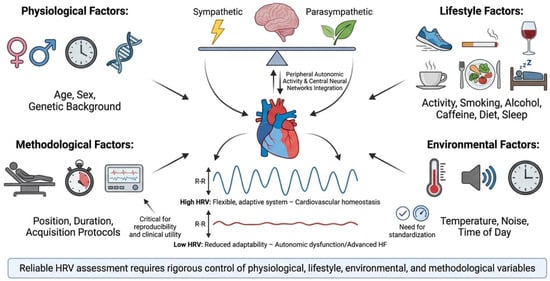
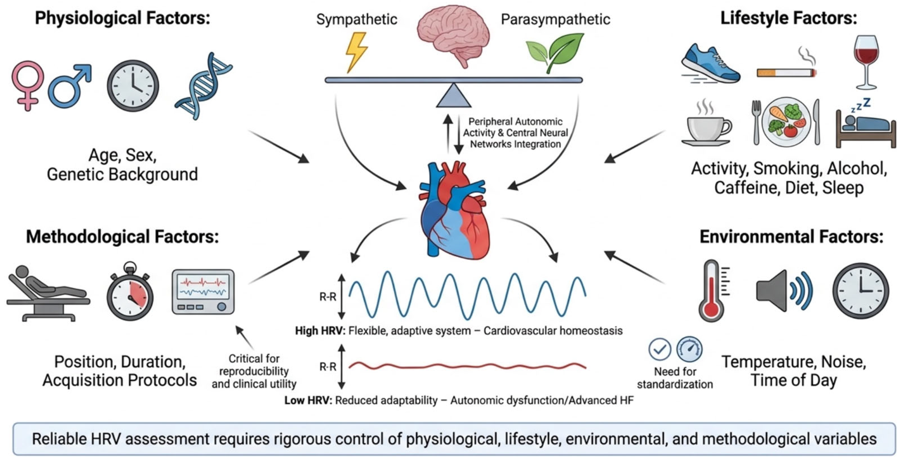
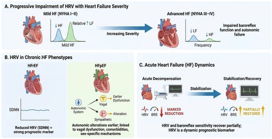
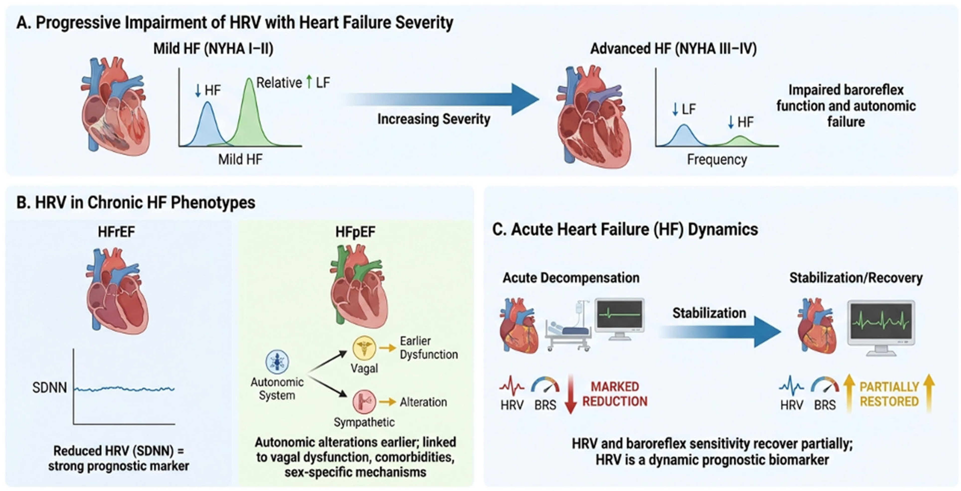
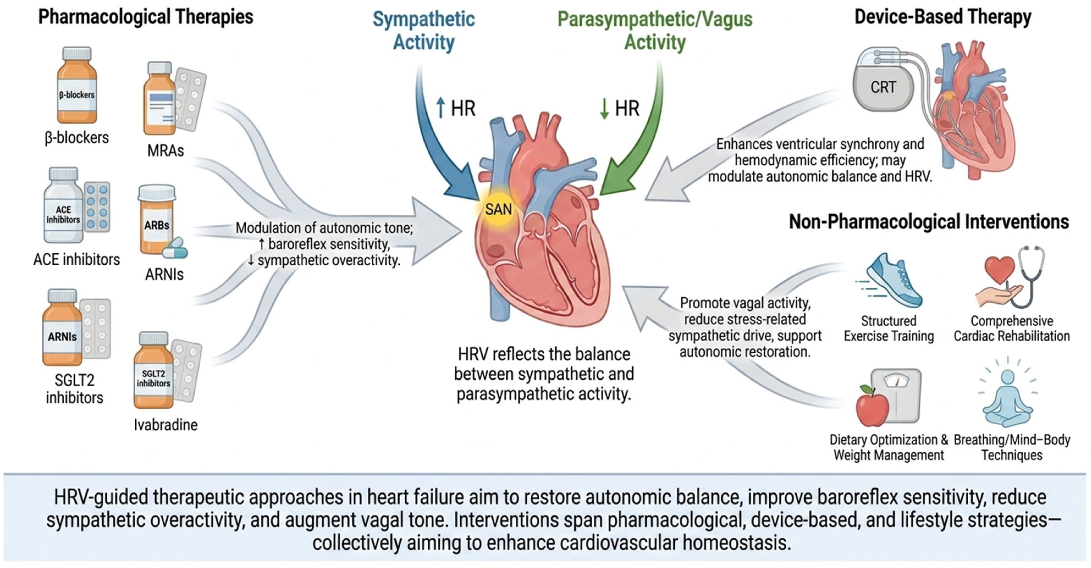

Disclaimer/Publisher’s Note: The statements, opinions and data contained in all publications are solely those of the individual author(s) and contributor(s) and not of MDPI and/or the editor(s). MDPI and/or the editor(s) disclaim responsibility for any injury to people or property resulting from any ideas, methods, instructions or products referred to in the content. |
© 2026 by the authors. Licensee MDPI, Basel, Switzerland. This article is an open access article distributed under the terms and conditions of the Creative Commons Attribution (CC BY) license.