


Summary
Plaque sealing is defined as angioplasty of an angiographically non-significant coronary stenosis in a strategically important vessel without previous infarction. In most patients qualifying for this approach, cardiac catheterisation is performed for atypical chest pain and stress tests are negative or not conclusive. Percutaneous coronary intervention (PCI) is performed in the hope of prevention of uncontrolled plaque rupture. Positive stress tests, on the other hand, convert the indication into a purely therapeutic one. PCI is suggested in combination with chemical plaque passivation by statins, since most of intramural plaques with equal potential of rupturing are not visualized by the angiographic "luminogram". Plaque sealing of a distinct coronary lesion, not significant in terms of visual estimate of diameter stenosis or flow measurements, provides a prognostically beneficial intervention without major additional effort and with low risk.
Zusammenfassung
Unter «plaque sealing» verstehen wir im engeren Sinn die perkutane Intervention an Koronarstenosen, die angiographisch bei visueller Analyse der Diameterreduktion als nicht signifikant Oder grenzwertig signifikant (40–60% Diameterreduktion) eingeschätzt werden. Insbesondere diskutieren wir diese Therapieoption bei strategisch wichtigen Gefässen, an denen bislang noch kein Infarkt abgelaufen ist. Diese Patienten kommen häufig mit atypischer Angina pectoris und negativer Ergometrie zur invasiven Diagnostik. Im Gegensatz dazu stellt ein objektiv positiver Ischämienachweis eine therapeutische Indikation dar. Das hinter "plaque sealing" stehende Konzept ist eine prognostische Indikation zur Verhinderung einer unkontrollierten Plaqueruptur. Kathetergestütztes "plaque sealing" wird zusammen mit pharmakologischer Stabilisierung durch Statine vorgeschlagen, da die Koronarangiographie als "Luminogramm" die Mehrzahl der intraluminalen Plaques nicht darstellen kann. Mit "plaque sealing" kann eine angiographisch objektivierte, hämodynamische noch nicht relevante, aber potentiell gefährliche Plaque durch perkutane Intervention während der invasiven Diagnostik ohne grossen Zeitaufwand und mit geringem Risiko neutralisiert werden.
Calculated probabilities of myocardial infarction and restenosis of a mild stenosis with or without angioplasty
The literature clearly indicates that about 80% or more of all infarctions result from superadded thrombosis on a lesion which by itself is not haemodynamically significant [1,2,3,4]. This does not mean that a mild stenosis is more dangerous in terms of producing an acute infarction than a severe stenosis. It rather implies that there is commonly a large number of mild stenoses to every single severe stenosis and it is the cumulative risk of all the nonsignificant stenoses that prevails [5]. According to the CASS (Coronary Artery Surgery Study) registry data, the risk of infarction over 3 years is 2% for a stenosis of less than 50%, 7% for a stenosis of 50 to 70%, 8% for a stenosis of 70 to 90%, 15% for a stenosis of 90 to 98%, and 7% for a functionally occlusive lesion [6]. This proves that, after all, the tight but not subtotal stenosis is the most dangerous one. Subtotal stenoses are usually either protected by collaterals or they represent lesions that have already been occluded before. A number of other factors also influence the risk of subsequent occlusion and infarction such as an irregular surface of the lesion or a lesion in a tortuous segment [6], a branching point stenosis [6,7] or a proximal location of the stenosis [2]. The latter criterion may be misleading. The more proximal a lesion, the more likely it is that the resultant occlusion will become manifest to the clinician or the pathologist.
What is the likely outcome of the mild or moderate lesion subjected to coronary angioplasty? Enough data have been accumulated to emphasise the fact that such lesions have a lower risk of acute occlusion and restenosis after angioplasty, which, nevertheless, has to be taken into account. In the era of modern angioplasty equipment and techniques, particularly with the use of stents for suboptimal results and for impending or acute occlusions, infarctions should occur in less than 3% in the acute phase. To this we have to add roughly a 0.5% incidence of infarctions during the next year (including the risk carried by repeat angioplasty in about 20% of patients), if we count only the ones being unequivocally attributable to the dilated site [8]. Beyond this period, spontaneous infarctions related to the dilated site are extremely rare. Even in case of restenosis, the highest incidence of related infarctions reported is 0.5% [9,10,11,12].
It has also been observed that the incidence of all late infarctions (related or unrelated to the dilated site) is independent of the occurrence of restenosis [13]. This indicates that these infarctions are not related to the dilated site itself but predominantly to other diseased segments (unsealed plaques). Although occlusions at the dilated site do occur in about 1% during long-term follow-up after angioplasty of nontotal lesions, these occlusions pertain typically to previously occluded or collateralised vessels and rarely cause a clinical event [14]. Though the above data largely pertain to significant stenoses, the figures for nonsignificant stenoses are bound to be similar, if not lower. In fact, one study of 415 patients who underwent angioplasty for moderately severe stenoses reported only one case of myocardial infarction [15]. Another small study addressing truly nonsignificant stenoses (<50% diameter stenosis assessed by quantitative coronary angiography), which had been dilated in the same session with a significant stenosis and had angiographic followup at 6 months, reported no major adverse cardiac events related to these stenoses during that period. However, no angiographic benefit at 6 months, i.e. the same nonsignificant diameter stenosis, was observed [16].
Definition of plaque sealing
Plaque sealing is defined as angioplasty of an angiographically non-significant coronary stenosis in a strategically important vessel without previous infarction [17]. This is based on the following rationale: coronary angioplasty typically induces an extensive splitting of the plaque or the border zone. This splitting engenders proliferation of new tissue layers covering the former plaque area, i.e. plaque sealing. The smooth muscle-rich intima observed over the first few months after angioplasty transforms into a collagen-rich layer subsequently [18]. Thus once the acute effect with its inherent risk of acute occlusion has passed, the subsequent risk of acute occlusion should be markedly reduced compared with that of an untreated plaque. The covering of the intimal wound by cell proliferation, caused by angioplasty, may well have the beneficial effect of plaque sealing but it is also a major component of restenosis. Restenosis is a significant shortcoming of coronary angioplasty but it can be dealt with effectively and is not irreversible. In contrast, acute occlusion due to plaque rupture may cause irreversible myocardial damage or even death [19].
Several clarifications need to be made around this working hypothesis. The reasons for a patient to undergo coronary angiography, the assessment of stenosis severity versus the symptoms, the risk profile of coronary artery disease, and the anticipated course of the disease from the presenting stage are to be considered. In our understanding, plaque sealing is a preventive action towards a possible future rupture of a coronary plaque that has been identified on the occasion of an angiography indicated by quite a variety of symptoms or associated disease conditions like e.g. preoperative risk stratification. In a typical scenario, the patient’s symptoms definitively are not due to a flow-limiting stenosis — defined by any means of morphological (angiography, intravascular ultrasound) or physiological (coronary flow reserve, fractional flow reserve) assessment, and thus the patient does not have typical angina pectoris or objective signs of ischaemia. If the patient has demonstrable angina pectoris (by stress test or scintigraphy), PTCA of an angiographically borderline stenosis would represent standard therapeutic angioplasty and plaque sealing would be a welcomed side product but not the primary intention. The second crucial point for this concept is a pragmatic one: angiography has been performed on the respective patient for whatever reason, there is vascular access and a coronary catheter in place. A lesion of <50% diameter stenosis by visual estimate or on-line quantitative coronary angiography has been identified in a proximal segment of the left anterior descending, left circumflex, or right coronary artery with a sufficiently large perfusion territory to cause a major myocardial infarction in case of occlusion. Given these circumstances, an immediate decision is warranted whether to proceed with a preventive angioplasty to transform this potentially vulnerable plaque mechanically into scar tissue by stimulating a healing response. Do we know anything about the potential of this specific plaque to undergo rupture? Do we know when this is likely to happen? Do we know about the natural course of nonsignificant plaques? Not really, because data are sparse [3]. Positive data exist as to the presence of numerous angiographically inapparent plaques with growth towards the lamina elastica externa and enlargement of the vessel circumference [20] in apparently, i.e. luminographically, normal coronary arteries. Next, the potential risk of performing angioplasty in this lesion must be taken into account.
Plaque morphology: impact for mechanical stabilisation
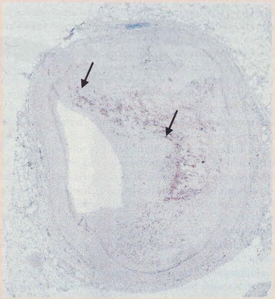
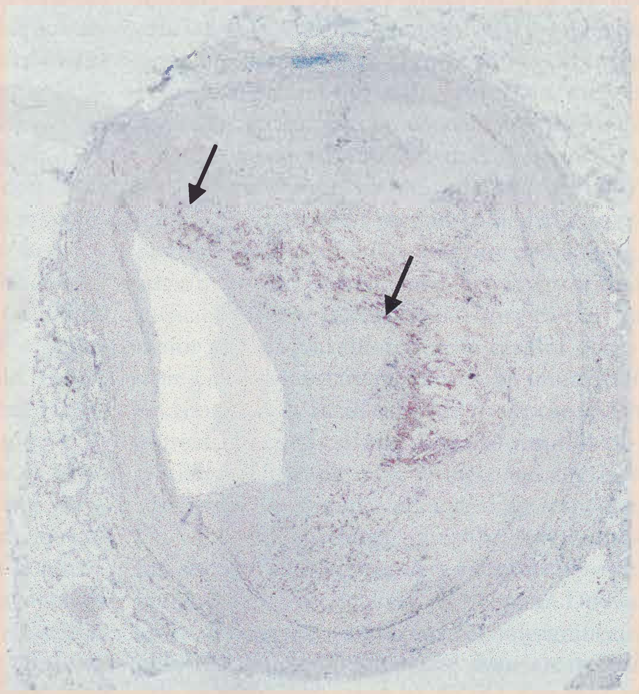
Figure 1 shows characteristics of the vulnerable and the stable plaque [21]. Atherosclerotic plaques typically consist of a lipid-rich core in the central portion of the eccentrically thickened intima. The lipid core is bonded on its luminal side by a fibrous cap. The thickness of the fibrous cap is one of the determinants of the probability of the plaque to undergo rupture. Figure 1 represents a non pressure-fixed crosssection of a coronary artery with a thick fibrous cap in the upper right side and a thin fibrous cap in the lower left side (arrows). Clinical data suggest that stable plaques more often show luminal narrowing detectable by angiography than do vulnerable plaques [21]. The central, lipid-rich core contains many lipid-laden macrophage foam cells derived from blood monocytes. In figure 1, the dark staining indicates CD 68 positive inflammatory macrophages, which extend close to the surface of the thin fibrous cap in the lower left margin. At the site of lesion disruption, smooth muscle cells are often activated and the inflammatory reaction is pronounced. Disruption of fibrous caps occurs at points of ongoing inflammation, indicated by the presence of the activated macrophages [22]. Macrophages can degrade extracellular matrix by phagocytosis or by secreting proteolytic enzymes such as plasminogen activators and matrix metalloproteinases, all capable of weakening the fibrous cap [23]. In this context, the concept of atherosclerosis as an inflammatory disease has emerged and inflammatory processes have been shown to play a role even in very early stages of the disease characterised by endothelial dysfunction and mediated by leucocyte adhesion molecules [24]. Several reports have shown an association between the incidence of atherosclerosis and the presence of infectious agents, among them chlamydia pneumoniae [25]. Interestingly, there are observations that chlamydial proteins can induce metalloproteinase production under experimental conditions and thus provide a link to plaque vulnerability [26]. There are several ongoing clinical studies addressing the role of infectious agents in acute ischaemic syndromes and restenosis, which might elucidate the possible role of antiinflammatory (antibiotic?) therapy in the context of pharmacological plaque passivation.
Figure 1.
(courtesy of Thomas Schaffner, MD, Pathology, University Hospital Berne): characteristics of the vulnerable and the stable plaque. Non pressure-fixed cross-section of a coronary artery with a thick fibrous cap in the upper right side and a thin fibrous cap in the lower left side (arrows). The dark staining indicates CD 68 positive inflammatory macrophages, which extend close to the surface of the thin fibrous cap in the lower left margin.
Only a minority of spontaneous plaque ruptures leads to total occlusion of the vessel and to infarction. The majority heals and may contribute to a more severe stenosis by an overzealous coating with neointimal layers. In analogy to the mechanical plaque sealing theory, it is unlikely that the very same site will fissure again in the future. Yet, these small fissures only lead to a very localised sealing of the plaque and the remainder of the plaque mass remains unsealed and therefore dangerous.
The mechanical approach by PTCA intends to convert the thin fibrous cap of lower left side into the thick fibrous septum of the upper right side in figure 1 by creating a stretch, tear, or even dissection of the vessel wall, thereby inducing the wound healing mechanisms.
Factors triggering plaque rupture
Considering mechanical aspects of plaque stabilisation, possible factors triggering plaque rupture [23] and the respective therapeutic options will be taken into account. Cap tension is determined in part by the circumferential tension in the wall, which relates to blood pressure and luminal diameter according to Laplace’s law. An additional important factor in this equation, however, is the tensile strength of the fibrous cap itself determined by its own consistency and that of the surrounding or underlying tissue, e.g. the atheromatous gruel of the lipid core. While blood pressure can easily be treated medically, the lipid content of the plaque might be influenced by aggressive lipid lowering. This is suggested by experimental studies [27,28] and the incidence of coronary events in large-scale statin studies [29,30], although not yet ultimately proven by direct morphologic evidence in humans. Mechanical plaque stabilisation remains to be considered upon angiographic identification of a plaque in a strategically important location until final evidence of pharmacological passivation exists. Heart contraction, especially axial vessel bending and stretching might be another trigger mechanism with few possibilities of interfering (beta-blockers?). Vasomotion and vasospasm are not within the scope of mechanical stabilisation and might be influenced pharmacologically (nitrates and calcium antagonists). Capillary bleeding within the plaque has been suggested as a possible trigger for which no therapeutic option is evident except elimination of the plaque. Wall shear stresses are usually smaller than blood and pulse pressure but exact calculation of regional wall stress within the human coronary arteries might represent a future option for determining loci minoris resistentiae within plaque-loaded coronary segments.
Identification of the vulnerable plaque?
Identification of a coronary plaque prone to rupture would be the most desirable approach with regard to intended mechanical stabilisation. However, currently no method exists that reliably discriminates vulnerable and stable plaques. Intravascular ultrasound (IVUS) in the present 2D mode detects coronary wall morphology [31,32] but has not been proven to identify vulnerable plaques [33]. In the future, complex approaches towards anatomically correct 3D-reconstuction of intravascular ultrasound using image fusion of IVUS and angiography [34] might provide indirect indicators for plaques at risk via cap/plaque volume ratio and shear stress calculations. Current technology does not yet allow to perform these analyses routinely in a practicable way. Coronary angioscopy has been shown to provide some criteria for identification of unstable plaques [33,35,36,37,38], but the method has been abandoned because of unrealistic complexity for routine purposes. Intracoronary temperature measurements, magnetic resonance imaging (MRI), or MR spectroscopy might offer theoretical potential for application in this respect, without any current clinical practicability, however. The development of a tool for intracoronary analysis of plaque vulnerability (detection of the inflammatory process?) would be most intriguing for the concept of mechanical plaque stabilisation.
Physiological assessment
There is no rationale from flow dynamics or structure-function relationships to dilate nonsignificant stenoses, since lesions with diameter reduction of 45% or less have an almost normal coronary flow reserve [39]. The concept of measuring coronary flow reserve or fractional flow reserve [40] only relates to functional lesion severity. The concept of plaque sealing is preventive and independent of functional measurements. Dilating nonsignificant stenoses with a reduced coronary flow reserve or fractional flow reserve means therapeutic angioplasty. Plaque sealing still occurs but it is not the principal reason for the procedure.
The ultimate equation
The ultimate equation opposes the risk of a spontaneous infarction based on a moderate (haemodynamically nonsignificant) lesion of 7% over 3 years to a 3% infarction rate during coronary angioplasty to which 0.5% has to be added for the next 4 to 5 years in those ⅔% of patients without restenosis and 2.5% in the remaining third with restenosis (factoring in a 2% infarction risk during the repeat coronary angioplasty). This yields a pessimistically calculated overall infarction risk of the hypothetical angioplasty group of 5% which represents a relative risk reduction of about 30% over 3 years and argues in favour of angioplasty for such lesions.
Mechanical and chemical plaque stabilisation
The merits of lipid lowering in slowing progression of coronary artery disease have been established already based on life-style changes and diet in small, well documented study groups [41]. Convincing evidence is provided by large-scale lipid lowering studies that lipid lowering reduces the incidence of coronary events [42,43] and may induce plaque passivation [30,44]. The effect may be still more impressive with aggressive lipid lowering and lead to the conclusion that the need for coronary intervention might be postponed or even obviated pharmacologically according to data from the AVERT trial [45]. However, there is no convincing proof up to now that coronary artery disease really can be put to a halt instead of interventional therapy being only deferred by a few years. Moreover, the reduction in events in the AVERT trial, which compares PTCA with conservative management under aggressive lipid lowering, is mainly due to reduction in minor events (e.g. repeat hospitalisation) in the medically treated group and not to reduction in hard endpoints like death or myocardial infarction during the first 18 months of follow-up. It is trivial that once an invasive strategy is engaged, it produces consecutive medical attendance and expenditure, in part solely due to the patient’s enhanced awareness of "a disease which could not be managed medically".
The need for plaque sealing is affected but not eliminated by aggressive lipid lowering. It only provides a solid background. We do not call for screening everyone over 50 with coronary angiography to detect functionally nonsignificant plaques and dilate them. But once a coronary angiogramme has been performed for whatever reasons and there is documentation of a nonsignificant coronary stenosis in a strategically important location without previous infarction, it is justified to proceed to preventive angioplasty according to the plaque sealing concept. In case of multiple borderline plaques, this concept is moot for obvious reasons. Diffuse nonsignificant coronary artery disease needs aggressive medical risk factor management. The plaque sealing patient additionally receives lipid lowering agents to profit from both the pharmacological and the mechanical approach. While working on a medical management to completely prevent plaque rupture or progression, mechanical stabilisation of the plaque by plaque sealing has a role.
References
- Hackett, D.; Davies, G.; Maseri, A. Pre-existing coronary stenoses in patients with first myocardial infarction are not necessarily severe. Eur. Heart J. 1988, 9, 1317–23. [Google Scholar] [CrossRef] [Scilit]
- Ambrose, J.A.; Tannenbaum, M.A.; Alexopoulos, D.; HjemdahlMonsen, C.E.; Leavy, J.; Weiss, M.; et al. Angiographic progression of coronary artery disease and the development of myocardial infarction. J. Am. Coll. Cardiol. 1988, 12, 56–62. [Google Scholar] [CrossRef] [Scilit] [PubMed]
- Little, W.C.; Constantinescu, M.; Applegate, R.J.; Kutcher, M.A.; Burrows, M.T.; Kahl, F.R.; et al. Can coronary angiography predict the site of a subsequent myocardial infarction in patients with mild-to-moderate coronary artery disease? Circulation 1988, 78, 1157–66. [Google Scholar] [CrossRef] [Scilit]
- Brown, B.G.; Gallery, C.A.; Badger, R.S.; Kennedy, J.W.; Mathey, D.; Bolson, E.L.; et al. Incomplete lysis of thrombus in the moderate underlying atherosclerotic lesion during intracoronary infusion of streptokinase for acute myocardial infarction: quantitative angiographic observations. Circulation 1986, 73, 653–61. [Google Scholar] [CrossRef] [Scilit]
- Giroud, D.; Li, J.M.; Urban, P.; Meier, B.; Rutishauer, W. Relation of the site of acute myocardial infarction to the most severe coronary arterial stenosis at prior angiography [see comments]. Am. J. Cardiol. 1992, 69, 729–32. [Google Scholar] [CrossRef] [Scilit]
- Ellis, S.; Alderman, E.; Cain, K.; Fisher, L.; Sanders, W.; Bourassa, M. Prediction of risk of anterior myocardial infarction by lesion severity and measurement method of stenoses in the left anterior descending coronary distribution: a CASS Registry Study. J. Am. Coll. Cardiol. 1988, 11, 908–16. [Google Scholar] [CrossRef] [Scilit] [PubMed]
- Taeymans, Y.; Theroux, P.; Lesperance, J.; Waters, D. Quantitative angiographic morphology of the coronary artery lesions at risk of thrombotic occlusion. Circulation 1992, 85, 78–85. [Google Scholar] [CrossRef] [Scilit]
- Ernst, S.M.; van der Feltz, T.A.; Bal, E.T.; van Bogerijen, L.; van den Berg, E.; Ascoop, C.A.; et al. Long-term angiographic follow-up, cardiac events, and survival in patients undergoing percutaneous transluminal coronary angioplasty. Br. Heart J. 1987, 57, 220–5. [Google Scholar] [CrossRef] [Scilit] [PubMed]
- Guiteras, P.; Tomas, L.; Varas, C.; Auge, J.M.; Masotti, M.; Crexells, C.; et al. Five years of angiographic and clinical followup after successful percutaneous transluminal coronary angioplasty. Eur. Heart J. 1989, 10 (Suppl), G42–8. [Google Scholar] [CrossRef] [Scilit]
- Kober, G.; Vallbracht, C.; Kadel, C.; Kaltenbach, M. Results of repeat angiography up to eight years following percutaneous transluminal angioplasty. Eur. Heart J. 1989, 10 (Suppl), G4953. [Google Scholar] [CrossRef] [Scilit]
- Dimas, A.P.; Grigera, F.; Arora, R.R.; Simpfendorfer, C.C.; Hollman, J.L.; Frierson, J.H.; et al. Repeat coronary angioplasty as treatment for restenosis. J. Am. Coll. Cardiol. 1992, 19, 1310–4. [Google Scholar] [CrossRef] [Scilit] [PubMed]
- Bottner, R.K.; Green, C.E.; Ewels, C.J.; Recientes, E.; Patrissi, G.A.; Kent, K.M. Recurrent ischemia more than 1 year after successful percutaneous transluminal coronary angioplasty. An analysis of the extent and anatomic pattern of coronary disease. Circulation 1989, 80, 1580–4. [Google Scholar] [CrossRef] [Scilit] [PubMed]
- Vlietstra, R.E.; Holmes, D.R., Jr.; Rodeheffer, R.J.; Bailey, K.R. Consequences of restenosis after coronary angioplasty. Int. J. Cardiol. 1991, 31, 143–7. [Google Scholar] [CrossRef] [Scilit]
- Kitazume, H.; Kubo, I.; Iwama, T.; Ageishi, Y. Long-term angiographic follow-up of lesions patent 6 months after percutaneous coronary angioplasty. Am. Heart J. 1995, 129, 441–4. [Google Scholar] [CrossRef] [Scilit]
- Rozenman, Y.; Gilon, D.; Welber, S.; Sapoznikov, D.; Wexler, D.; Lotan, C.; et al. Total coronary artery occlusion late after successful coronary angioplasty of moderately severe lesions: incidence and clinical manifestations. Cardiology 1994, 85, 222–8. [Google Scholar] [CrossRef] [Scilit]
- Hamon, M.; Bauters, C.; McFadden, E.P.; Lablanche, J.M.; Bertrand, M.E. Six-month quantitative angiographic followup of 50% diameter stenoses dilated during multilesion percutaneous transluminal coronary angioplasty. Am. J. Cardiol. 1993, 71, 1226–9. [Google Scholar] [CrossRef] [Scilit]
- Meier, B.; Ramamurthy, S. Plaque sealing by coronary angioplasty. Cathet Cardiovasc Diagn. 1995, 36, 295–7. [Google Scholar] [CrossRef] [Scilit]
- Inoue, K.; Nakamura, N.; Kakio, T.; Suyama, H.; Tanaka, S.; Goto, Y.; et al. Serial changes of coronary arteries after percutaneous transluminal coronary angioplasty: histopathological and immunohistochemical study. J. Cardiol. 1994, 24, 279–91. [Google Scholar]
- Virmani, R.; Farb, A.; Burke, A.P. Coronary angioplasty from the perspective of atherosclerotic plaque: morphologic predictors of immediate success and restenosis. Am. Heart J. 1994, 127, 163–79. [Google Scholar] [CrossRef] [Scilit]
- Glagov, S.; Weisenberg, E.; Zarins, C.K.; Stankunavicius, R.; Kolettis, G.J. Compensatory enlargement of human atherosclerotic coronary arteries. N Engl. J. Med. 1987, 316, 1371–5. [Google Scholar] [CrossRef] [Scilit] [PubMed]
- Libby, P. Molecular bases of the acute coronary syndromes. Circulation 1995, 91, 2844–50. [Google Scholar] [CrossRef] [Scilit] [PubMed]
- Falk, E.; Shah, P.K.; Fuster, V. Coronary plaque disruption. Circulation 1995, 92, 657–71. [Google Scholar] [CrossRef] [Scilit] [PubMed]
- Gronholdt, M.L.; Dalager-Pedersen, S.; Falk, E. Coronary atherosclerosis: determinants of plaque rupture. Eur. Heart J. 1998, 19 (Suppl C), C24–9. [Google Scholar]
- Ross, R. Atherosclerosis — an inflammatory disease. N Engl. J. Med. 1999, 340, 115–26. [Google Scholar] [CrossRef] [Scilit]
- Libby, P.; Egan, D.; Skarlatos, S. Roles of infectious agents in atherosclerosis and restenosis: an assessment of the evidence and need for future research. Circulation 1997, 96, 4095–103. [Google Scholar] [CrossRef] [Scilit] [PubMed]
- Kol, A.; Sukhova, G.K.; Lichtman, A.H.; Libby, P. Chlamydial heat shock protein 60 localizes in human atheroma and regulates macrophage tumor necrosis factor-alpha and matrix metalloproteinase expression. Circulation 1998, 98, 300–7. [Google Scholar] [CrossRef] [Scilit]
- Williams, J.K.; Sukhova, G.K.; Herrington, D.M.; Libby, P. Pravastatin has cholesterol-lowering independent effects on the artery wall of atherosclerotic monkeys. J. Am. Coll. Cardiol. 1998, 31, 684–91. [Google Scholar] [CrossRef] [Scilit]
- Libby, P.; Aikawa, M. New insights into plaque stabilisation by lipid lowering. Drugs 1998, 56, 9–13; discussion 33. [Google Scholar] [CrossRef] [Scilit]
- Kullo, I.J.; Edwards, W.D.; Schwartz, R.S. Vulnerable plaque: pathobiology and clinical implications. Ann. Intern Med. 1998, 129, 1050–60. [Google Scholar] [CrossRef] [Scilit]
- Brown, B.G.; Zhao, X.Q.; Sacco, D.E.; Albers, J.J. Lipid lowering and plaque regression. New insights into prevention of plaque disruption and clinical events in coronary disease. Circulation 1993, 87, 1781–91. [Google Scholar] [CrossRef] [Scilit]
- Hodgson, J.M.; Reddy, K.G.; Suneja, R.; Nair, R.N.; Lesnefsky, E.J.; Sheehan, H.M. Intracoronary ultrasound imaging: correlation of plaque morphology with angiography, clinical syndrome and procedural results in patients undergoing coronary angioplasty. J. Am. Coll. Cardiol. 1993, 21, 35–44. [Google Scholar] [CrossRef] [Scilit]
- Ge, J.; Erbel, R.; Gerber, T.; Gorge, G.; Koch, L.; Haude, M.; et al. Intravascular ultrasound imaging of angiographically normal coronary arteries: a prospective study in vivo. Br. Heart J. 1994, 71, 572–8. [Google Scholar] [CrossRef] [Scilit]
- de Feyter, P.J.; Ozaki, Y.; Baptista, J.; Escaned, J.; Di Mario, C.; de Jaegere, P.P.; et al. Ischemia-related lesion characteristics in patients with stable or unstable angina. A study with intracoronary angioscopy and ultrasound. Circulation 1995, 92, 1408–13. [Google Scholar] [CrossRef] [Scilit]
- Roelandt, J.R.; di Mario, C.; Pandian, N.G.; Wenguang, L.; Keane, D.; Slager, C.J.; et al. Three-dimensional reconstruction of intracoronary ultrasound images. Rationale, approaches, problems, and directions. Circulation 1994, 90, 1044–55. [Google Scholar] [CrossRef] [Scilit]
- Bauters, C.; Lablanche, J.M.; Renaud, N.; McFadden, E.P.; Hamon, M.; Bertrand, M.E. Morphological changes after percutaneous transluminal coronary angioplasty of unstable plaques. Insights from serial angioscopic follow-up [see comments]. Eur. Heart J. 1996, 17, 1554–9. [Google Scholar] [CrossRef] [Scilit]
- Lablanche, J.M.; Van Belle, E.; McFadden, E.; Gautier, L.; de Groote, P.; Bauters, C.; et al. Coronary angioscopy. Arch. Mal. Cœur Vaiss. 1997, 90, 29–33. [Google Scholar]
- Thieme, T.; Wernecke, K.D.; Meyer, R.; Brandenstein, E.; Habedank, D.; Hinz, A.; et al. Angioscopic evaluation of atherosclerotic plaques: validation by histomorphologic analysis and association with stable and unstable coronary syndromes. J. Am. Coll. Cardiol. 1996, 28, 1–6. [Google Scholar] [CrossRef] [Scilit] [PubMed]
- Kleber, F.X.; Dopfmer, S.; Thieme, T. Invasive strategies to discriminate stable and unstable coronary plaques. Eur. Heart J. 1998, 19 (Suppl C), C44–9. [Google Scholar]
- Gould, K.L. Coronary artery stenosis; Elsevier: New York, Amsterdam, London, 1991. [Google Scholar]
- Pijls, N.H.; De Bruyne, B.; Peels, K.; Van Der Voort, P.H.; Bonnier, H.J.; Bartunek, J.K.J.J.; et al. Measurement of fractional flow reserve to assess the functional severity of coronary-artery stenoses. N Engl. J. Med. 1996, 334, 1703–8. [Google Scholar] [CrossRef] [Scilit] [PubMed]
- Ornish, D.; Scherwitz, L.W.; Billings, J.H.; Gould, K.L.; Merritt, T.A.; Sparler, S.; et al. Intensive lifestyle changes for reversal of coronary heart disease. JAMA 1998, 280, 2001–7. [Google Scholar] [CrossRef] [Scilit]
- Randomised trial of cholesterol lowering in 4444 patients with coronary heart disease: the Scandinavian Simvastatin Survival Study (4S). Lancet 1994, 344, 1383–9. [CrossRef] [Scilit]
- Shepherd, J.; Cobbe, S.M.; Ford, I.; Isles, C.G.; Lorimer, A.R.; MacFarlane, P.W.; et al. Prevention of coronary heart disease with pravastatin in men with hypercholesterolemia. West of Scot land Coronary Prevention Study Group. N Engl J Med 1995, 333, 1301–7. [Google Scholar]
- Effect of simvastatin on coronary atheroma: the Multicentre Anti-Atheroma Study (MAAS). Lancet 1994, 344, 633–8. [CrossRef] [Scilit]
- Pitt, B.; Waters, D.; Brown, V.; McCormick, L.; Wagner, B.; Shurzinske, L.; et al. Results of the atorvastatin versus revascularization treatment (AVERT) study: an 18-month study on aggressive lipid lowering in patients with stable coronary artery disease indicated for catheter-based revascularization. Circulation 1998, 98 (Suppl I), 1–636. [Google Scholar]
© 1999 by the author. Attribution - Non-Commercial - NoDerivatives 4.0.