


Abstract
Congenital renal arteriovenous malformation (RAVM) is an extremely rare abnormality with about 200 reported cases. It may be asymptomatic and found incidentally, or present with cardiac or renal symptoms. Hence establishing the physiological and clinical significance of RAVM is crucial in identifying patients in need of treatment and selecting the appropriate treatment. Herein, we report the rare case of a patient presenting with accelerated hypertension, heart failure and a congenital RAVM. We discuss the approach to assessment and treatment of congenital RAVMs in the context of cardiac disease.
Introduction
Congenital renal arteriovenous malformation (RAVM), first described by Varela in 1928, is an uncommon abnormality and only about 200 cases have been reported to date [1,2,3,4,5,6]. It accounts for 14–25% of RAVMs, the rest being acquired malformations (trauma during percutaneous renal biopsy is the most frequent cause). A majority of RAVMs are symptomatic and manifest with local (haematuria) or systemic (high output heart failure and hypertension) symptoms [1,2]. Occasionally they may be detected incidentally. Assessing the physiological and clinical significance of RAVM is therefore crucial in identifying those with and without need for treatment.
Case summary
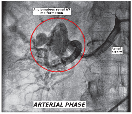
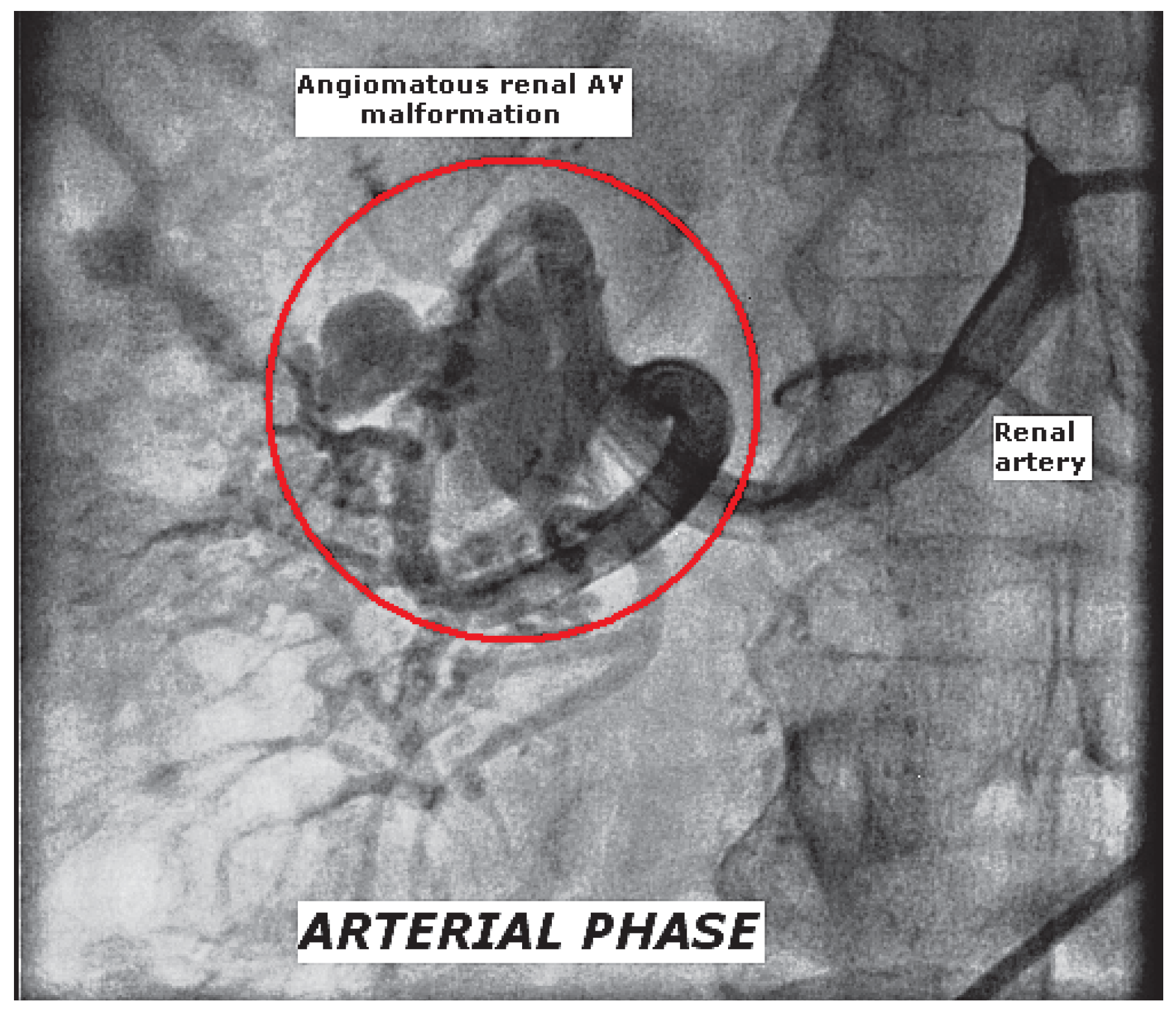
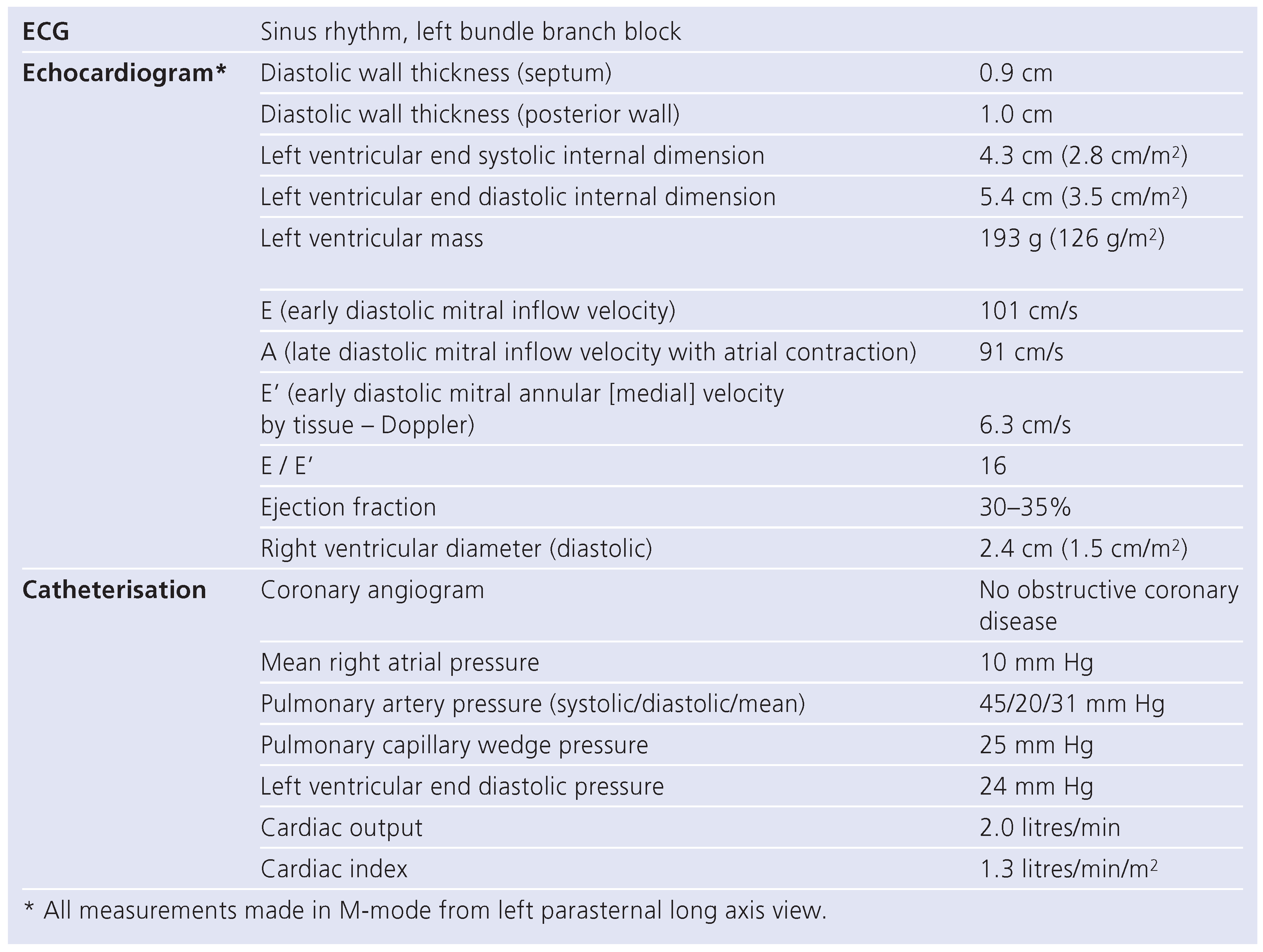
A 50-year-old female was transferred from an outlying facility for worsening dyspnoea and a first-time diagnosis of heart failure. She had longstanding hypertension which was sub-optimally controlled on atenolol, amlodipine and hydrochlorothiazide. She was a non-diabetic and denied use of tobacco, alcohol, or recreational drugs. She reported oedema and orthopnoea but denied chest pain, palpitations or syncope. Symptoms were progressive for one year with rapid worsening for 2–3 weeks prior to presentation. On physical examination the pulse was 110 per minute, blood pressure 210/110 mm Hg, respiration 22 per minute, BMI 22 kg/m2 and oxygen saturation 95% on 4 litres of O2 per minute. Notable findings included jugular vein distension, S3 gallop, bilateral basal crepitations and ankle oedema. Blood counts, biochemistry panel and thyroid profile were unremarkable. The echocardiogram revealed global hypokinesis of left ventricle (LV) with an ejection fraction of 30–35% and normal wall thickness. Cardiac catheterisation revealed normal epicardial coronaries. Selective injection of bilateral renal arteries was done in view of her poorly controlled hypertension. Renal artery stenosis was absent; a 3 × 4 cm angiomatous arteriovenous malformation (AVM) was noted in the upper pole of the right kidney (Figure 1). Retrospectively the patient reported no prior haematuria, colic, back pain, abdominal trauma or renal procedures. Diagnostic considerations at this point were (1.) high output failure due to RAVM; (2.) excessive renin secretion (secondary to regional renal ischaemia from the shunt) causing hypertension and heart failure; (3.) idiopathic dilated cardiomyopathy with incidental RAVM. Relevant findings from echocardiogram and right/left heart catheterisation are shown in Table 1.
Figure 1.
Arterial phase image of selective right renal arteriogram showing the arteriovenous malformation in the upper pole.
Table 1.
Summary of cardiac evaluation in the index patient.
Discussion
The exact pathogenesis of congenital RAVM is unknown. While some believe it results from erosion of a congenital renal artery aneurysm into a vein, others believe it is present at birth. Peak incidence is between 30 and 40 years, the right kidney is more frequently involved than the left, and women are affected three times as often as men [1,2]. In general, congenital lesions typically have a cirsoid (varix-like) appearance, and are more often located in the upper pole. Haematuria is the most common presenting symptom in patients with congenital RAVM, while those with acquired RAVM frequently present with cardiovascular manifestations [1,2]. In our patient absence of prior trauma or renal intervention/procedure, and location/appearance on the angiogram were all consistent with congenital RAVM.
Cardiovascular manifestations of RAVM include hypertension, hypertensive heart disease and high output state with or without heart failure. Hypertension is a result of increased stroke volume and excess renin secretion (secondary to regional renal ischaemia produced by the shunt). In addition, RAVM has been shown to produce systolic heart failure secondary to uncontrolled hypertension, with documented improvement in both LV function and blood pressure following embolisation [3,4,5,6]. In our patient, normal LV wall thickness and near normal LV mass index argued against hypertensive heart disease; significant coronary artery disease was excluded and a low cardiac index ruled out a high output state. We believe that our patient had idiopathic dilated cardiomyopathy with renin-mediated hypertension due to the associated congenital RAVM. Uncontrolled blood pressure due to the RAVM potentially aggravated her heart failure symptoms and led to progressive decompensation. We therefore decided to conduct a trial of medical management after consultation with an urologist. Embolisation was planned in case of refractory hypertension or heart failure. On a medical regimen consisting of carvedilol, enalapril, acetylsalicylic acid and furosemide our patient had a dramatic clinical improvement in blood pressure control and symptoms. At 6 months’ follow up there was significant improvement in functional capacity and blood pressures were well controlled; the echocardiogram showed improvement in LV function with an EF of 40% and mild mitral regurgitation. The dramatic improvement in blood pressure control and symptoms after initiation of enalapril with only modest improvement in LV function supported our diagnosis of idiopathic dilated cardiomyopathy aggravated by hypertension due to associated RAVM.
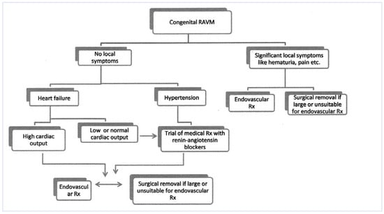
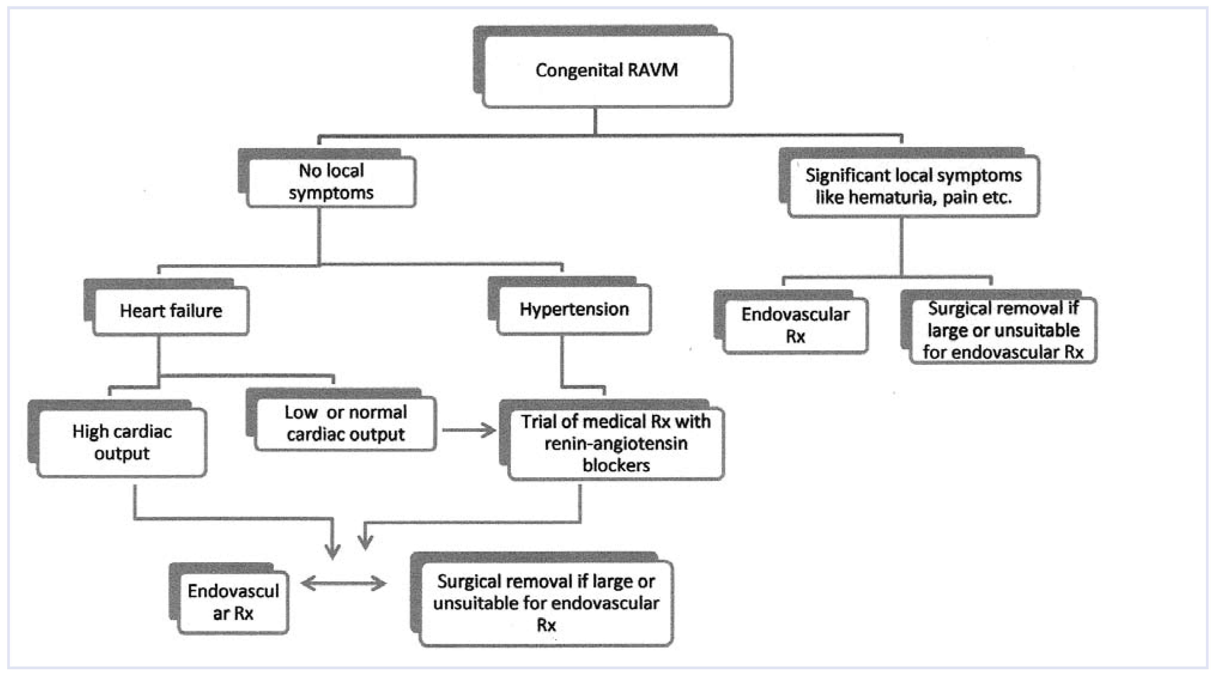
While renal angiography remains the diagnostic gold standard, noninvasive tests such as computed tomographic or magnetic resonance angiography have excellent sensitivity and specificity and are increasingly used [1,2]. Doppler ultrasonography is a useful initial test but has limited sensitivity, especially for smaller RAVMs. Though selective renal vein renin measurement has been proposed as a potential tool for assessing the functional significance of RAVM, we did not perform this measurement as many recent reports have suggested poor specificity and sensitivity of this test [1,2,3,4]. Wherever indicated, definitive therapy of RAVM involves transcatheter embolisation, alcohol ablation or surgical ligation [1,2,5,6]. Complications of transcatheter therapy include bleeding, pulmonary and paradoxical systemic embolism and non-target vessel embolisation. Partial or total nephrectomy is reserved for selected patients with extremely large RAVMs not amenable to transcatheter embolisation or surgical ligation. However, our case suggests that medical therapy with angiotensin-converting enzyme inhibitors might be a reasonable initial management strategy in selected patients with RAVM (normal or low cardiac index and no local symptoms) and hypertension. Embolisation will remain the therapy of choice for patients with high output failure, hypertension not controlled by optimal medical therapy (including a renin-angiotensin blocker) and local symptoms such as haematuria. Figure 2 depicts a suggested approach for the assessment and treatment of congenital RAVM based on our experience and a literature review.
Figure 2.
Flowchart depicting the suggested approach for evaluation and management of congenital renal arteriovenous malformation. Rx = therapy; RAVM = renal arteriovenous malformation.
In summary, congenital RAVM is extremely rare and can be a cause or aggravating factor in refractory hypertension and heart failure. Though embolisation provides a definitive cure and is necessary in many patients, selected patients might do well on medical therapy (renin-angiotensin blockers) alone. A diligent evaluation of the clinical/functional significance of RAVM and its effects on the underlying cardiac disorder is therefore crucial for selecting the appropriate treatment strategy and in avoiding unnecessary procedures with their attendant morbidity.
Conflicts of Interest
none.
References
- Bauer, S.B. Anomalies of the upper urinary tract. In Campbell-Walsh Urology, 9th ed.; Wein, A.J., Kavoussi, L.R., Novick, A.C., Eds.; Saunders Elsevier: Philadelphia, PA, 2007; pp. 3297–3298. [Google Scholar]
- Crotty, K.L.; Orihuela, E.; Warren, M.M. Recent advances in the diagnosis and treatment of renal arteriovenous malformations and fistulas. J Urol. 1993, 150, 1355–1359. [Google Scholar] [CrossRef] [Scilit] [PubMed]
- Tarif, N.; Mitwalli, A.H.; Al Samayer, S.A.; et al. Congenital renal arteriovenous malformation presenting as severe hypertension. Nephrol Dial Transplant. 2002, 17, 291–294. [Google Scholar] [CrossRef] [Scilit] [PubMed]
- Sasaki, N; Joashi, U.C.; Vergara, M.; Saland, J.M.; Love, B.A. Postrenal biopsy AVM leading to severe hypertension and dilated cardiomyopathy. Pediatr Nephrol. 2009, 24, 2459–2462. [Google Scholar] [PubMed]
- Lekuona, I.; Laraudogoitia, E.; Salcedo, A.; Sádaba, M. Congestive heart failure in a hypertensive patient (don’t forget the stethoscope). Lancet. 2001, 357, 358. [Google Scholar] [CrossRef] [Scilit] [PubMed]
- Zambrana, A.R.; Martín, F.L.; Blanco, J.Z.; Blanco, J.M.; Serrano, M.J.; Suárez, J.L. Arteriovenous renal congenital fistula. Actas Urol Esp. 2009, 33, 696–699. [Google Scholar] [PubMed]

© 2010 by the author. Attribution - Non-Commercial - NoDerivatives 4.0.