


Abstract
Hydroponic cultivation has emerged as a potential approach for producing plant-derived materials containing bioactive phytochemicals. However, whether hydroponically cultivated mulberry (Morus alba L.) roots exert anti-allergic activity remains unknown. Here, we evaluated the anti-allergic effects of hydroponically cultivated mulberry fibrous root extract and investigated the potential contribution of oxyresveratrol. HPLC analysis demonstrated that the hydroponically cultivated mulberry fibrous root extract contained measurable amounts of oxyresveratrol and several other polyphenols. Oral administration of the extract significantly attenuated passive systemic anaphylaxis and reduced sneezing in an ovalbumin-induced allergic rhinitis model without affecting allergen-specific IgE production. Oxyresveratrol, administered at an amount equivalent to that present in the extract, partially reproduced the protective effect against anaphylaxis. In mouse bone marrow-derived mast cells, oxyresveratrol inhibited IgE-mediated degranulation and suppressed the production of cysteinyl leukotrienes, IL-13, and IL-6. Oxyresveratrol also reduced hydrogen peroxide-induced intracellular ROS accumulation, consistent with its antioxidant activity. Collectively, these findings suggest that hydroponically cultivated mulberry fibrous root extract exerts anti-allergic effects and that oxyresveratrol may contribute to these effects, at least in part, through the suppression of IgE-mediated mast cell activation. These findings provide a basis for further investigation of hydroponically cultivated mulberry fibrous root extract as a potential functional food ingredient.
1. Introduction
Mulberry (Morus alba L.) has long been consumed both as a food and as a traditional herbal remedy [1,2]. Among its various plant parts, the dried root bark, commonly referred to as Morus root bark, has attracted attention because of its anti-inflammatory and antioxidant properties. These biological activities have been attributed, at least in part, to a variety of phytochemicals, including oxyresveratrol, prenylflavonoids, and benzofurans [3,4,5].
Hydroponic cultivation has emerged as a promising strategy for producing functional plant materials under controlled environmental conditions and ensuring stable production. We previously showed that hydroponically cultivated mulberry fibrous roots contain several polyphenolic constituents, including oxyresveratrol [6]. However, the biological properties of this plant material, particularly its potential anti-allergic activity, remain largely unknown.
Mast cells play a central role in the pathogenesis of IgE-mediated allergic diseases, including anaphylaxis and allergic rhinitis. Upon allergen-induced crosslinking of FcεRI-bound IgE, mast cells rapidly release histamine, lipid mediators, and pro-inflammatory cytokines, thereby initiating and amplifying allergic inflammation [7,8]. Numerous polyphenolic compounds have been reported to suppress IgE-mediated mast cell activation, highlighting their potential to modulate allergic responses [9]. Oxyresveratrol has also been shown to exert anti-inflammatory, antioxidant, and anti-allergic effects in several experimental settings [10,11].
Based on these observations, we hypothesized that hydroponically cultivated mulberry fibrous roots containing oxyresveratrol and other polyphenols may exert anti-allergic effects by suppressing mast cell activation. In the present study, we evaluated the effects of hydroponically cultivated mulberry fibrous root extract in murine models of IgE/mast cell-mediated allergic reactions. We further investigated the contribution of oxyresveratrol to these effects using a murine anaphylaxis model and in vitro mast cell assays.
2. Materials and Methods
2.1. Hydroponic Cultivation of Mulberry (Morus alba L.)
Mulberry (Morus alba L. cv. Ichinose) was cultivated using a hydroponic culture system developed by Morera Co., Ltd. (formerly Stream Co., Ltd., Nagano, Japan). Seeds were germinated in polyurethane foam for 30 days at 22 °C. Seedlings approximately 1–2 cm in height were transferred to styrofoam boards and placed in cultivation units equipped with stainless-steel pans. A nutrient solution containing a commercially available liquid fertilizer (Vegetable Life A; OAT Agrio Co., Ltd., Tokyo, Japan) was continuously circulated throughout the system. Plants were grown at 23 °C under a 12 h light/12 h dark cycle using white fluorescent lamps (Hydrofarm, Shoemakersville, PA, USA), as previously described [6].
2.2. Comparison of Selected Polyphenol Contents Between Hydroponically Cultivated Mulberry (Morus) Fibrous Roots and Commercially Available Morus Root Bark
Dried powders of hydroponically cultivated mulberry (Morus) fibrous roots and commercially available Morus root bark (Kojima Kampo Co., Ltd., Osaka, Japan) were extracted by immersion in 70% aqueous ethanol for 14 h at room temperature with occasional shaking. The extracts were filtered through a 0.45 μm membrane filter and analyzed using a Shimadzu HPLC system (LC-2050C 3D, Shimadzu Rika Co., Ltd., Tokyo, Japan) equipped with a reversed-phase column (Luna 3 μm C18(2), 100 Å, 150 × 2 mm). Separation was performed by gradient elution using 0.1% formic acid in water and 0.1% formic acid in methanol as the mobile phases. Diosmetin (Sigma-Aldrich, St. Louis, MO, USA, Cat. No. D7321) was used as an internal standard.
Eluted compounds were detected at 250 nm using a Shimadzu SPD-40V UV–Vis detector (Shimadzu Rika Co., Ltd., Tokyo, Japan). Peaks corresponding to chlorogenic acid, oxyresveratrol, kuwanon, and morusin were identified and quantified using the respective authentic standards purchased from Shimadzu Rika Co., Ltd., Tokyo, Japan.
2.3. Preparation of a Butylene Glycol Extract of Hydroponically Cultivated Mulberry (Morus) Fibrous Roots
Dried powder prepared from hydroponically cultivated mulberry (Morus) fibrous roots was extracted with 50% aqueous butylene glycol by Yamada Yakken Co., Ltd. (Osaka, Japan), according to the manufacturer’s protocol. Briefly, the dried plant material was macerated in 50% aqueous butylene glycol at room temperature for 1 week. The resulting preparation was sequentially filtered, with a final filtration step through a 0.45 μm filter. The amount of plant material used, solvent-to-material ratio, and extraction yield were not disclosed by the manufacturer.
The oxyresveratrol concentration in the resulting extract was determined using the HPLC method described in Section 2.2 and was 450.7 μg/mL. According to the manufacturer, 1 mL of the extract was equivalent to 50 mg of dried hydroponically cultivated Morus fibrous root material. Authentic oxyresveratrol used in the animal experiments was purchased from Tokyo Chemical Industry Co., Ltd. (Tokyo, Japan; Cat. No. O0373) and had a purity of >99%.
2.4. Animals
Six- to nine-week-old female BALB/c mice were purchased from Japan SLC (Tokyo, Japan). Mice were housed under a 12 h light/12 h dark cycle under standard laboratory conditions with free access to food and water and were allowed to acclimatize before the experiments. Mice were randomly allocated to experimental groups. No animals or data points were excluded from the analyses. Because this was an exploratory proof-of-concept study, no formal a priori sample-size calculation was performed; sample sizes were selected on the basis of previous studies using these models while minimizing the number of animals used [12,13,14]. The experimental unit was an individual mouse.
All procedures involving animals complied with institutional guidelines for animal care and use and were approved by the Institutional Animal Care and Use Committee of the University of Yamanashi (approval no. A3-40; approved 16 January 2026). No unexpected adverse events occurred during the experiments. Animals were monitored throughout the experimental procedures in accordance with the approved institutional animal-use protocol.
2.5. Passive Systemic Anaphylaxis (PSA)
The PSA model was induced as previously described [15]. Briefly, on days 1–4, mice were orally administered the hydroponically cultivated mulberry fibrous root extract once daily. The administered amount was equivalent to 7.5 mg of dried fibrous root material and contained 67.6 μg of oxyresveratrol. A separate group received 67.6 μg of purified oxyresveratrol (Tokyo Chemical Industry Co., Ltd., Tokyo, Japan; Cat. No. O0373). The administered amounts corresponded to approximately 340–360 mg dried fibrous root material equivalent/kg body weight and 3.1–3.2 mg oxyresveratrol/kg body weight. Purified oxyresveratrol was dissolved in 50% butylene glycol. Before administration, both the fibrous root extract preparation and purified oxyresveratrol solution were diluted 1:1 (v/v) with PBS, resulting in a final butylene glycol concentration of 25%. Each preparation was administered in a final volume of 300 μL per mouse. Control mice received an equivalent volume (300 μL) of vehicle containing 25% butylene glycol.
On day 3, mice were sensitized by intraperitoneal (i.p.) injection of 10 μg anti-TNP IgE (BD Biosciences, San Diego, CA, USA; Cat. No. 554118) in 100 μL PBS. On day 4, one hour after p.o., PSA was induced by i.p. injection of 50 μg TNP-BSA (LSL, Tokyo, Japan; Cat. No. LSL-LG-1117) in 100 μL PBS. Anaphylaxis was assessed by measuring changes in rectal temperature before and up to 60 min after i.p. injection using a digital thermometer (Natsume Seisakusho, Tokyo, Japan). The primary outcome of the PSA experiment was the change in rectal temperature following TNP-BSA challenge. Investigators assessing body temperature were blinded to treatment allocation.
2.6. Allergic Rhinitis Model
The allergic rhinitis model was established as previously described [16]. Mice were sensitized on days 1 and 8 by intradermal injection of 2.5 μg of ovalbumin (OVA; Sigma-Aldrich, St. Louis, MO, USA; Cat. No. A5503) adsorbed to aluminum hydroxide gel (InvivoGen, San Diego, CA, USA; Cat. No. vac-alu-50) in a total volume of 100 μL of PBS. On days 15–19 and 22–24, mice were orally administered once daily with an amount of hydroponically cultivated mulberry fibrous root extract equivalent to 7.5 mg of dried fibrous root material and containing 67.6 μg of oxyresveratrol (eight administrations in total). Based on the typical body weights of the mice, these amounts corresponded to approximately 340–360 mg dried fibrous root material equivalent/kg body weight and 3.1–3.2 mg oxyresveratrol/kg body weight.
The extract preparation was diluted 1:1 (v/v) with PBS to obtain a final butylene glycol concentration of 25% and was administered in a final volume of 300 μL per mouse. Control mice received an equivalent volume of vehicle containing 25% butylene glycol. Allergic rhinitis was elicited by intranasal challenge with 20 μL of OVA solution (50 mg/mL in PBS) on days 22–25. Immediately after the final challenge on day 25, the mice were video-recorded for 10 min, and the number of sneezes during this period was counted by investigators blinded to the treatment groups.
2.7. Measurement of OVA-Specific IgE
Serum levels of OVA-specific IgE were measured by an in-house ELISA. Briefly, 96-well ELISA plates were coated with OVA (Sigma Aldrich, St. Louis, MO, USA; Cat. No. A5503) and incubated overnight at 4 °C. After washing, diluted serum samples were added and incubated for 2 h at room temperature. Plates were then incubated with HRP-conjugated goat anti-mouse IgE secondary antibody (Invitrogen, Carlsbad, CA, USA; Cat. No. PA1-84764) for 2 h. After a final wash, TMB substrate solution (Invitrogen, Carlsbad, CA, USA; Cat. No. N301) was added, and optical density (OD) was measured using a microplate reader. Serum samples were collected 1 h after the final i.n. challenge.
2.8. Bone Marrow-Derived Mast Cell (BMMC) Culture
BMMCs were generated from mouse femoral bone marrow cells as previously described [17]. Briefly, BMMCs were cultured in RPMI1640 supplemented with 10% FBS, 1% penicillin/streptomycin, 1% sodium pyruvate, 1% non-essential amino acids, and 10 ng/mL mouse recombinant IL-3 and stem cell factor (Peprotech, Cranbury, NJ, USA; Cat. No. 213-13 and 250-03). After 5–6 weeks of culture, mature mast cells were identified by flow cytometric detection of cell-surface c-kit and FcεRIα using PE-conjugated anti-mouse c-Kit (BioLegend, San Diego, CA, USA, clone 2B8; Cat. No. 105807) and FITC-conjugated anti-mouse FcεRIα (BioLegend, San Diego, CA, USA, clone MAR-1; Cat. No. 134305) antibodies (purity > 95% c-Kit+FcεRIα+).
2.9. Cell Viability Assay
Cell viability was assessed using the Cell Counting Kit-8 (Dojindo Laboratories, Kumamoto, Japan; Cat. No. CK04-11) according to the manufacturer’s instructions. This colorimetric assay is based on the reduction of WST (water-soluble tetrazolium salt)-8 by cellular dehydrogenases to produce a water-soluble formazan dye, the amount of which is proportional to the number of viable cells. Cell viability was calculated as follows: cell viability (%) = (absorbance of the treatment group/absorbance of the vehicle control group) × 100.
2.10. Measurement of β-Hexosaminidase Release
The β-hexosaminidase release assay was performed as previously described [17]. Briefly, BMMCs were sensitized overnight with 1 μg/mL anti-TNP IgE (BD Biosciences, San Diego, CA, USA; Cat. No. 554118). The cells were then pretreated with oxyresveratrol (TCI, Tokyo, Japan; Cat. No. O0373) for 30 min and stimulated with 1 μg/mL anti-mouse IgE antibody (BD Biosciences, San Diego, CA, USA; Cat. No. 553416) in Tyrode’s buffer for 40 min. After stimulation, the supernatants were collected, and the remaining cells were lysed with 1% Triton X-100. The supernatants and cell lysates were incubated with p-nitrophenyl-N-acetyl-β-D-glucosaminide for 90 min, and the reactions were terminated by adding 0.2 M glycine. Absorbance was measured at 405 nm using a microplate reader (CORONA ELECTRIC, Tokyo, Japan). β-Hexosaminidase release was calculated as follows:
β-Hexosaminidase release (%) = [activity in the supernatant/(activity in the supernatant + activity in the cell lysate)] × 100
2.11. Measurement of Cysteinyl Leukotrienes and Cytokine
BMMCs (1 × 10⁶ cells) were sensitized overnight with 1 μg/mL anti-TNP mouse IgE monoclonal antibody and pretreated with oxyresveratrol for 30 min prior to stimulation with 1 μg/mL anti-mouse IgE antibody. After 6 h, culture supernatants were collected, and the concentrations of cysteinyl leukotrienes (LTC4/D4/E4), IL-13, and IL-6 were measured using a Cysteinyl Leukotriene ELISA Kit (Enzo Life Sciences, Farmingdale, NY, USA; Cat. No. ADI-900-070), a Mouse IL-13 ELISA Kit (Invitrogen, Carlsbad, CA, USA; Cat. No. 88-7137-88), and a Mouse IL-6 ELISA Kit (R&D Systems, Minneapolis, MN, USA; Cat. No. DY406), respectively.
2.12. Reactive Oxygen Species (ROS) Detection
Intracellular ROS levels were measured using CellROX Green Reagent (Invitrogen, Carlsbad, CA, USA; Cat. No.C10444). BMMCs were cultured in RPMI 1640 medium without 2-ME and incubated with 5 μM CellROX Green Reagent for 30 min at 37 °C according to the manufacturer’s instructions. After pretreatment with oxyresveratrol for 30 min, BMMCs were stimulated with hydrogen peroxide (Nacalai, Kyoto, Japan; Cat. No. 18412-15) for 5 min, washed with PBS, and analyzed using an BD Biosciences Accuri C6 flow cytometer (BD Biosciences, San Diego, CA, USA).
2.13. Statistical Analysis
Data were analyzed using GraphPad Prism 10 (GraphPad Software, Boston, MA, USA). Data are expressed as the mean ± SD. Two-group comparisons were performed using paired or unpaired Student’s t-tests, as appropriate. Multigroup comparisons were performed using one-way ANOVA followed by Dunnett’s multiple-comparisons test or two-way ANOVA followed by Šídák’s multiple-comparisons test, as appropriate. p < 0.05 was considered statistically significant. Detailed results of all statistical analyses are provided in Supplementary Table S1.
3. Results
3.1. Phytochemical Characterization of Hydroponically Cultivated Mulberry Fibrous Roots
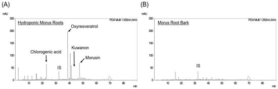
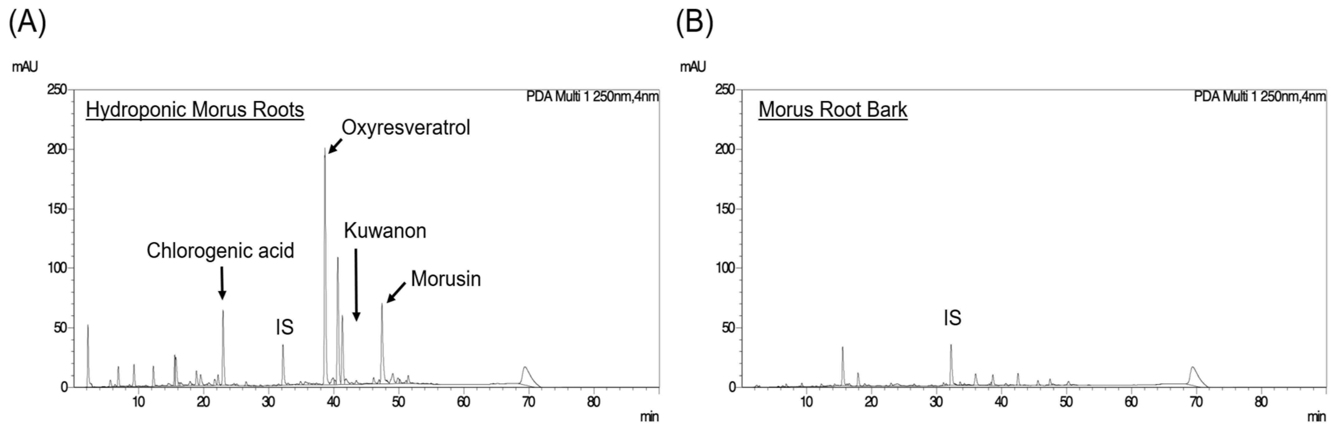
To assess the reproducibility of the phytochemical profile previously reported by our group [6], we performed HPLC analysis of an independently produced lot of hydroponically cultivated mulberry fibrous roots (Morus alba L.) (Figure 1A). Consistent with our previous findings, oxyresveratrol, chlorogenic acid, and morusin were detected and quantified in this independently produced lot. Although oxyresveratrol showed the most prominent peak at 250 nm, quantitative analysis using compound-specific calibration curves indicated that chlorogenic acid was the most abundant of the measured polyphenols, reflecting differences in UV absorbance among compounds.
Figure 1.
HPLC chromatograms of aqueous ethanol extracts of hydroponically cultivated mulberry fibrous roots and Morus root bark. Butylene glycol extracts were analyzed by reverse-phase HPLC using a gradient of 0.1% formic acid in water and 0.1% formic acid in methanol. (A) Hydroponically cultivated mulberry fibrous roots (B) Morus root bark. The peak labeled IS corresponds to diosmetin, which was used as the internal standard.
Quantitative comparison showed that the oxyresveratrol content was approximately 77-fold higher in the hydroponically cultivated mulberry fibrous roots than in the Morus root bark, and differences were also observed in the levels of the other polyphenols examined (Table 1). Because the two materials differed in both cultivation conditions and the part of the root analyzed, these differences cannot be attributed specifically to hydroponic cultivation. Nevertheless, the present analysis confirmed the characteristic quantitative profile of these four polyphenols in the hydroponically cultivated mulberry fibrous roots described in our previous report [6]. Although their absolute amounts varied among production lots, the same overall quantitative pattern was consistently observed across the lots examined.
Table 1.
Quantitative comparison of selected polyphenolic constituents in hydroponically cultivated mulberry fibrous roots and from Morus root bark.
A separate production lot was used in the subsequent animal experiments. Because the absolute oxyresveratrol content varied among lots, the oxyresveratrol concentration of the lot used for the animal studies was measured independently and used to determine the administered dose.
3.2. Hydroponically Cultivated Mulberry Fibrous Root Extract Attenuates Allergic Responses In Vivo
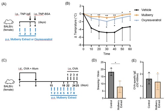
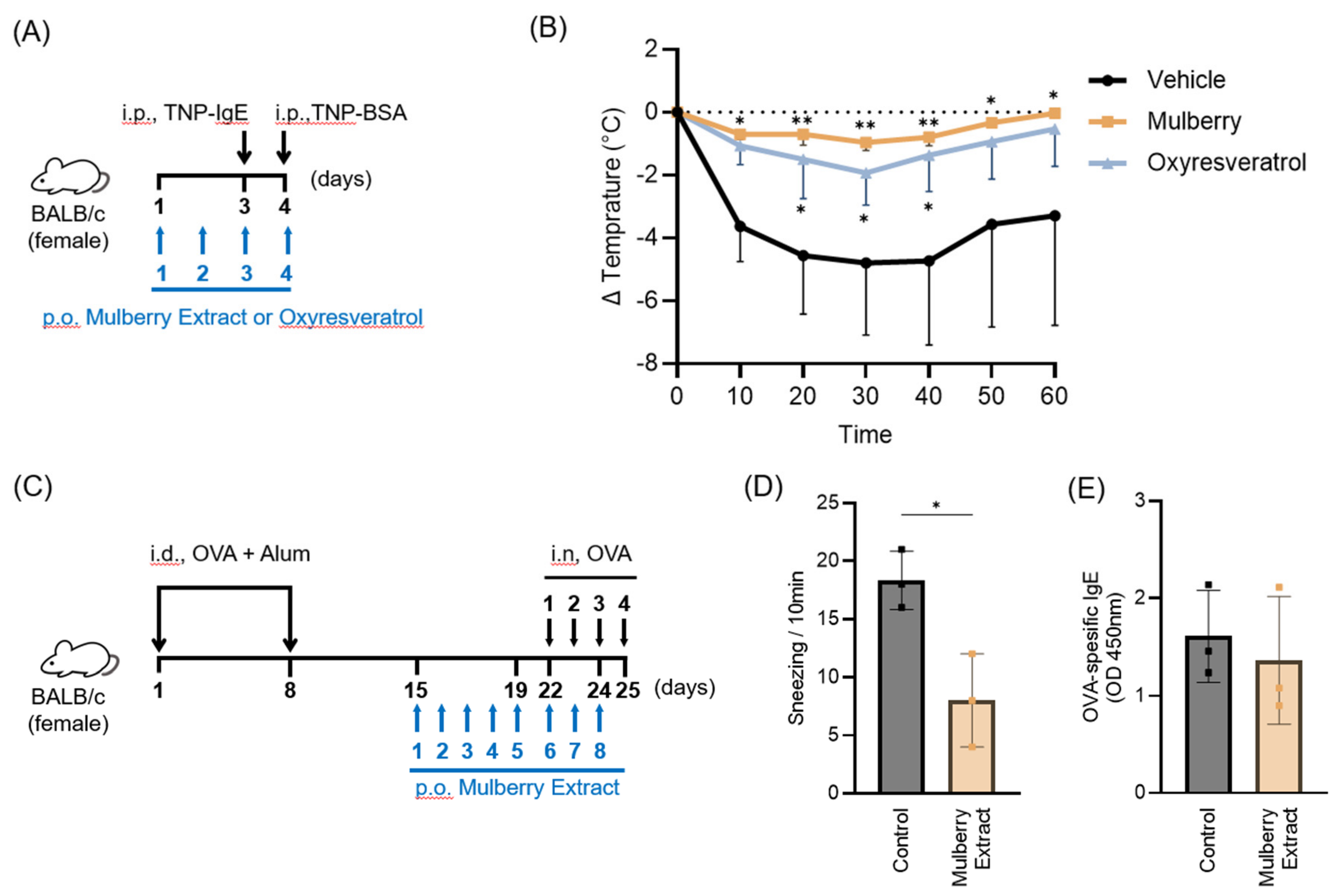
We next examined whether hydroponically cultivated mulberry fibrous root extract affects IgE/mast cell-mediated allergic responses in vivo using a passive systemic anaphylaxis (PSA) model in mice (Figure 2A). Oral administration of the extract at an amount equivalent to 7.5 mg of dried fibrous root material per mouse and containing 67.6 μg of oxyresveratrol in 300 μL of 25% butylene glycol significantly attenuated anaphylaxis-associated hypothermia compared with vehicle treatment (Figure 2B). Administration of an equivalent amount of purified oxyresveratrol (67.6 μg/mouse) also attenuated the decrease in body temperature (Figure 2B).
Figure 2.
Hydroponically cultivated mulberry fibrous root extract attenuates allergic responses in vivo. (A) Experimental scheme of the PSA mouse model. (B) Changes in core body temperature following TNP-BSA challenge in mice treated with hydroponically cultivated mulberry extract or oxyresveratrol. (C) Experimental scheme of the OVA-induced allergic rhinitis mouse model. (D) Number of sneezing episodes during the 10 min period following the final intranasal allergen challenge. (E) Serum levels of OVA-specific IgE. Data are presented as mean ± SD (n = 3). * p ≤ 0.05; ** p ≤ 0.01; Statistical analysis was performed using two-way repeated-measures ANOVA followed by Šídák’s multiple-comparisons test comparing each treatment group with the vehicle group (B) and unpaired t-tests (D,E).
To further evaluate the anti-allergic activity of the extract, we employed an ovalbumin (OVA)-induced allergic rhinitis model, another model involving IgE-mediated mast cell activation (Figure 2C). Oral administration of the same amount of the fibrous root extract, equivalent to 7.5 mg of dried fibrous root material per mouse and containing 67.6 μg of oxyresveratrol, significantly reduced the number of sneezes following intranasal OVA challenge (Figure 2D). In contrast, serum OVA-specific IgE levels were not significantly affected by extract treatment (Figure 2E).
Taken together, these findings demonstrate that hydroponically cultivated mulberry fibrous root extract attenuates allergic responses in two mouse models involving IgE-mediated mast cell activation. The finding that purified oxyresveratrol also attenuated hypothermia in the PSA model is consistent with the possibility that this polyphenol contributes, at least in part, to the anti-allergic activity of the extract.
3.3. Oxyresveratrol Suppresses IgE-Mediated Mast Cell Activation In Vitro
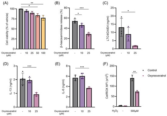
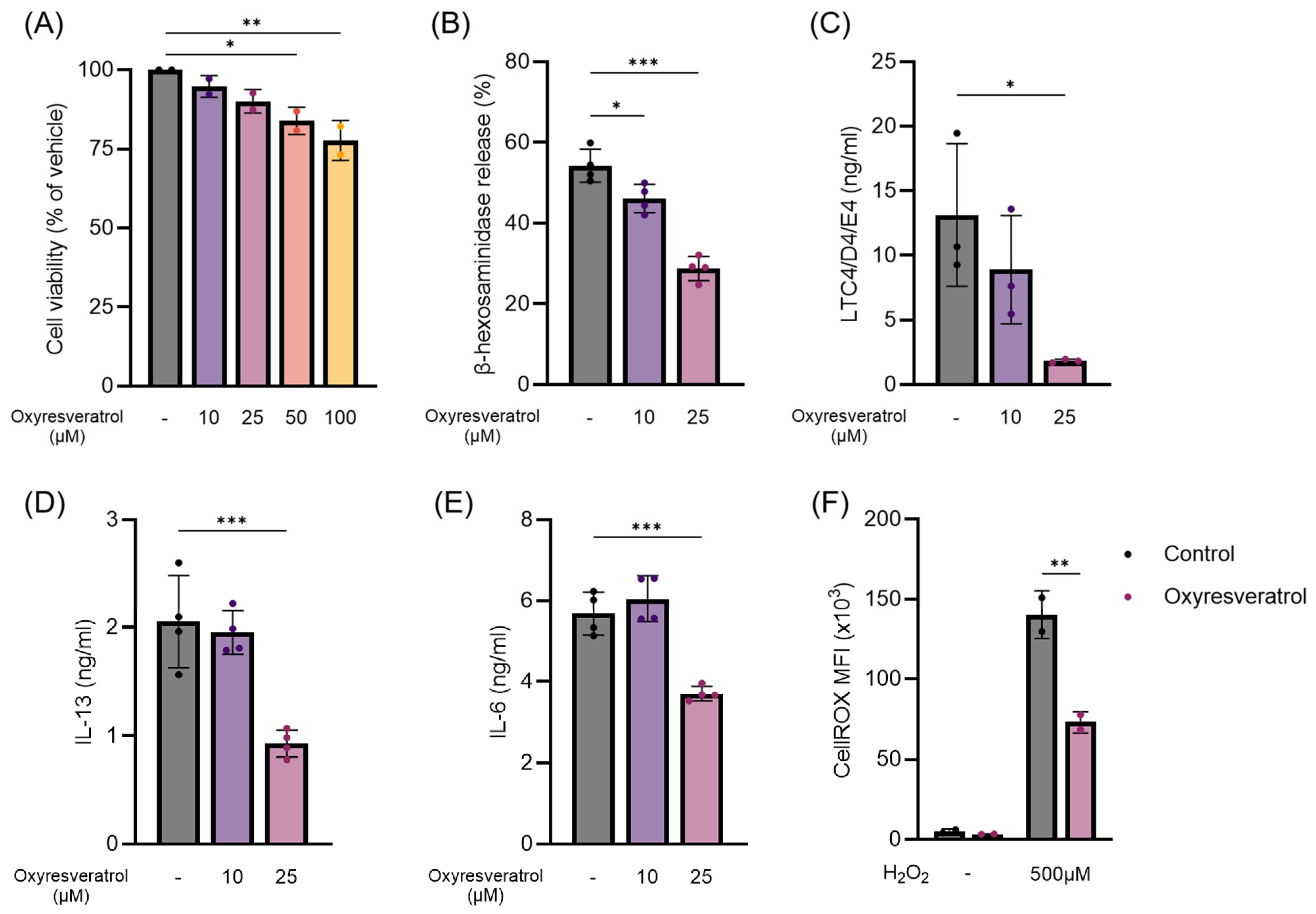
Based on the oxyresveratrol content of the hydroponically cultivated mulberry fibrous root extract and its previously reported anti-allergic properties [11], we next investigated the direct effects of oxyresveratrol on mast cell activation using mouse bone marrow-derived mast cells (BMMCs). As treatment with 50 and 100 μM oxyresveratrol reduced cell viability (Figure 3A), subsequent experiments were conducted using non-cytotoxic concentrations of 10 and 25 μM.
Figure 3.
Oxyresveratrol inhibits IgE-mediated mast cell activation in vitro. (A) Cell viability of BMMCs treated with the indicated concentrations of oxyresveratrol for 24 h. (B) β-Hexosaminidase release from BMMCs stimulated with anti-IgE antibody for 40 min in the presence or absence of oxyresveratrol. (C) Cysteinyl leukotriene (LTC4/D4/E4), (D) IL-13 and (E) IL-6 production by BMMCs stimulated with anti-IgE antibody for 6 h in the presence or absence of oxyresveratrol. (F) BMMCs were pretreated with 25 μM oxyresveratrol and subsequently stimulated with 500 μM hydrogen peroxide for 5 min. Intracellular ROS levels were assessed by measuring the mean fluorescence intensity (MFI) of CellROX. Data are presented as mean ± SD (n = 2–4). * p ≤ 0.05; ** p ≤ 0.01; *** p ≤ 0.001. Statistical analyses were performed using one-way ANOVA (A–E) and two-way ANOVA (F).
Pretreatment of BMMCs with oxyresveratrol significantly suppressed IgE-mediated degranulation, as assessed by β-hexosaminidase release, in a dose-dependent manner (Figure 3B). Consistent with this finding, oxyresveratrol significantly reduced the production of cysteinyl leukotrienes (LTC4/D4/E4), IL-13, and IL-6 following IgE-mediated activation (Figure 3C–E). Given the established contribution of reactive oxygen species (ROS) to FcεRI-mediated signaling and mast cell activation [18], we further examined the effects of oxyresveratrol on intracellular ROS generation. Oxyresveratrol significantly attenuated hydrogen peroxide-induced ROS accumulation in BMMCs (Figure 3F), supporting its antioxidant activity in these cells. These findings suggest that oxyresveratrol suppresses IgE-mediated mast cell activation in vitro. Further studies are required to determine whether ROS suppression directly contributes to its inhibitory effects on FcεRI-mediated mast cell activation.
4. Discussion
In the present study, we showed that (1) hydroponically cultivated mulberry fibrous root extract contained oxyresveratrol and exhibited a quantitative polyphenol profile distinct from that of Morus root bark, a traditionally known herbal medicine (Figure 1 and Table 1); (2) the hydroponically cultivated mulberry fibrous root extract attenuated allergic responses in two mouse models involving IgE-mediated mast cell activation, namely passive systemic anaphylaxis (PSA) and allergic rhinitis (Figure 2); and (3) oxyresveratrol directly inhibited IgE-mediated mast cell activation in vitro (Figure 3). Together, these findings indicate that hydroponically cultivated mulberry fibrous root extract possesses anti-allergic activity and suggest that oxyresveratrol may be one of the constituents contributing to this activity through the suppression of IgE-mediated mast cell activation.
Although the anti-allergic effects of oxyresveratrol and mulberry leaves have been reported previously [11,19], the anti-allergic properties of hydroponically cultivated mulberry fibrous roots have not been well characterized. To our knowledge, this is the first study to show that an extract from hydroponically cultivated mulberry fibrous roots containing oxyresveratrol attenuates IgE/mast cell-mediated allergic responses in vivo. These findings identify hydroponically cultivated mulberry fibrous roots as a potential source of bioactive polyphenols with anti-allergic properties.
Purified oxyresveratrol attenuated PSA to an extent similar to that observed with the hydroponically cultivated mulberry fibrous root extract. However, in addition to oxyresveratrol, the hydroponically cultivated mulberry fibrous root extract contained other polyphenols, including chlorogenic acid and morusin, both of which have been reported to exert anti-allergic effects [20,21,22]. Although their estimated amounts in the administered extract were lower than the doses or concentrations examined in previous studies, their contribution to the observed anti-allergic effects cannot be excluded.
Several limitations of this study should be acknowledged. First, the molecular mechanisms by which the hydroponically cultivated mulberry fibrous root extract and oxyresveratrol suppress IgE-mediated mast cell activation remain incompletely understood. Second, although the polyphenol contents of the hydroponically cultivated fibrous root extract and Morous root bark extract were quantitatively compared, their anti-allergic activities were not directly compared. Third, purified oxyresveratrol was not evaluated in the allergic rhinitis model, limiting our ability to determine its specific contribution to the protective effect of the extract in this model. Fourth, because this was an exploratory proof-of-concept study, no formal a priori sample-size calculation was performed, and the number of animals in each experimental group was small. The limited sample size may reduce the precision and generalizability of the estimated treatment effects. In addition, only female BALB/c mice were examined; therefore, whether these findings extend to male mice, other mouse strains, or humans remains unknown. Nevertheless, inhibitory effects were observed in both in vivo and in vitro experimental settings. Future studies using larger sample sizes and directly comparable plant materials are required to validate these findings and clarify the influence of cultivation conditions on polyphenol composition and anti-allergic activity.
5. Conclusions
Collectively, our findings show the anti-allergic activity of hydroponically cultivated mulberry fibrous root extract in experimental models and suggest that oxyresveratrol contributes, at least in part, to these effects. These findings support further investigation of the extract as a potential functional food ingredient.
6. Patents
Regarding intellectual property and commercialization, there are no granted patents or pending patent applications related to this study, including the hydroponic cultivation system or the resulting mulberry product.
Supplementary Materials
The following supporting information can be downloaded at: https://www.mdpi.com/article/10.3390/nutraceuticals6040068/s1. Table S1: The individual data analysis.
Author Contributions
The study was designed by Y.K., T.K., T.F. and A.N., Y.K., K.N., N.Q.V.T. and Y.N. performed the experiments and analyzed the data. T.K., T.F. and A.N. supervised experimental procedures and study concepts. Y.K., T.K. and A.N. wrote the manuscript and A.N. finalized the manuscript. All authors have read and agreed to the published version of the manuscript.
Funding
This research was funded in part by a grant from Morera Co., Ltd. to A.N. The funder had no role in the analysis or interpretation of the data, preparation of the manuscript, or decision to submit the manuscript for publication.
Informed Consent Statement
Not applicable.
Data Availability Statement
The data that support the findings of this study are available on request from the corresponding author.
Acknowledgments
The authors thank Yukino Fukasawa and Maiko Aihara for their valuable assistance. During the preparation of this manuscript, the authors used ChatGPT 5.2 for the purposes of improving language and readability. The authors have reviewed and edited the output and take full responsibility for the content of this publication.
Conflicts of Interest
Koki Nishime is employed by Kankyo-Hozen Labortory Inc. (Yamanashi, Japan). Dr. Tetsuya Konishi is employed by Morera Co., Ltd. (Nagano, Japan). Tomoko Fukushima is the president of Morera Co., Ltd. (Nagano, Japan). The remaining authors declare no conflicts of interest. Employees of the funder participated in the study design, data analysis, and manuscript writing, as detailed in the Author Contributions section. The funder has been collaborating with Dr. Nakao to promote and disseminate scientific knowledge regarding hydroponic cultivation. The primary aim of the present study was to investigate the potential biomedical benefits of hydroponic cultivation, rather than to develop or commercialize the hydroponic system or the resulting mulberry product. Neither Dr. Konishi nor Morera Co., Ltd., nor any of the other authors, has any financial interest in the commercialization of the hydroponic system or the resulting mulberry product. We would also like to clarify that all raw data used in this study were generated and obtained by the academic investigators. The academic investigators had unrestricted access to all raw data and full independence in the analysis and interpretation of the data, as well as in the decision to prepare and submit the manuscript for publication. The funder had no control over the analysis, interpretation, or publication of the results.
References
- Chan, E.W.; Phui-Yan, L.Y.; Siu-Kuin, W.O. Phytochemistry, pharmacology, and clinical trials of Morus alba. Chin. J. Nat. Med. 2016, 14, 17–30. [Google Scholar] [PubMed]
- Fatima, M.; Dar, M.A.; Dhanavade, M.J.; Abbas, S.Z.; Bukhari, M.N.; Arsalan, A.; Liao, Y.; Wan, J.; Shah Syed Bukhari, J.; Ouyang, Z. Biosynthesis and Pharmacological Activities of the Bioactive Compounds of White Mulberry (Morus alba): Current Paradigms and Future Challenges. Biology 2024, 13, 506. [Google Scholar] [CrossRef] [Scilit] [PubMed]
- Wu, Y.-X.; Kim, Y.J.; Kwon, T.H.; Tan, C.P.; Son, K.H.; Kim, T. Anti-inflammatory effects of mulberry (Morus alba L) root bark and its active components. Nat. Prod. Res. 2020, 34, 1786–1790. [Google Scholar] [CrossRef] [Scilit] [PubMed]
- Langeder, J.; Koch, M.; Schmietendorf, H.; Tahir, A.; Grienke, U.; Rollinger, J.M.; Schmidtke, M. Correlation of bioactive marker compounds of an orally applied Morus alba root bark extract with toxicity and efficacy in BALB/c mice. Front. Pharmacol. 2023, 14, 1193118. [Google Scholar] [CrossRef] [Scilit] [PubMed]
- Kavitha, Y.; Geetha, A. Anti-inflammatory and preventive activity of white mulberry root bark extract in an experimental model of pancreatitis. J. Tradit. Complement. Med. 2018, 8, 497–505. [Google Scholar] [CrossRef] [Scilit] [PubMed]
- Sakurai, M.; Sato, S.; Fukushima, T.; Konishi, T. Characteristics of Morus alba L. Cultured by In-Room Hydroponics. Am. J. Plant Sci. 2022, 13, 91–108. [Google Scholar]
- Galli, S.J.; Gaudenzio, N.; Tsai, M. Mast Cells in Inflammation and Disease: Recent Progress and Ongoing Concerns. Annu. Rev. Immunol. 2020, 38, 49–77. [Google Scholar] [CrossRef] [Scilit] [PubMed]
- Dispenza, M.C.; Metcalfe, D.D.; Olivera, A. Research Advances in Mast Cell Biology and Their Translation Into Novel Therapies for Anaphylaxis. J. Allergy Clin. Immunol. Pract. 2023, 11, 2032–2042. [Google Scholar] [CrossRef] [Scilit] [PubMed]
- Li, Y.; Prabhakaran, P.; Wang, Y.; Li, D.; Sun-Waterhouse, D.; Li, F. Insights into food allergy mechanisms and the anti-allergic potential of natural polyphenols in regulating FcεRI-mediated mast cell degranulation. Food Chem. 2025, 490, 145099. [Google Scholar] [CrossRef] [Scilit] [PubMed]
- Likhitwitayawuid, K. Oxyresveratrol: Sources, Productions, Biological Activities, Pharmacokinetics, and Delivery Systems. Molecules 2021, 26, 4212. [Google Scholar] [CrossRef] [Scilit] [PubMed]
- Ashraf, M.I.; Shahzad, M.; Shabbir, A. Oxyresveratrol ameliorates allergic airway inflammation via attenuation of IL-4, IL-5, and IL-13 expression levels. Cytokine 2015, 76, 375–381. [Google Scholar] [CrossRef] [Scilit] [PubMed]
- Li, P.; Hon, S.S.-M.; Tsang, M.S.-M.; Kan, L.L.-Y.; Lai, A.Y.-T.; Chan, B.C.-L.; Leung, P.-C.; Wong, C.-K. Integrating 16S rRNA Sequencing, Microflora Metabolism, and Network Pharmacology to Investigate the Mechanism of SBL in Alleviating HDM-Induced Allergic Rhinitis. Int. J. Mol. Sci. 2024, 25, 8655. [Google Scholar] [CrossRef] [Scilit] [PubMed]
- Rui, X.; Ruan, S.; Zhang, Y.; Fu, R.; Sun, P.; Lamu, D.; Wang, W. PPARgamma Modulates CD4+ T-Cell Differentiation and Allergic Inflammation in Allergic Rhinitis: A Potential Therapeutic Target. Biomedicines 2025, 13, 1616. [Google Scholar] [CrossRef] [Scilit] [PubMed]
- Li, P.; Tsang, M.S.-M.; Kan, L.L.-Y.; Hou, T.; Hon, S.S.-M.; Chan, B.C.-L.; Chu, I.M.-T.; Lam, C.W.-K.; Leung, P.-C.; Wong, C.-K. The Immuno-Modulatory Activities of Pentaherbs Formula on Ovalbumin-Induced Allergic Rhinitis Mice via the Activation of Th1 and Treg Cells and Inhibition of Th2 and Th17 Cells. Molecules 2022, 27, 239. [Google Scholar] [CrossRef] [Scilit] [PubMed]
- Nakamura, Y.; Ishimaru, K.; Tahara, Y.; Shibata, S.; Nakao, A. Disruption of the suprachiasmatic nucleus blunts a time of day-dependent variation in systemic anaphylactic reaction in mice. J. Immunol. Res. 2014, 474217. [Google Scholar] [CrossRef] [Scilit] [PubMed]
- Miyata, M.; Hatsushika, K.; Ando, T.; Shimokawa, N.; Ohnuma, Y.; Katoh, R.; Suto, H.; Ogawa, H.; Masuyama, K.; Nakao, A. Mast cell regulation of epithelial TSLP expression plays an important role in the development of allergic rhinitis. Eur. J. Immunol. 2008, 38, 1487–1492. [Google Scholar] [CrossRef] [Scilit] [PubMed]
- Kobayashi, Y.; Sakai, K.; Tran, N.Q.V.; Ishimaru, K.; Sato, T.; Nakamura, Y.; Nakagomi, D.; Tanaka, S.; Koizumi, S.; Nakao, A. IL-33 Sensitizes Mast Cells to PIEZO1 Stimulation Leading to Degranulation. Allergy 2024, 79, 3517–3520. [Google Scholar] [CrossRef] [Scilit] [PubMed]
- Chelombitko, M.A.; Chernyak, B.V.; Fedorov, A.V.; Zinovkin, R.A.; Razin, E.; Paruchuru, L.B. The Role Played by Mitochondria in FcεRI-Dependent Mast Cell Activation. Front. Immunol. 2020, 11, 584210. [Google Scholar] [CrossRef] [Scilit] [PubMed]
- Jiang, S.; Luo, T.; Huang, Y.; Yang, Z.; Yang, Z.; Meng, X. Mulberry leaf polyphenols alleviate food allergy and moderate intestinal flora and lipid metabolism in allergic mice. J. Future Foods, 2026; in press.
- Shi, Z.; Jiang, W.; Chen, X.; Xu, M.; Wang, J.; Lai, Y.; Zha, D. Chlorogenic acid ameliorated allergic rhinitis-related symptoms in mice by regulating Th17 cells. Biosci. Rep. 2020, 40, BSR20201643. [Google Scholar] [CrossRef] [Scilit] [PubMed]
- Qin, H.D.; Shi, Y.Q.; Liu, Z.H.; Li, Z.G.; Wang, H.S.; Wang, H.; Liu, Z.P. Effect of chlorogenic acid on mast cell-dependent anaphylactic reaction. Int. Immunopharmacol. 2010, 10, 1135–1141. [Google Scholar] [CrossRef] [Scilit] [PubMed]
- Jin, S.E.; Ha, H.; Shin, H.-K.; Seo, C.-S. Anti-Allergic and Anti-Inflammatory Effects of Kuwanon G and Morusin on MC/9 Mast Cells and HaCaT Keratinocytes. Molecules 2019, 24, 265. [Google Scholar] [CrossRef] [Scilit] [PubMed]

Disclaimer/Publisher’s Note: The statements, opinions and data contained in all publications are solely those of the individual author(s) and contributor(s) and not of MDPI and/or the editor(s). MDPI and/or the editor(s) disclaim responsibility for any injury to people or property resulting from any ideas, methods, instructions or products referred to in the content. |
© 2026 by the authors. Licensee MDPI, Basel, Switzerland. This article is an open access article distributed under the terms and conditions of the Creative Commons Attribution (CC BY) license.