


Abstract
Background/Objectives: Stevia rebaudiana Bertoni, a perennial plant of the Asteraceae family, is widely used as a natural sweetener owing to its steviol glycosides, but it also contains phenolic compounds and diterpenes reported to exhibit antioxidant, anti-inflammatory and antiproliferative effects. Heterogeneous treatment protocols and the use of different cell viability assays, however, hinder reliable cross-study comparison of their antiproliferative potential. This systematic review and meta-analysis assesses the in vitro antiproliferative activity of stevia constituents and crude extracts across diverse cancer cell lines based on cell viability assay data. Methods: A comprehensive literature search was performed following PRISMA guidelines. Data were synthesized through meta-analysis and meta-regression using IC50 values as the effect size. Key variables included assay type (MTT, SRB), incubation time (8, 24, 48, 72 h), constituent class (phenols, glycosides, diterpenes, crude extracts) and cancer cell type. Results: Of the 250 articles initially identified, seven met eligibility criteria, yielding 39 effect estimate datasets. A meta-regression of IC50 values across MTT and SRB assays revealed no statistically significant differences at 24 h (p = 0.080) and 48 h (p = 0.456), suggesting that values from the two assays are broadly comparable. Antiproliferative potency increased with incubation time, with IC50 decreasing from 3481.20 μg/mL at 8 h to 125.22 μg/mL at 72 h. Among constituent classes, phenols showed the highest potency (IC50 = 97.15 μg/mL at 48 h), exceeding that of glycosides (229.73 μg/mL) and diterpenes (171.55 μg/mL). Neuronal cancer cells were the most sensitive (IC50 = 21.00 μg/mL at 48 h), markedly more than breast (58.90 μg/mL) and gynecological (238.35 μg/mL) cancer cells. Conclusions: Stevia constituents, and particularly phenols, demonstrate promising in vitro antiproliferative activity that is both dose- and time-dependent. Furthermore, the lack of a significant difference between MTT- and SRB-derived results supports their combination in meta-analytical syntheses of antiproliferative data.
1. Introduction
Stevia rebaudiana Bertoni, commonly known as stevia, is a perennial plant belonging to the Asteraceae family, native to South America. Beyond its widespread use as a natural sweetener due to the presence of steviol glycosides, stevia has attracted considerable scientific interest due to its diverse biological activities, including antioxidant, anti-inflammatory, antimicrobial, and anticancer effects [1,2,3]. The primary bioactive constituents of stevia crude leaf extracts, besides the steviol glycosides stevioside and rebaudioside A, are phenolic compounds including flavonoids and phenolic acids, and diterpenes such as steviol and isosteviol [4]. Stevia leaf extracts have been reported to exhibit anticancer effects by targeting multiple stages of carcinogenesis, such as initiation and promotion [1,5]. Several mechanisms have been proposed in studies conducted on various cancer cell lines to explain how stevia may exert its anticancer action, including antioxidant activity via neutralization of reactive oxygen species (ROS), induction of apoptosis through caspase activation, cell cycle arrest at the G0/G1 phase, inhibition of DNA synthesis, and enhancement of antioxidant enzyme activity [2].
The diversity of stevia compounds and extracts tested across numerous cancer cell lines necessitates a rigorous quantitative assessment of their antiproliferative effects. However, significant variations in treatment protocols regarding compound concentrations and incubation periods complicate comparisons across studies. These methodological inconsistencies currently preclude a reliable evaluation of the relative efficacy of various stevia constituents in both in vitro and in vivo experimental models [6].
Another parameter that further shapes the complexity arises from the wide range of cell viability and antiproliferative assays employed to assess the biological effects of stevia-derived compounds. Researchers often rely on different biochemical principles to evaluate the survival of cancer cells post-treatment, which can lead to divergent results. Among the most commonly used methods is the Sulforhodamine B (SRB) assay, which estimates cell biomass through the binding of the SRB dye to basic amino acids of cellular proteins, with absorbance measured at 490–510 nm. Although protein content is present in both living and dead cells, dead and apoptotic cells progressively lose their adhesion to the culture surface and detach into the medium. Because the assay protocol involves removal of the medium prior to fixation, non-adherent dead cells are effectively excluded. As a result, the SRB signal reflects viable, adherent cell biomass [7]. The MTT assay is based on the reduction of tetrazolium salts by mitochondrial dehydrogenases in metabolically active cells, leading to the formation of insoluble formazan crystals quantified spectrophotometrically at 570 nm; since this enzymatic reduction depends on active cellular metabolism, it occurs exclusively in living cells, as dead cells lacking functional dehydrogenases do not contribute to the signal [8]. Because these assays rely on different biochemical and metabolic principles, they may differentially reflect cellular metabolic activity, viability, cytotoxicity, or proliferation. Furthermore, treatment duration, typically ranging from 8 to 72 h, may influence whether the measured response primarily reflects acute cytotoxicity, reduced cell viability, or antiproliferative activity. The outcomes of these assays are commonly reported as IC50 (half-maximal inhibitory concentration) values, defined as the concentration of a compound required to reduce the assay signal by 50% relative to untreated controls [9].
The emerging interest in specific natural compounds and medicinal plant extracts with potential health benefits underscores the need to systematically synthesize, compare, and summarize the available data regarding the antiproliferative activity of stevia-derived constituents. This can be effectively achieved through a systematic review and meta-analysis, ensuring statistical rigor and comprehensive evaluation [10]. Meta-analysis constitutes a component of evidence-based scientific practice and systematic review methodology, integrating results from multiple studies to identify methodological patterns and sources of variability across scientific disciplines [11]. Such approaches have become increasingly important in biomedical research in response to concerns regarding reproducibility, transparency, and methodological standardization [12]. Notably, a meta-regression–based approach [13] has previously proven effective for integrating heterogeneous assay data on plant-derived compounds, both for the antioxidant activity of stevia leaf extracts [3] and for the antiproliferative activity of hop-derived compounds across diverse cancer cell lines [14].
The objective of the present study is to statistically synthesize the existing body of literature to evaluate the antiproliferative potential of various stevia constituents across a comprehensive spectrum of cancer cell lines. This meta-analysis seeks to quantitatively integrate available evidence and establish a clear summary of the effects observed. Furthermore, by employing meta-regression, this analysis explores whether data derived from different cell viability assays can be combined for cross-study comparisons in stevia research, while acknowledging the different chemical principles underlying these assays.
2. Materials and Methods
2.1. Literature Search Strategy and Selection Criteria
An extensive literature search was conducted in the PubMed (https://pubmed.ncbi.nlm.nih.gov/ accessed on 30 December 2025) and Scopus (https://www.scopus.com/ accessed on 30 December 2025) databases to retrieve potentially all research articles evaluating the antiproliferative effects of stevia compounds on cancer cell lines. The last search was performed on 30 December 2025 using the keywords “Stevia” AND (“steviol” OR “glycosides” OR “Phenols” OR “flavonoids” OR “terpenoids” OR “tannins”) AND (“cancer” OR “carcinoma” OR “anticancer” OR “tumor” OR “antitumor” OR “proliferation” OR “antiproliferative” OR “antineoplastic” OR “apoptosis”) according to the guidelines and reporting standards for systematic reviews and meta-analyses (PRISMA, Preferred Reporting Items for Systematic Reviews and Meta-Analyses) (http://www.prisma-statement.org/ (accessed on 25 January 2026)) and best practices [15,16]. The completed PRISMA 2020 checklists are provided in the Supplementary Materials (Tables S1 and S2). In order to minimize the likelihood of publication bias and the effects of gray literature, articles written in languages other than English were not excluded [17].
The review protocol was registered in OSF Registries (https://osf.io/9erc7, accessed on 28 June 2026; DOI: 10.17605/OSF.IO/9ERC7).
The eligibility criteria for inclusion of articles in the meta-analysis were studies that evaluated cell viability through SRB and MTT assays expressed as IC50 values, in various cancer or non-cancerous cell lines following treatment with stevia extracts or chemical compounds for various incubation times, including 8, 24, 48 and 72 h. Studies that did not provide sufficient or reliable information and data for conducting the meta-analysis were excluded to ensure the validity of the results and the reliability of conclusions [18].
Initially, article titles and abstracts were screened, and relevant articles were further examined for adherence to inclusion or exclusion criteria. The results of the literature search were evaluated by two independent researchers (M.P. and A.T.), while any disagreements that arose during the search process were resolved through discussion with three other researchers (G.G.B., P.G.B. and P.I.K.), and article categorization was decided by consensus.
2.2. Data Extraction
Data extraction was performed on a predetermined Microsoft Excel® (Microsoft Corporation, Redmond, WA, USA) sheet. From each study, the following information was recorded: surname of the first author, year of publication, assay type, IC50 values along with standard error of the mean (SEM), incubation time, cell line, cell type, cancer or non-cancerous model, and the chemical classification of the tested compound or stevia extract used.
In studies where standard deviation (SD) was reported, the following formula was used to determine the standard error:
where n is the number of replicates. The omission of studies without reported standard deviations (SDs) can introduce selection bias, as their effect estimates remain informative. Furukawa et al. [19] showed that SDs for identical outcomes rarely vary substantially, supporting variance imputation when data are missing. To remain conservative, we replaced missing values with the highest observed SD among studies evaluating the same compound–cell line pair. This keeps all available studies in the meta-analysis while appropriately down-weighting those with imputed variance [19].
SEM = SD/√n
2.3. Statistical Analysis
The meta-analysis was performed according to the DerSimonian-Laird random effects meta-analysis model [20] in order to synthesize the results of all studies and account for potential heterogeneity between them due to differences in extraction procedures, types of cancer cell lines, and various antiproliferative activity assays. The weighted mean of IC50 values, expressed in μg/mL units with 95% confidence intervals, was used as the effect size of the meta-analysis. Where purified compounds were originally reported in molar units, these values were converted to μg/mL using the corresponding molecular weights in order to express all effect estimates on a single, common scale. Although molar units (μM) would be preferable for purified compounds, a mass-based scale was adopted because it was the only unit applicable to crude extracts, for which a defined mean molecular weight is not available; this is acknowledged as a limitation.
Additionally, a meta-regression analysis was conducted to compare IC50 estimates across different assay types and to assess whether assay type was associated with effect size [21]. Heterogeneity between studies was assessed using the I2 index and estimation of τ2 based on Cochran’s Q test [22]. To assess the existence of potential publication bias, the Egger test was used [23,24]. Furthermore, subgroup stratification analysis was performed, categorizing studies according to their characteristics, such as assay type, incubation time, category of chemical compounds or type of stevia extract, cell line classification (cancerous or non-cancerous), and cancer type.
The meta-analysis was performed using Stata13 statistical software (StataCorp LP, College Station, TX, USA) [25,26] with the commands “metan”, “metareg”, “metabias”. Statistical significance was set at p-value ≤ 0.05. Meta-analysis was performed only when two or more studies were available.
3. Results
3.1. Study Selection and Characteristics
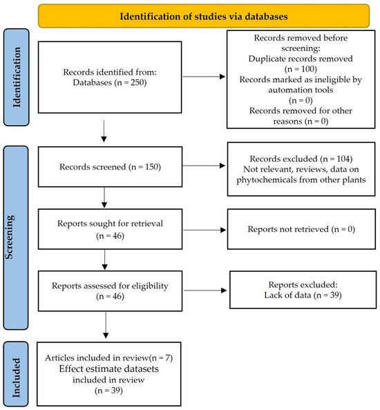
The extensive literature search for studies on antiproliferative activity of stevia compounds retrieved a total of 250 articles, using the search terms “Stevia” AND (“steviol” OR “glycosides” OR “Phenols” OR “flavonoids” OR “terpenoids” OR “tannins”) AND (“cancer” OR “carcinoma” OR “anticancer” OR “tumor” OR “antitumor” OR “proliferation” OR “antiproliferative” OR “antineoplastic” OR “apoptosis”). After screening the titles and abstracts of the articles according to eligibility criteria and compliance with PRISMA guidelines (http://www.prisma-statement.org/ (accessed on 25 January 2026)), seven articles [27,28,29,30,31,32,33] were identified that included 39 effect estimate datasets reporting on cell viability tests in various cancer or non-cancerous cell lines after treatment with stevia chemical compounds or extracts. The following PRISMA flow diagram graphically illustrates the article selection steps of the present systematic literature review (Figure 1).
Figure 1.
PRISMA (Preferred Reporting Items for Systematic Reviews and Meta-Analyses) flow diagram of the present study.
Among the 39 effect estimate datasets of the seven articles included in this systematic review, cell viability was predominantly assessed using the MTT assay (n = 36), followed by the SRB assay (n = 3). The majority of these effect estimate datasets (n = 33) focused on cancer cell lines, while six effect estimate datasets evaluated non-cancerous cell models. The identified cancer types spanned several categories: gastrointestinal (Caco-2, HCT-8, HCT-116, MKN-45, MGC-803, T84, and HT29), gynecological (HeLa), nervous system (C6 and SH-SY5Y), breast (MCF-7), and pancreatic (PaCa-2). Additionally, non-cancerous models included gastrointestinal lines (IEC-18), immortalized keratinocytes (HaCaT), and fibroblasts (NIH-3T3). Non-cancerous cell lines (HaCaT, NIH-3T3, CHO-K1 and IEC-18) were included to enable assessment of the selectivity of stevia constituents, that is, their capacity to inhibit cancer cell proliferation while sparing normal cells. Cancer and non-cancerous cell lines were analyzed separately in all subsequent analyses.
The present meta-analysis evaluates the effects of crude stevia leaf extracts in parallel with isolated phytochemical classes, specifically phenols, glycosides, and diterpenes, on cancer cell viability. This dual focus allows for a comprehensive comparison between the antiproliferative activity of the whole plant matrix and its individual bioactive components. To ensure methodological consistency, the meta-analysis synthesized data obtained at 8, 24, 48, and 72 h incubation time points. The characteristics and parameters of the included effect estimate datasets are systematically presented and delineated in Table 1, along with quantitative outcomes.
Table 1.
Characteristics of 39 effect estimate datasets included in the meta-analysis.
3.2. Meta-Analysis of Antiproliferative Activity Data of Stevia Constituents and Crude Extracts on Cancer Cells
3.2.1. Comparability of IC50 Values Across MTT and SRB Assays
To determine whether the choice of cell viability assay influenced the reported potency of stevia phytoconstituents, study-level estimates were stratified by assay type (MTT vs. SRB) across available exposure durations (Table 2). Significant statistical heterogeneity was observed across all subsets (I2 = 100%), justifying the application of random-effects models. Within each assay, assay-specific IC50 values decreased consistently with longer incubation, indicating higher potency at extended exposure. For instance, at 48 h the mean IC50 ranged between 58.9 and 151.34 μg/mL across assays, whereas at 8 h the value was substantially higher (3481.20 μg/mL in the MTT assay). By 72 h, a further decrease was observed, with the IC50 reaching 125.22 μg/mL in the MTT assay (Table 2).
Table 2.
Meta-analysis of IC50 from assays (MTT, SRB) according to incubation time (8, 24, 48, 72 h) in cancer cells.
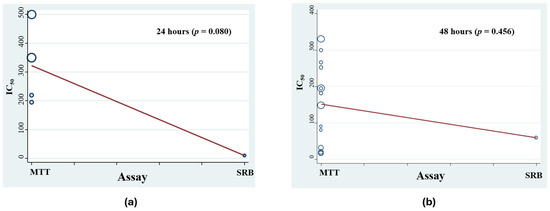
At time points where both assays were available (24 and 48 h), meta-regression analysis revealed no statistically significant differences in pooled IC50 values between MTT and SRB readouts (p = 0.080 at 24 h; p = 0.456 at 48 h) (Figure 2). Due to the limited representation of the SRB assay in the current literature (n = 1 dataset at 24 h and n = 1 at 48 h), these comparisons are exploratory and indicate broad numerical comparability rather than formal assay equivalence.
Figure 2.
Meta-regression of MTT and SRB assays for incubation times: (a) 24 h and (b) 48 h. The size of each circle represents the weight of each study.
Assessment of publication bias across the overall dataset using Egger’s test revealed no evidence of funnel plot asymmetry (p = 0.671).
3.2.2. Association Between Incubation Duration and IC50 Values
To establish an initial exploratory benchmark of overall antiproliferative capacity, datasets across all constituent categories and both assay types were pooled, followed by stratified analyses to dissect specific kinetic and chemical subgroups. In this pooled analysis (Table 3), summary IC50 estimates progressively decreased with longer exposure times, ranging from 3481.20 μg/mL (95% CI: 659.68, 6302.71) at 8 h (Table 2) to 270.88 μg/mL (95% CI: 168.01, 373.75) at 24 h, 145.17 μg/mL (95% CI: 86.82, 203.54) at 48 h, and 125.22 μg/mL (95% CI: 57.52, 192.91) at 72 h (Table 3).
Table 3.
Stratification meta-analysis of IC50 values for stevia-derived extracts and phytoconstituents across 24, 48, and 72 h incubation periods in cancer cell lines.
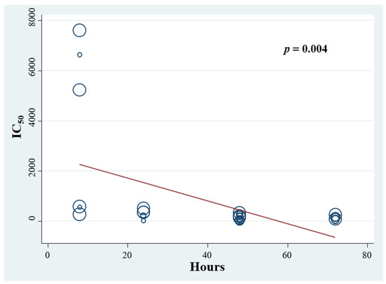
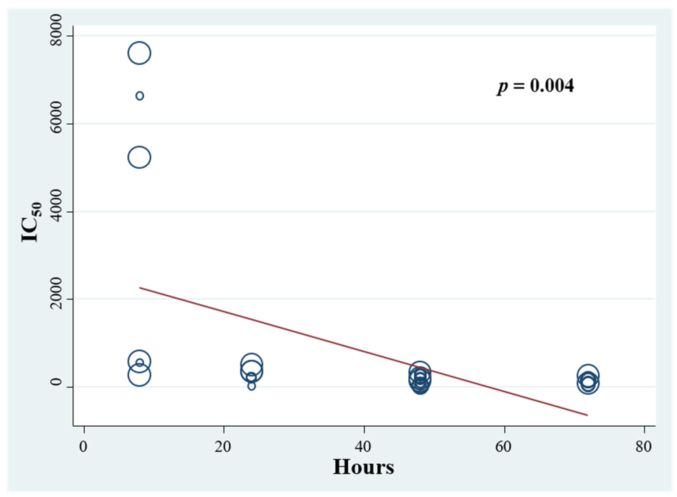
In an exploratory meta-regression across all time points, a significant negative association was observed between incubation duration and IC50 (β = −45.46 μg/mL per hour; 95% CI: −75.60, −15.33; p = 0.004) (Figure 3). Because incubation periods were predominantly segregated across individual publications—with all 8 h data derived from one article and all 72 h data from another—these cross-study comparisons remain exploratory and subject to study-level confounding (e.g., variations in cell lines, extract compositions, and assay conditions). Notably, within the single study evaluating multiple exposure intervals under uniform laboratory conditions (Chen et al. [29], 24 h vs. 48 h), IC50 values consistently declined across all tested lines, providing within-study support for this time-dependent pattern.
Figure 3.
Meta-regression analysis of IC50 values (obtained from MTT and SRB assays) for different incubation times (8, 24, 48 and 72 h).
3.2.3. Antiproliferative Potential of Stevia Phenols, Glycosides, Diterpenes and Crude Leaf Extracts
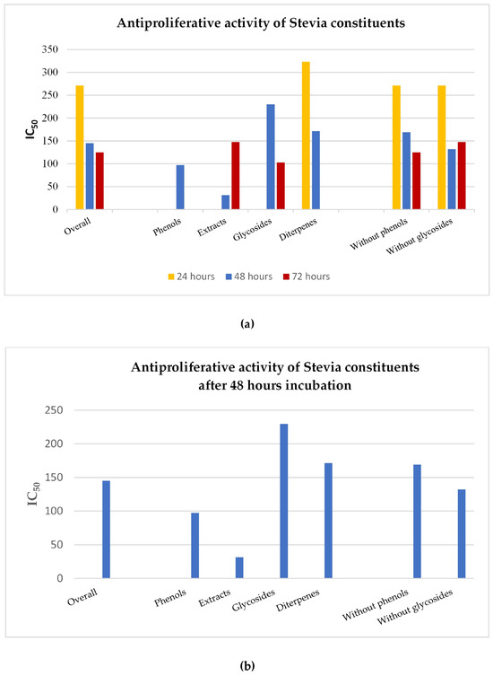
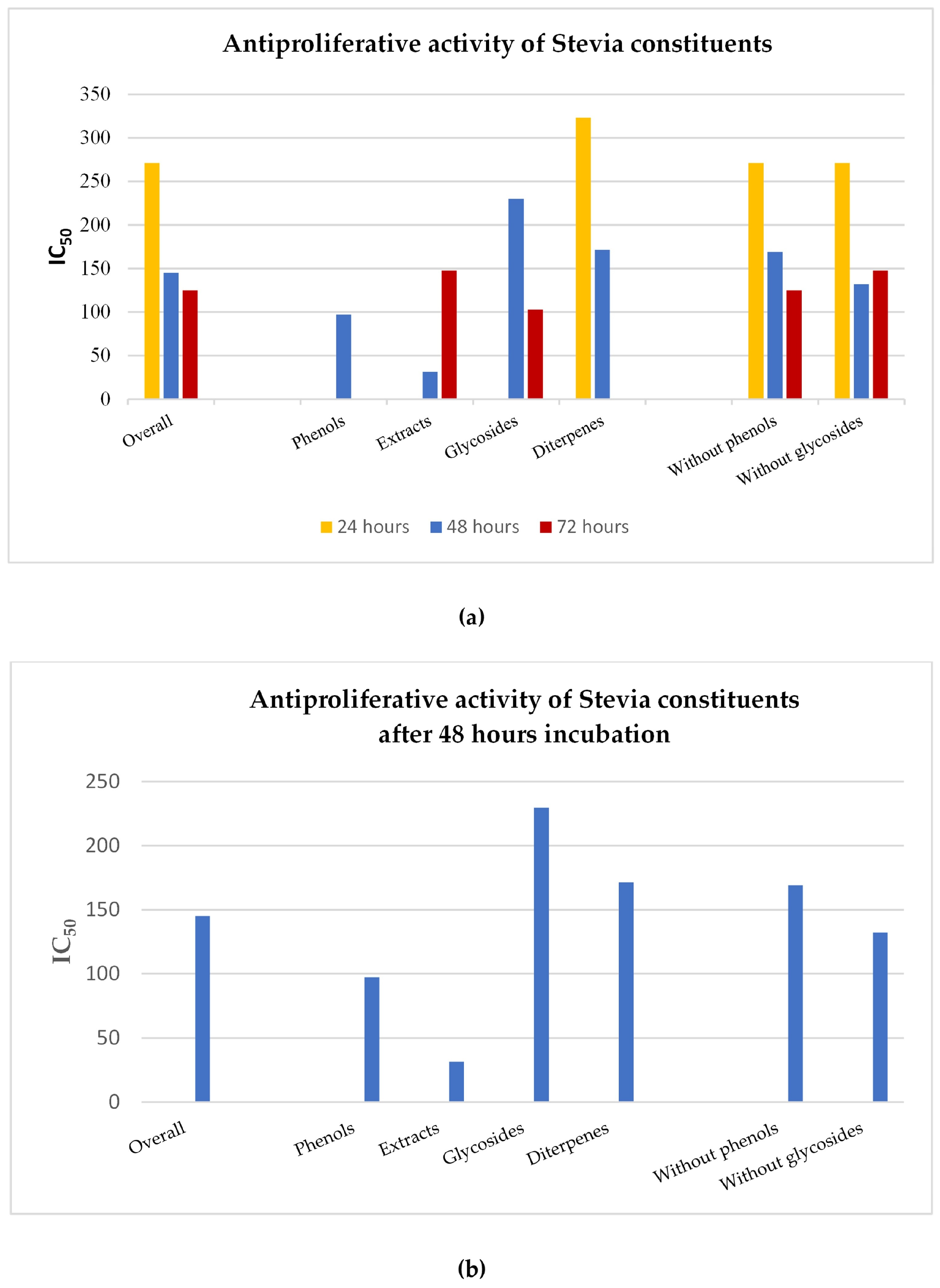
Subsequently, a stratification analysis was performed according to the groups of constituents (phenols, glycosides, diterpenes) and stevia extracts at incubation times of 24, 48 and 72 h (Table 3, Figure 4a). Fractions rich in phenolic compounds and flavonoids were grouped together under the “phenols” category, as flavonoids constitute a subclass of phenolic compounds.
Figure 4.
(a): IC50 of stevia constituents at three different incubation times (24, 48, 72 h), (b): at 48 h.
For the 48 h incubation, the pooled IC50 values were: for phenols 97.15 μg/mL (95% CI: 8.32, 186.28), for glycosides 229.73 μg/mL (95% CI: 160.48, 298.99), and for diterpenes 171.55 μg/mL (95% CI: 95.70, 247.41). For crude extracts at 48 h, only a single effect estimate was available (31.62 μg/mL), precluding pooled meta-analytic estimation and direct statistical comparison with isolated phytochemical classes. Accordingly, extracts were not considered in the potency ranking below. Among the constituent classes with multiple contributing datasets (n ≥ 2), phenolic compounds exhibited the highest antiproliferative potency, displaying markedly lower summary IC50 values than both diterpenes and glycosides (Table 3, Figure 4b).
A clearer picture regarding the contribution of phenolic constituents and glycosides to the antiproliferative activity of stevia emerged after stratification analysis. Specifically, a meta-analysis was performed on data from all groups except phenols (i.e., studies with data from extracts, glycosides, and diterpenes), thus creating the “Without phenols” group. Additionally, a meta-analysis was performed on all groups except glycosides (i.e., studies with data from phenols, extracts, and diterpenes), thus creating the “Without glycosides” group.
This analysis revealed that the absence of phenolic compounds showed a higher IC50 value (169.19 μg/mL; 95% CI: 103.62, 234.76) compared to the absence of glycosides (132.17 μg/mL; 95% CI: 67.99, 196.36) for an incubation time of 48 h (Figure 4b). These results reinforce the previous conclusion that the antiproliferative activity of stevia is mainly attributed to the presence of phenolic compounds and not to glycosides.
The antiproliferative activity at 8 h incubation time was very weak with IC50 values of 3481.20 μg/mL (95% CI: 659.68, 6302.71). It is worth noting that at 8 h, the antiproliferative activity on cancer cells was higher under diterpenes treatment, with an IC50 value of 469.52 μg/mL (95% CI: 234.25, 704.78), compared to the effect of glycosides, whose IC50 value was very high, at 6492.87 μg/mL (95% CI: 4740.01, 8245.72). Meta-analysis for the effect of phenols and extracts at 8 h was not performed due to lack of data (Table 4).
Table 4.
Stratification meta-analysis of IC50 according to stevia constituents/extracts at an 8 h incubation time in cancer cells.
3.2.4. Antiproliferative Activity of Stevia Phytoconstituents Depends on the Type of Cancer
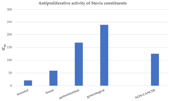
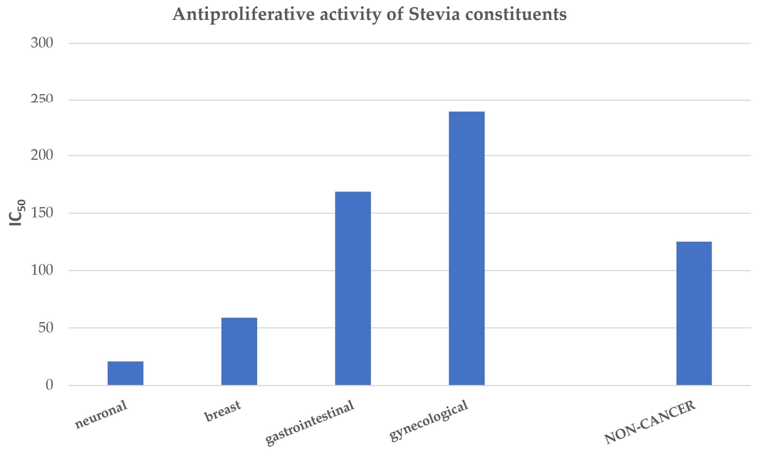
To assess whether the antiproliferative effect of stevia phytoconstituents is lineage-dependent, a stratified meta-analysis was performed according to cancer type at 48 h of exposure, which represents the duration with the most comprehensive data coverage (Table 5, Figure 5). Among malignant cell types, stevia components exerted the highest antiproliferative potency in nervous system cancer cells with an IC50 of 21.00 μg/mL (95% CI: 13.85, 28.16). Breast cancer cells exhibited intermediate sensitivity with an IC50 of 58.90 μg/mL, whereas gastrointestinal cancer (IC50 of 168.62 μg/mL, 95% CI: 81.43, 255.81) and gynecological cancer (IC50 of 238.35 μg/mL, 95% CI: 195.07, 281.63) showed considerably lower responsiveness.
Table 5.
Stratification meta-analysis of IC50 according to cancer type and non-cancerous cells (48 h incubation time).
Figure 5.
IC50 of stevia constituents for various cancer types and non-cancer cells (48 h incubation time).
To provide a preliminary reference point for basal cytotoxicity, non-cancerous cell lines (HaCaT, NIH-3T3, IEC-18, and CHO-K1) were analyzed and presented separately, yielding a pooled IC 50 of 125.15 μg/mL (95% CI: 20.88, 229.42). Although non-cancerous lines showed lower sensitivity (higher IC50) than the most responsive tumor lineages (nervous system and breast), computing a formal pooled Selectivity Index (“SI”) was deemed methodologically uninformative and biologically implausible. The available non-cancer models represent diverse tissue origins (epidermal, fibroblastic, intestinal, ovarian) and cross-species sources rather than matched tissue controls for the specific cancer types evaluated. Furthermore, the limited number of non-cancer datasets (n = 4) relative to cancer studies (n = 15) limits direct statistical comparison. Accordingly, these non-cancer values are reported purely as exploratory reference points.
4. Discussion
This systematic review and meta-analysis constitute the first comprehensive quantitative synthesis of published data evaluating the antiproliferative and cytotoxic potential of stevia constituents and extracts across multiple cancer cell lines. By integrating data from 39 effect estimate datasets encompassing various assay types (MTT, SRB), incubation times (8, 24, 48, and 72 h), and chemical categories (phenols, glycosides, diterpenes, and crude extracts), our analysis offers critical insights into the in vitro antiproliferative properties of stevia phytoconstituents.
The apparent time-dependent enhancement of antiproliferative activity, with summary values decreasing markedly from 3481.20 μg/mL at 8 h to 125.22 μg/mL at 72 h, suggests that stevia phytoconstituents require sustained cellular exposure to exert maximal cytotoxicity. Mechanistically, this progression is hypothesized to reflect time-dependent intracellular uptake, prolonged induction of oxidative stress, or cumulative apoptotic signaling pathways, although these downstream cellular events were not directly assessed in the present meta-analysis. For instance, the high IC50 values observed at the earliest time point (8 h) may correspond to an initial lag phase required for efficient membrane permeation or the metabolic deglycosylation of complex glycosides into bioactive aglycones such as steviol [29,34]. However, as highlighted in our results, incubation duration is confounded with primary study origin across several time points. These temporal patterns should therefore be considered exploratory, emphasizing the need for future preclinical investigations to systematically standardize exposure intervals [35].
The pronounced antiproliferative activity of phenolic fractions (IC50: 97.15 μg/mL at 48 h) compared to diterpenes (171.55 μg/mL) and glycosides (229.73 μg/mL) aligns well with the known multimodal bioactivity of plant polyphenols. While downstream molecular cascades were not directly captured by this meta-analysis, these observed differences plausibly reflect the distinct physicochemical and redox behaviors of these chemical classes. Plant phenolics are well recognized to exert concentration-dependent redox modulation—neutralizing reactive oxygen species (ROS) at physiological baselines while paradoxically promoting intracellular pro-oxidant states and intrinsic apoptotic signaling in malignant phenotypes [2,36,37]. This dual behavior provides a plausible biochemical basis for the differential cytotoxicity observed between transformed and non-transformed models, where non-cancerous control lines displayed generally lower sensitivity, though this trend remains exploratory given the limited non-cancer datasets available. Furthermore, the amphiphilic structure of phenolic backbones frequently facilitates enhanced passive membrane permeation and cell cycle checkpoint modulation relative to larger, hydrophilic glycosylated entities [38,39].
The variation in sensitivity across cancer lineages represents a particularly important finding of this study. Neuronal cancer cells exhibited marked susceptibility (IC50: 21.00 μg/mL), approximately three-fold lower than breast cancer cells (58.90 μg/mL) and nearly twelve-fold lower than gynecological cancer cells (238.35 μg/mL). This differential sensitivity is hypothesized to reflect lineage or tissue-specific differences in cellular uptake, transporter expression, or metabolic turnover, though these parameters were not directly measured in the present meta-analysis. Neuronal cancer models may possess distinct membrane permeability characteristics toward lipophilic phenolic compounds, which could render them more vulnerable to phenolic compound-induced cytotoxicity [40,41]. Additionally, neuronal cancer cells may present elevated baseline susceptibility to oxidative stress pathways [42], which could plausibly contribute to the heightened cytotoxicity observed. Conversely, the relatively lower responsiveness of gynecological cancer cells may relate to hormone-mediated survival pathways or distinct metabolic pathways that confer relative resistance [43]. Our observations highlight the importance of lineage-specific investigations and provide a rationale for future mechanistic and in vivo studies to experimentally test these proposed pathways.
A key strength of this work lies in its direct examination of a fundamental methodological question: whether data derived from different cell viability assays can be reliably combined for cross-study comparisons, given their distinct biochemical principles. Our meta-regression analysis revealed no statistically significant difference between MTT- and SRB-derived results, suggesting that IC50 values from the two assays are broadly comparable and that assay type was not a significant moderator of the effect estimates, although the small number of SRB studies limits the strength of this inference. It nonetheless suggests that results from different methodologies may be cautiously integrated to address growing concerns about reproducibility and transparency in biomedical research. Although the number of studies utilizing the SRB assay in the present dataset is limited (), the absence of marked divergence between MTT- and SRB-derived values is consistent with previous large-scale meta-analytic findings evaluating over 130 natural compound datasets phytoconstituents [14]. Together, these data support the notion that MTT and SRB serve as reliable, concordant readout platforms for assessing in vitro phytoconstituent antiproliferative activity, though larger matched datasets are warranted to formally establish assay equivalence in terms of stevia phytoconstituents.
Several limitations warrant consideration when interpreting these findings. The heterogeneity in data quality and completeness across included studies, combined with variations in extraction procedures and treatment protocols, introduces inherent variability. This is reflected in the very high statistical heterogeneity (I2 up to 100%); to account for it, a DerSimonian–Laird random-effects model was applied throughout and subgroup analyses (Table 2, Table 3, Table 4 and Table 5) were used to explore its sources, so pooled IC50 values should be interpreted with caution, as indicative rather than precise estimates. Additionally, the 39 effect estimate datasets were derived from seven independent articles, which introduces potential clustering of observations within individual studies due to shared laboratory conditions and experimental designs. Notably, the included studies represent the entirety of the currently available data, and this heterogeneity is itself a finding, reflecting the absence of standardized conditions and the need for guided, protocol-based studies to generate harmonized, comparable evidence. The diversity of cancer cell lines studied, while providing comprehensive coverage, prevented cell line-specific meta-analyses due to insufficient replication across studies. Furthermore, the reliance on in vitro data limits direct extrapolation to in vivo conditions, since the interplay of factors such as bioavailability, metabolism, distribution, and tumor microenvironment that interact in a living organism cannot be captured in cell culture models [44]. The lack of standardized stevia extract preparation methods across studies may also contribute to variability in reported IC50 values [45].
Future research should prioritize several key directions. First, the isolation, identification, and characterization of individual phenolic compounds responsible for the observed antiproliferative activity would enable structure-activity relationship studies and rational drug design [46]. Second, mechanistic investigations employing molecular techniques (gene expression profiling, proteomics, metabolomics) are needed to elucidate the precise pathways through which stevia phenolic compounds exert their effects [47]. Third, given the established antioxidant properties of phenolic compounds, clarifying the relationship between antioxidant capacity and antiproliferative activity requires targeted experimental designs that distinguish between ROS-dependent and ROS-independent mechanisms [37]. Fourth, the evaluation of stevia constituents in combination with conventional chemotherapeutic agents may reveal potential synergistic antiproliferative effects, which would first need to be established in preclinical models [48]. Finally, although the in vivo antioxidant effects of stevia leaf extracts have already been documented in animal models [49], their antiproliferative potential remains to be confirmed in vivo; progression to preclinical in vivo models and, eventually, clinical studies would be required to assess the safety, pharmacokinetics, and potential activity of stevia phytoconstituents.
Collectively, addressing these research priorities would advance the translation of stevia phytoconstituents from preliminary in vitro findings toward future preclinical and, ultimately, clinical evaluation.
Supplementary Materials
The following supporting information can be downloaded at: https://www.mdpi.com/article/10.3390/nutraceuticals6040067/s1. Table S1. PRISMA 2020 Checklist. Table S2. PRISMA 2020 for Abstracts Checklist.
Author Contributions
Conceptualization, G.G.B.; methodology, M.P., P.I.K., P.G.B. and G.G.B.; software, M.P., P.I.K., P.G.B. and G.G.B.; validation, M.P., P.I.K., P.G.B. and G.G.B.; formal analysis, M.P., A.T., P.I.K., P.G.B. and G.G.B.; investigation, M.P., A.T. and G.G.B.; resources, P.G.B. and G.G.B.; data curation, M.P., A.T., P.I.K., P.G.B. and G.G.B.; writing—original draft preparation, M.P. and G.G.B.; writing—review and editing, M.P., A.T., P.I.K., P.G.B. and G.G.B.; visualization, M.P. and G.G.B.; supervision, G.G.B.; project administration, G.G.B.; funding acquisition, P.G.B. and G.G.B. All authors have read and agreed to the published version of the manuscript.
Funding
This research received no external funding.
Institutional Review Board Statement
Not applicable.
Informed Consent Statement
Not applicable.
Data Availability Statement
All data used or produced in this research are included in this manuscript’s tables and figures.
Conflicts of Interest
The authors declare that the research was conducted in the absence of any commercial or financial relationships that could be construed as a potential conflict of interest.
Abbreviations
The following abbreviations are used in this manuscript:
| SRB | Sulforhodamine B |
| MTT | 3-(4,5-Dimethylthiazol-2-yl)-2,5-diphenyltetrazolium bromide |
References
- Chatsudthipong, V.; Muanprasat, C. Stevioside and Related Compounds: Therapeutic Benefits beyond Sweetness. Pharmacol. Ther. 2009, 121, 41–54. [Google Scholar] [CrossRef] [Scilit] [PubMed]
- Ghanta, S.; Banerjee, A.; Poddar, A.; Chattopadhyay, S. Oxidative DNA Damage Preventive Activity and Antioxidant Potential of Stevia rebaudiana (Bertoni) Bertoni, a Natural Sweetener. J. Agric. Food Chem. 2007, 55, 10962–10967. [Google Scholar] [CrossRef] [Scilit] [PubMed]
- Papaefthimiou, M.; Kontou, P.I.; Bagos, P.G.; Braliou, G.G. Integration of Antioxidant Activity Assays Data of Stevia Leaf Extracts: A Systematic Review and Meta-Analysis. Antioxidants 2024, 13, 692. [Google Scholar] [CrossRef] [Scilit] [PubMed]
- Carakostas, M.C.; Curry, L.L.; Boileau, A.C.; Brusick, D.J. Overview: The History, Technical Function and Safety of Rebaudioside A, a Naturally Occurring Steviol Glycoside, for Use in Food and Beverages. Food Chem. Toxicol. 2008, 46, S1–S10. [Google Scholar] [CrossRef] [Scilit] [PubMed]
- Yasukawa, K.; Kitanaka, S.; Seo, S. Inhibitory Effect of Stevioside on Tumor Promotion by 12-O-Tetradecanoylphorbol-13-Acetate in Two-Stage Carcinogenesis in Mouse Skin. Biol. Pharm. Bull. 2002, 25, 1488–1490. [Google Scholar] [CrossRef] [Scilit] [PubMed]
- Iatridis, N.; Kougioumtzi, A.; Vlataki, K.; Papadaki, S.; Magklara, A. Anti-Cancer Properties of Stevia rebaudiana; More than a Sweetener. Molecules 2022, 27, 1362. [Google Scholar] [CrossRef] [Scilit] [PubMed]
- Riss, T.L.; Moravec, R.A.; Niles, A.L.; Duellman, S.; Benink, H.A.; Worzella, T.J.; Minor, L. Cell Viability Assays. Available online: https://www.ncbi.nlm.nih.gov/books/NBK144065/ (accessed on 1 July 2016).
- Kumar, P.; Nagarajan, A.; Uchil, P.D. Analysis of Cell Viability by the MTT Assay. Cold Spring Harb. Protoc. 2018, 2018, pdb-prot095505. [Google Scholar] [CrossRef] [Scilit] [PubMed]
- Sánchez-Díez, M.; Romero-Jiménez, P.; Alegría-Aravena, N.; Gavira-O’Neill, C.E.; Vicente-García, E.; Quiroz-Troncoso, J.; González-Martos, R.; Ramírez-Castillejo, C.; Pastor, J.M. Assessment of Cell Viability in Drug Therapy: IC50 and Other New Time-Independent Indices for Evaluating Chemotherapy Efficacy. Pharmaceutics 2025, 17, 247. [Google Scholar] [CrossRef] [Scilit] [PubMed]
- Siddaway, A.P.; Wood, A.M.; Hedges, L.V. How to Do a Systematic Review: A Best Practice Guide for Conducting and Reporting Narrative Reviews, Meta-Analyses, and Meta-Syntheses. Annu. Rev. Psychol. 2019, 70, 747–770. [Google Scholar] [CrossRef] [Scilit] [PubMed]
- Gambrill, E. Evidence-Based Practice and Policy: Choices Ahead. Res. Soc. Work Pract. 2006, 16, 338–357. [Google Scholar] [CrossRef] [Scilit]
- Ioannidis, J.P.A. Why Most Published Research Findings Are False. PLoS Med. 2005, 2, e124. [Google Scholar] [CrossRef] [Scilit] [PubMed]
- Baker, W.L.; Michael White, C.; Cappelleri, J.C.; Kluger, J.; Coleman, C.I.; From the Health Outcomes, Policy, and Economics (HOPE) Collaborative Group. Understanding Heterogeneity in Meta-analysis: The Role of Meta-regression. Int. J. Clin. Pract. 2009, 63, 1426–1434. [Google Scholar] [CrossRef] [Scilit] [PubMed]
- Tsionkis, G.; Andronidou, E.M.; Kontou, P.I.; Tamposis, I.A.; Tegopoulos, K.; Pergantas, P.; Grigoriou, M.E.; Skavdis, G.; Bagos, P.G.; Braliou, G.G. Humulus Lupulus (Hop)-Derived Chemical Compounds Present Antiproliferative Activity on Various Cancer Cell Types: A Meta-Regression Based Panoramic Meta-Analysis. Pharmaceuticals 2025, 18, 1139. [Google Scholar] [CrossRef] [Scilit] [PubMed]
- Page, M.J.; McKenzie, J.E.; Bossuyt, P.M.; Boutron, I.; Hoffmann, T.C.; Mulrow, C.D.; Shamseer, L.; Tetzlaff, J.M.; Akl, E.A.; Brennan, S.E.; et al. The PRISMA 2020 Statement: An Updated Guideline for Reporting Systematic Reviews. BMJ 2021, 372, n71. [Google Scholar] [CrossRef] [Scilit] [PubMed]
- Moher, D.; Liberati, A.; Tetzlaff, J.; Altman, D.G.; The PRISMA Group. Preferred Reporting Items for Systematic Reviews and Meta-Analyses: The PRISMA Statement. PLoS Med. 2009, 6, e1000097. [Google Scholar] [CrossRef] [Scilit] [PubMed]
- Hopewell, S.; McDonald, S.; Clarke, M.J.; Egger, M. Grey Literature in Meta-Analyses of Randomized Trials of Health Care Interventions. Cochrane Database Syst. Rev. 2007, 2007, MR000010. [Google Scholar] [CrossRef] [Scilit] [PubMed]
- Delgado-Rodríguez, M.; Sillero-Arenas, M. Systematic Review and Meta-Analysis. Med. Intensiv. 2018, 42, 444–453. [Google Scholar] [CrossRef] [Scilit] [PubMed]
- Furukawa, T.A.; Barbui, C.; Cipriani, A.; Brambilla, P.; Watanabe, N. Imputing Missing Standard Deviations in Meta-Analyses Can Provide Accurate Results. J. Clin. Epidemiol. 2006, 59, 7–10. [Google Scholar] [CrossRef] [Scilit] [PubMed]
- DerSimonian, R.; Laird, N. Meta-Analysis in Clinical Trials Revisited. Contemp. Clin. Trials 2015, 45, 139–145. [Google Scholar] [CrossRef] [Scilit] [PubMed]
- Harbord, R.M.; Higgins, J.P.T. Meta-Regression in Stata. Stata J. 2008, 8, 493–519. [Google Scholar] [CrossRef] [Scilit]
- Higgins, J.P.T.; Thompson, S.G.; Deeks, J.J.; Altman, D.G. Measuring Inconsistency in Meta-Analyses. BMJ 2003, 327, 557–560. [Google Scholar] [CrossRef] [Scilit] [PubMed]
- Egger, M.; Smith, G.D.; Schneider, M.; Minder, C. Bias in Meta-Analysis Detected by a Simple, Graphical Test. BMJ 1997, 315, 629–634. [Google Scholar] [CrossRef] [Scilit] [PubMed]
- Begg, C.B.; Mazumdar, M. Operating Characteristics of a Rank Correlation Test for Publication Bias. Biometrics 1994, 50, 1088–1101. [Google Scholar] [CrossRef] [Scilit]
- StataCorp. Stata Multilevel Mixed-Effects Reference Manual; StataCorp LP: College Station, TX, USA, 2013; Volume 9. [Google Scholar]
- StataCorp. Stata User’s Guide Release 13; StataCorp LP: College Station, TX, USA, 2013. [Google Scholar]
- Khatun, M.C.S.; Muhit, M.A.; Hossain, M.J.; Al-Mansur, M.A.; Rahman, S.M.A. Isolation of Phytochemical Constituents from Stevia rebaudiana (Bert.) and Evaluation of Their Anticancer, Antimicrobial and Antioxidant Properties via in Vitro and in Silico Approaches. Heliyon 2021, 7, e08475. [Google Scholar] [CrossRef] [Scilit] [PubMed]
- Pacifico, S.; Piccolella, S.; Nocera, P.; Tranquillo, E.; Dal Poggetto, F.; Catauro, M. New Insights into Phenol and Polyphenol Composition of Stevia rebaudiana Leaves. J. Pharm. Biomed. Anal. 2019, 163, 45–57. [Google Scholar] [CrossRef] [Scilit] [PubMed]
- Chen, J.; Xia, Y.; Sui, X.; Peng, Q.; Zhang, T.; Li, J.; Zhang, J. Steviol, a Natural Product Inhibits Proliferation of the Gastrointestinal Cancer Cells Intensively. Oncotarget 2018, 9, 26299–26308. [Google Scholar] [CrossRef] [Scilit] [PubMed]
- Gupta, E.; Kaushik, S.; Purwar, S.; Sharma, R.; Balapure, A.K.; Sundaram, S. Anticancer Potential of Steviol in MCF-7 Human Breast Cancer Cells. Pharmacogn. Mag. 2017, 13, 345–350. [Google Scholar] [CrossRef] [Scilit] [PubMed]
- López, V.; Pérez, S.; Vinuesa, A.; Zorzetto, C.; Abian, O. Stevia rebaudiana Ethanolic Extract Exerts Better Antioxidant Properties and Antiproliferative Effects in Tumour Cells than Its Diterpene Glycoside Stevioside. Food Funct. 2016, 7, 2107–2113. [Google Scholar] [CrossRef] [Scilit] [PubMed]
- Mann, T.S.; Agnihotri, V.K.; Kumar, D.; Pal, P.K.; Koundal, R.; Kumar, A.; Padwad, Y.S. In Vitro Cytotoxic Activity Guided Essential Oil Composition of Flowering Twigs of Stevia rebaudiana. Nat. Prod. Commun. 2014, 9, 1934578X1400900535. [Google Scholar] [CrossRef] [Scilit]
- Boonkaewwan, C.; Ao, M.; Toskulkao, C.; Rao, M.C. Specific Immunomodulatory and Secretory Activities of Stevioside and Steviol in Intestinal Cells. J. Agric. Food Chem. 2008, 56, 3777–3784. [Google Scholar] [CrossRef] [Scilit] [PubMed]
- Renwick, A.G.; Tarka, S.M. Microbial Hydrolysis of Steviol Glycosides. Food Chem. Toxicol. 2008, 46, S70–S74. [Google Scholar] [CrossRef] [Scilit] [PubMed]
- Munafò, M.R.; Nosek, B.A.; Bishop, D.V.M.; Button, K.S.; Chambers, C.D.; Percie Du Sert, N.; Simonsohn, U.; Wagenmakers, E.J.; Ware, J.J.; Ioannidis, J.P.A. A Manifesto for Reproducible Science. Nat. Hum. Behav. 2017, 1, 0021. [Google Scholar] [CrossRef] [Scilit] [PubMed]
- NavaneethaKrishnan, S.; Rosales, J.L.; Lee, K.Y. ROS-Mediated Cancer Cell Killing through Dietary Phytochemicals. Oxid. Med. Cell. Longev. 2019, 2019, 9051542. [Google Scholar] [CrossRef] [Scilit] [PubMed]
- Mileo, A.M.; Miccadei, S. Polyphenols as Modulator of Oxidative Stress in Cancer Disease: New Therapeutic Strategies. Oxid. Med. Cell. Longev. 2016, 2016, 6475624. [Google Scholar] [CrossRef] [Scilit] [PubMed]
- Chimento, A.; De Luca, A.; D’Amico, M.; De Amicis, F.; Pezzi, V. The Involvement of Natural Polyphenols in Molecular Mechanisms Inducing Apoptosis in Tumor Cells: A Promising Adjuvant in Cancer Therapy. Int. J. Mol. Sci. 2023, 24, 1680. [Google Scholar] [CrossRef] [Scilit] [PubMed]
- Stromsnes, K.; Lagzdina, R.; Olaso-Gonzalez, G.; Gimeno-Mallench, L.; Gambini, J. Pharmacological Properties of Polyphenols: Bioavailability, Mechanisms of Action, and Biological Effects in In Vitro Studies, Animal Models, and Humans. Biomedicines 2021, 9, 1074. [Google Scholar] [CrossRef] [Scilit] [PubMed]
- Oteiza, P.I.; Erlejman, A.G.; Verstraeten, S.V.; Keen, C.L.; Fraga, C.G. Flavonoid-Membrane Interactions: A Protective Role of Flavonoids at the Membrane Surface? Clin. Dev. Immunol. 2005, 12, 19–25. [Google Scholar] [CrossRef] [Scilit] [PubMed]
- Stompor, M.; Uram, Ł.; Podgórski, R. In Vitro Effect of 8-Prenylnaringenin and Naringenin on Fibroblasts and Glioblastoma Cells-Cellular Accumulation and Cytotoxicity. Molecules 2017, 22, 1092. [Google Scholar] [CrossRef] [Scilit] [PubMed]
- Liu, Z.; Zhou, T.; Ziegler, A.C.; Dimitrion, P.; Zuo, L. Oxidative Stress in Neurodegenerative Diseases: From Molecular Mechanisms to Clinical Applications. Oxidative Med. Cell. Longev. 2017, 2017, 2525967. [Google Scholar] [CrossRef] [Scilit] [PubMed]
- Brasseur, K.; Gévry, N.; Asselin, E. Chemoresistance and Targeted Therapies in Ovarian and Endometrial Cancers. Oncotarget 2017, 8, 4008–4042. [Google Scholar] [CrossRef] [Scilit] [PubMed]
- Cardoso, B.D.; Castanheira, E.M.S.; Lanceros-Méndez, S.; Cardoso, V.F. Recent Advances on Cell Culture Platforms for In Vitro Drug Screening and Cell Therapies: From Conventional to Microfluidic Strategies. Adv. Healthc. Mater. 2023, 12, e2202936. [Google Scholar] [CrossRef] [Scilit] [PubMed]
- Sun, S.; Yu, Y.; Jo, Y.; Han, J.H.; Xue, Y.; Cho, M.; Bae, S.J.; Ryu, D.; Park, W.; Ha, K.T.; et al. Impact of Extraction Techniques on Phytochemical Composition and Bioactivity of Natural Product Mixtures. Front. Pharmacol. 2025, 16, 1615338. [Google Scholar] [CrossRef] [Scilit] [PubMed]
- Mazumder, K.; Aktar, A.; Roy, P.; Biswas, B.; Hossain, M.E.; Sarkar, K.K.; Bachar, S.C.; Ahmed, F.; Monjur-Al-Hossain, A.S.M.; Fukase, K. A Review on Mechanistic Insight of Plant Derived Anticancer Bioactive Phytocompounds and Their Structure Activity Relationship. Molecules 2022, 27, 3036. [Google Scholar] [CrossRef] [Scilit] [PubMed]
- Kwon, Y.W.; Jo, H.S.; Bae, S.; Seo, Y.; Song, P.; Song, M.; Yoon, J.H. Application of Proteomics in Cancer: Recent Trends and Approaches for Biomarkers Discovery. Front. Med. 2021, 8, 747333. [Google Scholar] [CrossRef] [Scilit] [PubMed]
- Talib, W.H.; Awajan, D.; Hamed, R.A.; Azzam, A.O.; Mahmod, A.I.; AL-Yasari, I.H. Combination Anticancer Therapies Using Selected Phytochemicals. Molecules 2022, 27, 5452. [Google Scholar] [CrossRef] [Scilit] [PubMed]
- Papaefthimiou, M.; Kontou, P.I.; Bagos, P.G.; Braliou, G.G. Antioxidant Activity of Leaf Extracts from Stevia rebaudiana Bertoni Exerts Attenuating Effect on Diseased Experimental Rats: A Systematic Review and Meta-Analysis. Nutrients 2023, 15, 3325. [Google Scholar] [CrossRef] [Scilit] [PubMed]

Disclaimer/Publisher’s Note: The statements, opinions and data contained in all publications are solely those of the individual author(s) and contributor(s) and not of MDPI and/or the editor(s). MDPI and/or the editor(s) disclaim responsibility for any injury to people or property resulting from any ideas, methods, instructions or products referred to in the content. |
© 2026 by the authors. Licensee MDPI, Basel, Switzerland. This article is an open access article distributed under the terms and conditions of the Creative Commons Attribution (CC BY) license.