


Abstract
Phenolic compounds exhibit important biological properties; however, their solubility affects bioavailability when they are consumed by oral administration. Enzymatic glycosylation of these molecules improves solubility and bioavailability; however, their stability under gastrointestinal conditions must be considered for functional food applications. In this study, the digestive stability of phlorizin, phloretin, and their enzymatically synthesized glycosides—phlorizin-4-O-monofructofuranoside (4PMF) and phloretin 4′-O-α-D-glucopyranoside—was evaluated using a static in vitro digestion model, simulating stomach and small intestine conditions (SC and SIC respectively). 4PMF showed high susceptibility to hydrolysis under SC and SIC (58 ± 2% and 99.5 ± 0.9%, respectively), whereas phloretin 4′-O-α-D-glucopyranoside exhibited significantly greater stability (5.7 ± 0.6% and 14 ± 1%, respectively). Statistical analysis confirmed significant differences between compounds and digestion phases (p < 0.05). Additional analysis revealed that β-fructosyl bonds are acid-labile, whereas α-O-glucosidic bonds are resistant to acidic conditions. Residual glycosidase activities were observed in the commercial digestive enzymes used under SC and SIC. These findings support a dual mechanism that affects the stability of phenolic glycosides during in vitro digestion under SC and SIC, with glycosidic bond type also playing a role.
1. Introduction
Phenolic compounds are secondary metabolites found in many plant species which have a wide variety of chemical structures and biological activities. Due to their known antioxidant properties, some exhibit the capacity to control blood sugar, such as phlorizin and its aglycone, phloretin. Phlorizin is a naturally occurring β-glucosylated dihydrochalcone found in apples, while phloretin is its aglycone; both compounds have been studied extensively. Their antioxidant properties and ability to inhibit glucose transport at kidney level supports their antidiabetic effects [1,2]. Considering these favorable biological activities, substantial research has focused on modifying these compounds to enhance their physicochemical attributes and functional efficacy, to improve absorption and pharmacology, or as an ingredient for functional foods development [2,3]. Like most phenolic compounds, their complex molecular structure results in poor solubility in water. These characteristics can limit their absorption in the digestive system [4,5]. To resolve these limitations, enzymatic glycosylation has emerged as a powerful biocatalytic strategy to improve both the solubility and stability of phenolic compounds [6].
Enzymatic modification approaches of native phenolic molecular structure have recently enabled the synthesis of novel glycosylated derivatives, such as β-D-fructofuranosyl-(2→6)-phlorizin and phloretin 4′-O-α-D-glucopyranoside, which exhibit improved aqueous solubility and superior functional properties compared to their parent compounds [7,8]. Furthermore, a levansucrase derived from Paraburkholderia phymatum (KesL) has been found to synthesize phlorizin polyfructosides, with 4-O-monofructosyl phlorizin (4PMF) being the predominant product, expanding the spectrum of enzymatically glycosylated derivatives available for subsequent functional assessment [9].
However, even though enzymatic glycosylation is effective in creating new phenolic derivatives with improved physical and chemical properties, the stability of these enzymatically synthesized molecules in the gastrointestinal tract has not been evaluated [10]. Understanding the digestive behavior of these modified molecules is essential for evaluating their potential as functional ingredients, because the biological activity of phenolic molecules may depend on their structural integrity or the controlled release of their aglycone forms during digestion [5,11]. During gastrointestinal digestion of phenolic glycosides, glycosidic bonds may be hydrolyzed via chemical and enzymatic mechanisms. O-glucosides, like phlorizin, are known to be substrates of lactase–phlorizin hydrolase, which is a brush-border enzyme that can break glycosidic bonds before they are absorbed in the small intestine [12]. On the other hand, various glycosidic linkages may exhibit unique chemical susceptibilities based on their structural characteristics. Specifically, β-fructosyl bonds in fructosylated derivatives are recognized to be vulnerable to acid-catalyzed hydrolysis in low-pH environments [13]. Dynamic digestion systems have been proposed to more accurately replicate physiological settings; nevertheless, static in vitro digestion models such as the standardized INFOGEST protocol remain prevalent because of their reproducibility, ease of use, and absence of ethical issues [4,5]. These models allow for analyzing individual factors such as pH or enzymatic activity, which can influence the phenolic compounds’ breakdown. However, the presence of non-specific enzymatic activities in commercial digestion reagents can influence the interpretation of results obtained from in vitro digestion models. Several studies have shown that commercial enzyme preparations—including pepsin, pancreatin, lipases, and bile extracts—may contain trace glycosidase activities capable of hydrolyzing carbohydrate linkages [14,15,16]. More recent characterizations of digestion systems have shown the complex enzymatic composition of commercial preparations used in gastrointestinal simulations [17]. Therefore, the apparent hydrolysis of phenolic glycosides observed during in vitro digestion may result not only from chemical instability at different pH values, but also from residual enzymatic activities present in the digestion reagents themselves. Despite growing interest in the gastrointestinal behavior of enzymatically synthesized phenolic glycosides, a direct comparison of how structurally distinct glycosidic linkages—specifically β-fructosyl and α-O-glucosidic bonds—determine digestive stability within the same experimental system has not been previously reported. Most available studies have examined individual glycosides or focused on pH effects in isolation, without systematically distinguishing between acid-catalyzed and enzyme-mediated hydrolysis mechanisms. Furthermore, while residual glycosidase activities have been partially documented in certain commercial digestion preparations, a comparative characterization across all major reagents employed in a single digestion protocol including pepsin, pancreatin, bile salts, and pancreatic lipase remains absent from the literature.
Based on these knowledge gaps, and on existing evidence regarding the chemical lability of β-fructosyl bonds under acidic conditions and the susceptibility of α-O-glucosidic bonds to enzymatic hydrolysis, it was hypothesized that these two linkage types would display fundamentally different stability profiles during simulated gastrointestinal digestion—with β-fructosyl bonds being predominantly cleaved through acid-catalyzed mechanisms during the gastric phase, and α-O-glucosidic bonds being more susceptible to enzyme-mediated hydrolysis during the intestinal phase. It was further hypothesized that residual glycosidase activities present in commercial digestion reagents may contribute to the observed hydrolysis, and that their systematic characterization is necessary for accurate mechanistic interpretation of in vitro digestion outcomes. The purpose of this work was to investigate the digestive stability of phlorizin, phloretin, and their enzymatically glycosylated derivatives—phlorizin-4-O-monofructofuranoside (4PMF) and phloretin 4′-O-α-D-glucopyranoside—utilizing a controlled static in vitro digestion model. This study sought to (i) assess the impact of glycosidic linkage type on hydrolysis behavior in gastric and intestinal environments and (ii) differentiate between acid-catalyzed and enzyme-facilitated hydrolysis by measuring residual glycosidase activity in the digestion reagents used.
2. Materials and Methods
Phloretin was purchased from Hunan MT Health Inc. (Changsha, China). Phlorizin was supplied by Merck (Darmstadt, Germany). Sucrose, dimethyl sulfoxide, formic acid, acetonitrile, hydrochloric acid, and sodium hydroxide were obtained from Sigma-Aldrich Inc. (St. Louis, MO, USA). All reagents were of high purity grade (≥98%). Acetonitrile was HPLC grade.
2.1. Phenolic Compounds and Glycosylated Derivatives
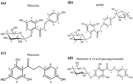
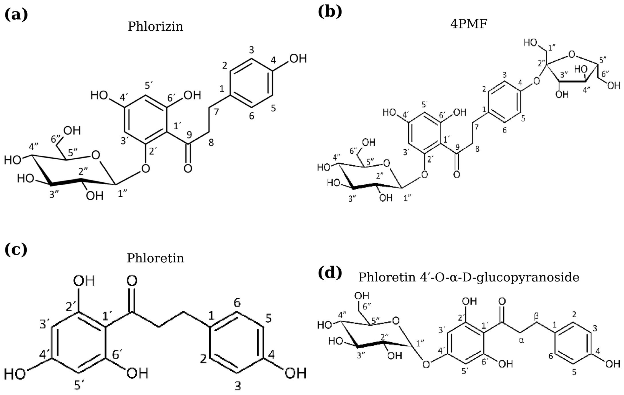
The molecular structures of the phenolic compounds used in this study—phlorizin, phloretin, and their glycosylated derivatives—are shown in Figure 1.
Figure 1.
Molecular structures of the phenolic compounds and their glycosylated derivatives used in this study: (a) phlorizin, (b) 4PMF, (c) phloretin, and (d) phloretin 4′-O-α-D-glucopyranoside.
Phlorizin polyfructosides (PPFs) were previously synthesized by enzymatic fructosylation using KesL as described by [9]. The resulting reaction mixture contained 4PMF as the predominant component, together with minor fructosylated derivatives. The fraction enriched in 4PMF was used in the present study and was kindly provided by the authors of the original work.
Phloretin α-glucoside (phloretin 4′-O-α-D-glucopyranoside) was previously synthesized using the R134A mutant of sucrose 6′-phosphate phosphorylase from Thermoanaerobacter thermosaccharolyticum (TtSPP_R134A) according to the method described by [8]. The purified compound was supplied by the authors for use in this study.
2.2. Static In Vitro Digestion
Phenolic compounds and their glycosylated derivatives were subjected to simulated gastric and small intestinal digestion using a static in vitro digestion model in the absence of microbiota or food matrices.
For each experiment, 50 mg of each compound was dissolved in 200 mL of sterile deionized water. The pH was adjusted to 2.0 using 5 M HCl to simulate stomach conditions (SC). Once the pH stabilized, porcine gastric mucosal pepsin (330 mg; 1:10,000 activity, Hycel®, Zapopan, Mexico) was added and the mixture was incubated for 2 h under constant agitation.
At the end of the SC, the pH was adjusted to 5.0–5.5 using 3 M of NaOH to simulate the small intestinal conditions (SIC). Pancreatin (0.19 g; CREON®, North Chicago, IL, USA), lipase (1 mg), and bile salts (1 g; Difco®, Franklin Lakes, NJ, USA) were then added. The mixture was incubated at 37 °C under constant agitation (150 rpm) for 4 h.
Samples were collected before digestion and after 2 h of SC. During the SIC, samples were taken immediately after pH adjustment and enzyme addition (t = 0 h) and after 4 h of incubation. The concentrations of the phenolic compounds were determined by HPLC–UV.
2.3. Effect of pH on the Stability of Phenolic Glycosides
To evaluate the effect of pH alone on the stability of phenolic glycosides, in the absence of digestive enzymes, 50 mg of each compound was dissolved in 200 mL of sterile deionized water (initial pH ≈ 6.0), and an initial sample was collected. The pH of the same solution was then adjusted to 2.0 using 5 M of HCl, and the mixture was incubated at 37 °C under agitation (280 rpm) for 2 h, after which a second sample was collected. Samples were analyzed using the HPLC method described below.
2.4. HPLC–UV Analysis
Samples obtained during the in vitro digestion experiments were analyzed by HPLC–UV based on the method described by [18] with slight modifications. Analyses were performed using a Waters ACQUITY Arc chromatograph (Milford, MA, USA) equipped with a Nova-Pak® C18 column (3.9 mm × 150 mm, 4 μm).
A gradient system composed of solvent A (water containing 0.05% formic acid, v/v) and solvent B (acetonitrile containing 0.05% formic acid, v/v) was used at a flow rate of 0.8 mL min−1 and a column temperature of 40 °C. The gradient program started at 10% B (0 min) and increased to 50% B over 20 min, followed by an increase to 95% B over the next 5 min. All compounds were detected by UV absorbance at 254 nm. Prior to injection, aliquots of each digestion sample were diluted 1:1 (v/v) with dimethyl sulfoxide, which helped maintain the phenolic compounds and their glycosylated derivatives, compounds with limited aqueous solubility, fully dissolved immediately prior to injection, thereby preventing analyte precipitation or peak broadening/tailing during chromatographic separation. The percentage of hydrolysis for each glycosylated derivative was calculated based on the disappearance of its corresponding HPLC peak area relative to the initial peak area (t = 0), according to the following equation:
where Peak areat is the peak area of the glycosylated derivative at a given digestion time point, and Peak areat=0 is the peak area of the same compound before digestion.
2.5. Residual Glycosidase Activity
Residual glycosidase activity in the commercial digestion reagents was evaluated using sucrose as a model substrate. Solutions of pepsin from porcine gastric mucosa, pancreatin, lipase from porcine pancreas, and dehydrated bile salts (Oxgall) were prepared separately at a concentration of 100 mg mL−1 in distilled water.
For each assay, an aliquot of enzyme solution was mixed with a sucrose solution (50 mg mL−1) and incubated at 37 °C for 10 min. The reaction was stopped, and the release of reducing sugars was quantified using the dinitrosalicylic acid method according to [19]. One unit of residual glycosidase activity was defined as the amount of enzyme releasing 1 μmol of reducing sugar equivalents per minute under the assay conditions and was expressed as units per gram (U g−1) of commercial reagent.
Sucrose was used as a model substrate to provide an integrative estimate of residual glycosidase activity rather than to discriminate between specific α- or β-glycosidase activities, allowing comparison of the overall hydrolytic potential among the digestion reagents. All assays were performed in triplicate.
2.6. Statistical Analysis
All experiments were performed as three independent biological replicates (n = 3), each corresponding to a separate digestion assay conducted with freshly prepared reagents and samples, and results are expressed as mean ± standard deviation. Statistical analysis was carried out using one-way analysis of variance (ANOVA) followed by Tukey’s post hoc test to evaluate significant differences among conditions. Differences were considered statistically significant at p < 0.05. Statistical analyses were applied to the hydrolysis percentages obtained for the evaluated glycosides during the gastric and intestinal phases, as well as to the residual glycosidase activity data obtained for the digestion reagents. Given the reduced sample size per group (n = 3), formal normality tests (e.g., Shapiro–Wilk) were not performed, as such tests have limited statistical power to detect deviations from normality under these conditions. One-way ANOVA is generally considered robust to moderate departures from normality, particularly in balanced experimental designs such as the one used in this study.
3. Results and Discussion
3.1. In Vitro Digestion
In vitro digestion models enable us to assess the stability of bioactive compounds under maintained simulated gastrointestinal conditions. This results in a feasible way to examine both pH- and enzyme-induced effects without the confounding effects of food matrices. This method is effective for assessing glycosylated phenolic compounds since the type of glycosidic bond has a big effect on how they are digested. In this case, chromatographic profiles analyzed under SC and SIC provide us with a firsthand view of the breakdown of phlorizin, 4PMF, phloretin, and phloretin 4′-O-α-D-glucopyranoside (Figure 1).
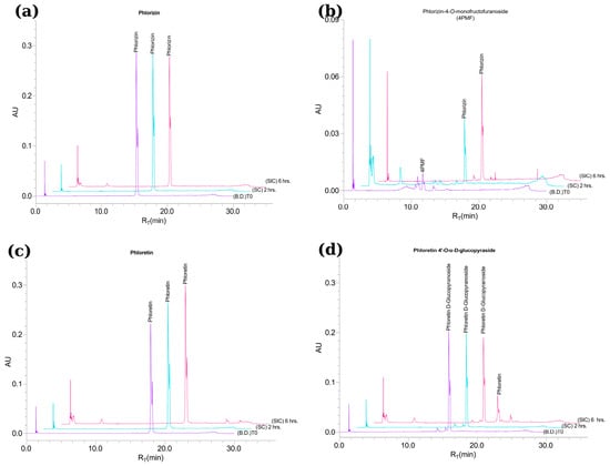
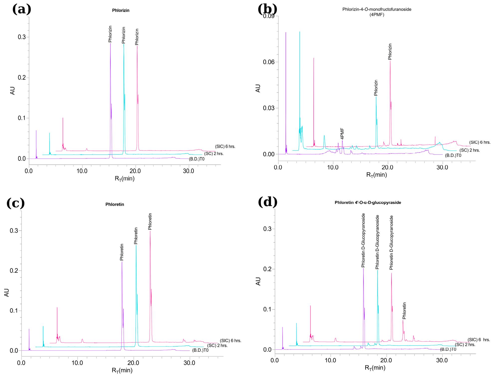
According to this approach, chromatographic profiles (Figure 2) showed unique hydrolysis patterns, depending on both phenolic glycoside and digestion phase. No conversion of phlorizin into phloretin was detected under SIC used in this study (Figure 2a), in contrast to previous reports describing phlorizin hydrolysis into its aglycone during gastrointestinal digestion [20,21,22]. These discrepancies with respect to previous studies may be attributed to variations in experimental conditions, such as enzyme activity, pH, and incubation period. Notably, previous research demonstrated that specific glycosylated derivatives of phlorizin may withstand simulated gastrointestinal conditions. Jakobek et al. [23] indicated that phloretin-2′-glucoside from apple pulp could withstand all stages of digestion, demonstrating consistent recovery across phases. The fructosylated sample used in this study consisted of a mixture of phlorizin polyfructosides, with 4PMF as the predominant compound, as confirmed by HPLC analysis. Based on chromatographic resolution and retention time, 4PMF was clearly distinguished from minor components, allowing reliable monitoring of its hydrolysis during digestion experiments. 4PMF eluted at a mean retention time of 10.93 min, whereas minor unidentified fructosylated impurities eluted between approximately 8 and 12 min. The hydrolysis product, phlorizin, eluted at 15.29 min (Figure 2b).
Figure 2.
Chromatographic profiles of molecules for in vitro digestion under stomach conditions (SC) and small intestine conditions (SIC) at 0, 2 and 6 h for (a) phlorizin, (b) 4PMF, (c) phloretin, and (d) phloretin 4′-O-α-D-glucopyranoside. Detection wavelength: 254 nm. Traces are vertically offset for visual clarity; retention times are not affected. Chromatograms shown are representative of three independent digestion experiments (n = 3).
Consequently, the progressive decrease in fructosylated compound signals was accompanied by a corresponding increase in the phlorizin signal, supporting the cleavage of β-fructosyl linkages under both SC and SIC, which were hydrolyzed by 58 ± 2% and 99.5 ± 0.9%, respectively (Table 1). These findings are consistent with previous research showing acid-mediated cleavage of fructosyl linkages in fructan-type oligosaccharides [13,24] and support the formation of phlorizin as an intermediate. As expected, phloretin was not hydrolyzed under SC and SIC (Figure 2c), with a retention time of approximately 17.92 min (Table 1). This outcome is consistent with its chemical nature as the aglycone of phlorizin, which lacks a glycosidic bond and is therefore not susceptible to either acid-catalyzed or enzyme-mediated hydrolysis under the conditions employed in this study. It has been reported that polyphenols exhibit improved stability under acidic conditions, as phenolic hydroxyl groups remain largely protonated and less prone to oxidative degradation at low pH [25,26,27]. This is consistent with the enhanced stability observed for phloretin under the acidic conditions used in this study.
Table 1.
Percentage of hydrolysis of glycosylated derivatives of phlorizin and phloretin during simulated stomach conditions (SC) and small intestine conditions (SIC) during in vitro digestion.
In the particular case of phloretin, since phloretin lacks glycosidic moieties in its molecular structure, it is intrinsically immune to hydrolytic cleavage, thus excluding any degradation via glycosidic bond hydrolysis. In agreement, an earlier pharmacokinetic study on Sprague-Dawley rats indicated that phloretin shows significant acid stability despite its limited oral bioavailability [28]. Additionally, Mariadoss et al. [29] demonstrated that phloretin’s stability is enhanced in acidic environments when encapsulated in chitosan-based nanoparticles. Further research has demonstrated that both phlorizin and phloretin are detectable following extended intestinal digestion [30]. Recent investigations utilizing the standardized INFOGEST model have validated the stability of phloretin after simulated gastrointestinal digestion [5,23]. Additional evidence from hydrogel and protein-emulsion systems containing phloretin reinforces its structural stability before intestinal release [31,32]. On the contrary, the glycosylated derivative, phloretin 4′-O-α-D-glucopyranoside, showed hydrolysis exclusively under SIC with 14 ± 1% of hydrolysis (Figure 2d, Table 1).
This compound, with a retention time of approximately 15.87 min (Table 1), revealed an appearance of a phloretin peak found in its chromatogram profile, indicating partial cleavage of the α-glycosidic bond.
The stability of this O-glucoside under SC and its subsequent hydrolysis during SIC corresponds to previous reports on structurally analogous molecules. Specifically, O-glycosides like apigenin-7-O-glucoside showed higher stability compared to C-glycosides during simulated small intestine digestion [33], whereas additional research indicates that flavonoid glycosides experience considerable alterations during the small intestine phase [34]. Lactase–phlorizin hydrolase, located at the intestinal brush border, is known as a key enzyme that catalyzes the hydrolysis of β-O-glucosidic bonds in dietary phenolic glycosides [35]. Although this enzyme is absent in the current in vitro system, its activity provides a valuable physiological reference for interpreting the enzyme-associated hydrolysis observed under SIC. In this context, it has been observed in other works that residual glycosidase activities present in commercial enzyme preparations may contribute to the hydrolysis of phenolic glycosides during simulated intestinal digestion, which could account for the hydrolytic behavior observed for both 4PMF and phloretin 4′-O-α-D-glucopyranoside in the current study [23,36,37]. Despite this, hydrolysis is not always prevalent, as the stability of glycosylated phenolics appears to depend strongly on the type of glycosidic bond involved. Xie et al. [38] demonstrated that the O-glycosidic bond of isoquercitrin was cleaved during both the gastric and intestinal phases of in vitro digestion, whereas the C-glycosidic bond of orientin remained intact throughout the entire process, owing to the inherently greater stability of the C–C bond relative to the C–O bond.
In addition to glycosidase activity, pH fluctuations through the gastrointestinal tract can significantly affect phenolic stability. Numerous investigations have documented alterations in total and specific phenolics during digestion, attributed to pH-dependent alterations and the liberation from conjugated forms. As explained above, commercial enzyme preparations employed in digestion models have been recognized to exhibit unexpected side activities. Enzymatic characterization shows that peptidase, lipase, and pectinase preparations possess measurable glycosidase activities, such as β-glucosidase and β-galactosidase [14,15,16], while pancreatin preparations exhibited a heterogeneous enzymatic composition depending on the production batch [17]. These observations suggest that acidic pH and residual glycosidase activity may promote the hydrolysis of phenolic glycosides during in vitro digestion. To elucidate possible causes, further digestions with all phenolic compounds were performed under controlled pH conditions, and glycosidase activity was independently assessed on all digestion reagents to study the effect of pH and glycosidase activity on the hydrolysis of phenolic glycosides during in vitro digestion.
3.2. Effect of pH on the Stability of Phloretin and Phlorizin and Their Respective Glycosides
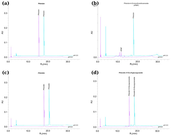
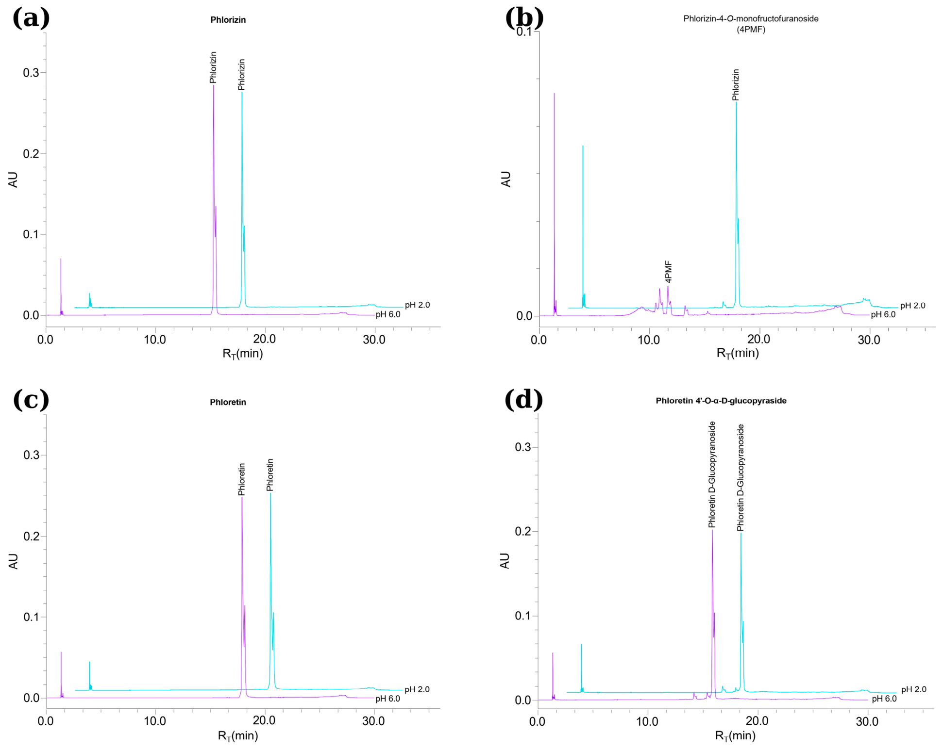
The pH level of the gastrointestinal tract has a significant impact on the stability of phenolic compounds, as pH variations during digestion can cause structural changes and degradation [39,40,41]. In this study, the stability of phloretin, phlorizin, and their glycosides in controlled acidic and near-neutral conditions without adding digestive enzymes was assessed. This experiment was performed in order to separate changes caused by pH from those caused by enzymatic side activities. Figure 3 shows the chromatographic profiles that were made after changing the pH. These profiles show whether the compounds stayed the same or changed only because of the gastric-like pH environment.
Figure 3.
Effect of hydrochloric acid addition on the stability of (a) phlorizin, (b) 4PMF, (c) phloretin, and (d) phloretin 4′-O-α-D-glucopyranoside. Samples were dissolved in deionized water (pH 6.0; purple trace) and subsequently acidified to pH 2.0 with HCl and incubated for 2 h (pH 2.0; cyan trace). Detection wavelength: 254 nm. Chromatograms shown are representative of three independent digestion experiments (n = 3).
All evaluated compounds (Figure 3), including their glycosylated forms, remained stable under near-neutral conditions (pH 6.0–7.0). In the particular case of 4PMF, it showed no signal of hydrolysis or formation of secondary peaks, thus reinforcing the fact that β-fructosyl linkages are not susceptible to spontaneous cleavage at neutral pH and remain intact in the absence of enzymatic activity. The observed stability agrees with previous studies reporting limited fructan hydrolysis under near-neutral conditions [24].
A similar behaviour was observed by the authors of [27], who reported that dicaffeoylquinic acids maintained molecular stability when exposed to nearly neutral pH levels, indicating that in a phenolic system, hydrolysis is induced mainly by acidic conditions rather than being caused by molecular stability. Similar observations were found in chromatographic profiles obtained at every evaluated in vitro digestion stage (Figure 2), with β-fructosyl linkages showing higher susceptibility to hydrolysis relative to α-O-glucosidic linkages. These observations suggest that phenolic glycoside compounds would be chemically stable under neutral-like conditions in vitro and would not be altered chemically until exposed to acidic conditions or enzymatic activities at specific phases during the in vitro digestion simulation. Only under acidic conditions (pH 2.0), 4PMF exhibited partial hydrolysis (Figure 3b), while the other compounds showed no appreciable degradation. This indicates that the acidic environment alone is enough to facilitate the cleavage of β-fructosyl bonds, thereby supporting their acid-labile behavior. Comparable acid-induced transformations have been described in polyphenolic systems; for instance, the procyanidin polymers exhibit depolymerization into epicatechin in gastric-like conditions via proton-catalyzed bond cleavage [42].
The pH-dependent stability of phenolic compounds has been documented in the literature; Friedman and Jürgens [25] demonstrated that phenolic acids, including caffeic, chlorogenic, and gallic acids, remained intact at acidic pH, consistent with the chemical resistance observed in this study for phlorizin, phloretin, and phloretin 4′-O-α-D-glucopyranoside at pH 2.0 (Figure 3). In this context, and in line with the idea that pH-induced alterations in electronic distribution can influence the stability of phenolic rings [26], the susceptibility of β-fructosyl linkages in 4PMF to acid-catalyzed hydrolysis is particularly notable, as the other compounds in the study exhibited no significant degradation under the same non-enzymatic acidic conditions.
The experiments mentioned above had the objective of identifying the effect of digestive pH-like conditions and, therefore, were conducted without digestive enzymes; the potential influence of side glycosidase activity present in commercial digestive reagents, including digestive enzyme preparations, must be considered when assessing in vitro digestion of phenolic glycosides. The literature reports that enzyme mixtures commonly utilized in gastrointestinal model simulations show low, although detectable, levels of α- and β-glycosidase activities capable of hydrolyzing glycosidic bonds [43]. This may complicate assigning the cause of observed hydrolysis exclusively to chemical mechanisms. In this context, this study included experiments with the independent measurement of residual glycosidase activity in each digestion reagent, which has the objective of helping to differentiate between pH-driven and enzyme-mediated cleavage of the type of glycosidic bond present in each of the phenolic compounds and glycoside derivatives assessed in this study.
3.3. Effect of Residual Glycosidase Activity in Commercial Pepsin and Pancreatin on the Stability of Phloretin and Phlorizin and Their Respective Glycosides
The evidence presented in Section 3.2 suggests acidic conditions as the primary driver of β-fructosyl bond cleavage in 4PMF, confirming the inherent acid lability of this linkage under gastric-like pH environments. The picture looks rather different for phloretin 4′-O-α-D-glucopyranoside, whose hydrolysis appears to be less sensitive to pH and more contingent on the residual glycosidase activity present in the commercial digestion reagents used throughout this study. To test this hypothesis, the residual glycosidase activity of each commercial enzyme preparation and reagent used in the in vitro digestion assays was directly quantified (Table 2).
Table 2.
Residual glycosidase activity of gastric enzymes and bile salts employed for digestion.
The statistically significant differences recorded across compounds and digestion phases (p < 0.05) make it difficult to attribute the observed hydrolytic patterns to chance—pointing instead to the influence of specific structural features and environmental conditions on the stability of each glycosidic linkage. Rather than searching for specific α- or β-glycosidase activities, sucrose was chosen as a model substrate to obtain a wider approach to the overall hydrolytic potential of each digestive reagent and its possible contribution to glycoside transformation during in vitro digestion. It should be noted that this sucrose-based assay offers an indirect and non-specific approximation of overall glycosidase activity, rather than direct evidence that the tested reagents act on phlorizin, 4PMF, phloretin, or phloretin 4′-O-α-D-glucopyranoside themselves; the phenolic α-O-glucosidic and β-fructosyl linkages in these compounds could differ in their susceptibility relative to the enzymes responsible for sucrose hydrolysis. Therefore, the enzymatic contribution proposed throughout this study is best regarded as a plausible, indirectly supported hypothesis rather than a directly demonstrated mechanism. As data in Table 2 reveals, pepsin is the most active preparation, exhibiting the highest residual glycosidase activity (191 ± 4 U g−1), followed at some distance by bile salts (84 ± 3 U g−1), pancreatic lipase (52 ± 4 U g−1), and pancreatin (14 ± 2 U g−1). Taken together, these figures reveal that commercial digestion reagents—despite not being formulated for carbohydrate hydrolysis—display measurable glycosidase activities that could meaningfully influence the apparent stability of phenolic glycosides during in vitro digestion assays. Although pepsin showed the highest residual glycosidase activity, this side activity did not impact the cleavage of glycosidic bonds of any of the glycosides assessed during the gastric phase; this is consistent with its well-known property as a protease adapted to highly acidic conditions (pH ≈ 2), where it exhibits limited flexibility and low affinity to non-protein substrates [44,45]. In addition, its active site is specifically designed for peptide bond hydrolysis, which probably restricts the interactions with phenolic glycosides [46]. Nevertheless, the detection of quantifiable glycosidase activity in this preparation aligns with previous API-ZYM studies reporting side activities, including glycosidases, in commercial enzyme mixtures. These observations highlight the importance of assessing side enzymatic activities before using such reagents in in vitro digestion protocols [16].
By contrast, pancreatin constitutes a more complex multienzymatic system involving proteases, lipases, and α-amylase. However, α-amylase catalyzes the hydrolysis of internal α-(1→4) glycosidic bonds in glucans [17,47], a substrate specificity that is structurally distinct from the phenolic α-O-glucosidic bond present in phloretin 4′-O-α-D-glucopyranoside; therefore, α-amylase activity cannot be assumed to account for the hydrolysis observed for this compound. The residual glycosidase activity detected in pancreatin (Table 2) more plausibly reflects the presence of non-target, uncharacterized glycosidase side-activities in this commercial multienzymatic preparation, consistent with previous API-ZYM reports of such side activities in digestive enzyme mixtures [16]. Confirming which specific enzyme(s) are responsible for the hydrolysis of phloretin 4′-O-α-D-glucopyranoside during SIC would require direct assays using this compound as substrate, which is beyond the scope of the present study. The literature establishes that intestinal glycosidases, such as lactase–phlorizin hydrolase and various α- and β-glycosidases, facilitate the hydrolysis of phenolic O-glycosides [48,49]. This is consistent with several studies; for instance, a recent investigation utilizing rat small intestine extract demonstrated that mammalian digestive glycosidases catalyze the deglycosylation of β-O-glycosides and α-O-glycosides in different food matrices [43]. Furthermore, existing reports indicate that flavonoid O-glycosides undergo cleavage within the small intestine before absorption [50,51]. Collectively, the findings of the present study, supported by the current literature, suggest that the glycosidase activity of digestive enzymes plays a relevant role in the hydrolysis of O-glycosidic linkages in phenolic glycosides, rather than the luminal pH levels under physiological intestinal conditions. The present results altogether support a global dual mechanism governing the hydrolysis of phenolic glycosides during in vitro digestion. While β-fructosyl linkages are essentially cleaved through acid-catalyzed reactions during the gastric phase, O-glucosidic linkages appear to be more dependent on enzymatic activity under intestinal conditions. This distinction is critical for interpreting digestion results, as residual glycosidase activity in commercial enzyme preparations may lead to an overestimation of compound instability. These statistically supported differences further reinforce the distinction between acid-driven and enzyme-mediated hydrolysis pathways. From the perspective of the methodological procedure during in vitro digestion for phenolic glycosides, the results presented in this study emphasize the necessity to characterize digestive reagents or preparations prior to their use in simulated digestion studies. Residual enzymatic activities can significantly influence the apparent stability of glycosylated phenolic compounds. Several recent studies examining gastrointestinal digestion of glycosylated compounds have notably overlooked this factor [38,52,53,54,55], highlighting the necessity of integrating enzymatic profiling into digestion protocols for precise mechanistic interpretation.
Whether the contrasting stability patterns observed in this study—the high susceptibility of 4PMF to acid-catalyzed hydrolysis versus the greater stability of phloretin 4′-O-α-D-glucopyranoside—would be similarly evident under dynamic conditions, such as those simulated by systems like the TNO gastrointestinal model (TIM-1), which incorporate continuous secretion and physical mixing, remains to be established [56].
4. Conclusions
The type of glycosidic bond appears to play an important role in the digestive stability of enzymatically synthesized phenolic glycosides. 4PMF, bearing a β-fructosyl linkage, was highly susceptible to acid-catalyzed hydrolysis during the gastric phase (58 ± 2%) and was nearly completely degraded intestinally (99.5 ± 0.9%). In contrast, phloretin 4′-O-α-D-glucopyranoside, bearing an α-O-glucosidic bond, remained largely stable under acidic conditions (5.7 ± 0.6%) and was hydrolyzed mainly during the intestinal phase (14 ± 1%), consistent with an enzyme-driven rather than chemical mechanism. These results are consistent with a possible dual hydrolysis mechanism, in which acid catalysis may govern β-fructosyl bond cleavage in the stomach, while enzymatic activity may drive α-O-glucosidic bond cleavage in the intestine, although this mechanistic interpretation would benefit from further confirmatory studies. It should also be noted that, since the compared derivatives differ not only in glycosidic bond type but also in aglycone structure, glycosidic bond type could not be isolated as the sole experimental variable in this study; these findings should therefore be interpreted as an observed association rather than as evidence of an isolated causal effect. Future studies comparing derivatives that share the same aglycone but differ only in glycosidic bond type would help to more directly isolate the specific contribution of bond type to digestive stability.
Residual glycosidase activity was detected in all digestion reagents tested, with pepsin showing the highest activity (191 ± 4 U g−1) despite being a protease. This finding highlights a methodological consideration for in vitro digestion studies: non-target enzymatic activities in commercial reagents can confound the interpretation of glycoside stability data and should be characterized before use. This pattern, combined with the distinct hydrolysis behavior observed for 4PMF and phloretin 4′-O-α-D-glucopyranoside, suggests that glycosidic bond type may be an important structural determinant of the digestive stability of these synthesized derivatives, offering a preliminary mechanistic basis that could help guide the future design of more digestion-resistant glycosylated phenolic derivatives for food and nutraceutical applications.
Some limitations of this study should be acknowledged. Glycosylated derivatives were identified and monitored by HPLC–UV on the basis of retention time and chromatographic resolution, without confirmatory structural characterization by NMR or MS, and minor co-eluting fructosylated components were not individually identified. In addition, a static in vitro digestion model was employed which, while reproducible and widely used, does not fully capture dynamic physiological conditions such as peristaltic mixing and continuous, gradual changes in pH and enzyme secretion, which are better approximated by dynamic gastrointestinal simulators. Addressing these aspects in future work, through dynamic digestion systems and complete structural characterization of the glycosylated derivatives, would further strengthen the mechanistic conclusions drawn here. It should also be noted that digestive stability represents only one of several factors governing the overall bioavailability of these compounds; other processes such as intestinal permeability, metabolic transformation, and efflux transport may also substantially influence their bioaccessibility and physiological relevance.
Supplementary Materials
The following supporting information can be downloaded at: https://www.mdpi.com/article/10.3390/nutraceuticals6030061/s1, Table S1: One-way ANOVA for hydrolysis percentages; Table S2: Tukey’s test for pairwise comparisons of hydrolysis percentages; Table S3: One-way ANOVA for residual glycosidase activity; Table S4: Tukey’s test for pairwise comparisons of residual glycosidase activity; Table S5: Raw HPLC peak-area data used for hydrolysis percentage calculations; Table S6: Mean hydrolysis percentages used for statistical analysis.
Author Contributions
Conceptualization, M.G.-Á. and J.A.; Formal Analysis, C.F.-C.; Investigation, C.F.-C.; Resources, J.L.G.-A., F.J.P. and M.G.-Á.; Writing—Original Draft Preparation, C.F.-C.; Writing—Review and Editing, C.F.-C., J.L.G.-A., F.J.P., C.G., L.A.-D. and M.G.-Á.; Supervision, C.G., L.A.-D. and J.A.; Funding Acquisition, J.A. All authors have read and agreed to the published version of the manuscript.
Funding
This research was funded by the Fondo de Desarrollo Científico de Jalisco para Atender Retos Sociales (FODECIJAL: 10233-2022). César Femat-Castañeda acknowledges the Secretaría de Ciencia, Humanidades, Tecnología e Innovación (SECIHTI) for the PhD scholarship (No. 2021-000001-01NACF-14424).
Institutional Review Board Statement
Not applicable.
Informed Consent Statement
Not applicable.
Data Availability Statement
The data presented in this study are available upon request from the corresponding author.
Acknowledgments
The authors gratefully acknowledge the Ex vivo Digestion Laboratory at the Centro de Investigación y Asistencia en Tecnología y Diseño del Estado de Jalisco, A.C. (CIATEJ, Guadalajara, Jalisco, Mexico) for providing access to their facilities, which made it possible to carry out the experimental work reported in this study. During the preparation of this manuscript, the author(s) used Claude-Anthropic (Claude Sonnet 5 version) for the purposes of language editing and improving clarity, fluency, and scientific style of the text. The authors have reviewed and edited the output and take full responsibility for the content of this publication.
Conflicts of Interest
The authors declare no conflicts of interest. The funders had no role in the design of the study; in the collection, analyses, or interpretation of data; in the writing of the manuscript; or in the decision to publish the results.
Abbreviations
The following abbreviations are used in this manuscript:
| 4PMF | Phlorizin-4-O-monofructofuranoside |
| SC | Simulated stomach conditions |
| SIC | Simulated small intestine conditions |
| ANOVA | Analysis of variance |
| HPLC–UV | High-performance liquid chromatography with ultraviolet detection |
References
- Abouelenein, D.; Caprioli, G.; Mustafa, A.M. Phloridzin: Advances on Resources, Biosynthetic Pathway, Bioavailability, Bioactivity, and Pharmacology. In Handbook of Dietary Flavonoids; Springer International Publishing: Cham, Switzerland, 2023; pp. 1–29. [Google Scholar]
- Sarkar, D.; Christopher, A.; Shetty, K. Phenolic Bioactives from Plant-Based Foods for Glycemic Control. Front. Endocrinol. 2022, 12, 727503. [Google Scholar] [CrossRef] [Scilit] [PubMed]
- Jung, H.; Oh, J.W.; Kwon, Y.; Kang, W.; Seo, M.; Seol, Y.; Park, J.W. Enzymatic Biosynthesis of Simple Phenolic Glycosides as Potential Anti-Melanogenic Antioxidants. Antioxidants 2022, 11, 1396. [Google Scholar] [CrossRef] [Scilit] [PubMed]
- Dantas, A.M.; Fernandes, F.G.; Magnani, M.; da Silva Campelo Borges, G. Gastrointestinal Digestion Assays for Evaluating the Bioaccessibility of Phenolic Compounds in Fruits and Their Derivates: An Overview. Food Res. Int. 2023, 170, 112920. [Google Scholar] [CrossRef] [Scilit] [PubMed]
- Pais, A.C.S.; Coscueta, E.R.; Pintado, M.M.; Silvestre, A.J.D.; Santos, S.A.O. Exploring the Bioaccessibility and Intestinal Absorption of Major Classes of Pure Phenolic Compounds Using In Vitro Simulated Gastrointestinal Digestion. Heliyon 2024, 10, e28894. [Google Scholar] [CrossRef] [Scilit] [PubMed]
- Xu, L.; Qi, T.; Xu, L.; Lu, L.; Xiao, M. Recent Progress in the Enzymatic Glycosylation of Phenolic Compounds. J. Carbohydr. Chem. 2016, 35, 1–23. [Google Scholar] [CrossRef] [Scilit]
- Herrera-González, A.; Núñez-López, G.; Núñez-Dallos, N.; Amaya-Delgado, L.; Sandoval, G.; Remaud-Simeon, M.; Morel, S.; Arrizon, J.; Hernández, L. Enzymatic Synthesis of Phlorizin Fructosides. Enzym. Microb. Technol. 2021, 147, 109783. [Google Scholar] [CrossRef] [Scilit] [PubMed]
- Gonzalez-Alfonso, J.L.; Ubiparip, Z.; Jimenez-Ortega, E.; Poveda, A.; Alonso, C.; Coderch, L.; Jimenez-Barbero, J.; Sanz-Aparicio, J.; Ballesteros, A.O.; Desmet, T.; et al. Enzymatic Synthesis of Phloretin α-Glucosides Using a Sucrose Phosphorylase Mutant and Its Effect on Solubility, Antioxidant Properties and Skin Absorption. Adv. Synth. Catal. 2021, 363, 3079–3089. [Google Scholar] [CrossRef] [Scilit]
- Torres-González, O.R.; Arrizon, J.; Herrera-González, A.; Olvera-Carranza, C.; Sánchez-Hernández, I.M.; Padilla-Camberos, E.; González-Garibay, A.S. 4-O-Mono-Fructosyl Phlorizin-Enriched Fraction and Its Interaction with Carbohydrate Digestive Enzymes: In Vitro and In Silico Studies. Appl. Sci. 2026, 16, 2072. [Google Scholar] [CrossRef] [Scilit]
- Andreu, A. Enzymatic Glycosylation Strategies in the Production of Bioactive Compounds. Catalysts 2023, 13, 1359. [Google Scholar] [CrossRef] [Scilit]
- Monroy-García, I.N.; González-Galván, L.L.; Leos-Rivas, C.; Treviño-Garza, M.Z.; Sánchez-García, E.; Viveros-Valdez, E. In Vitro Gastrointestinal Bioaccessibility of the Phenolic Fraction from Agave inaequidens Flower. Foods 2025, 14, 2375. [Google Scholar] [CrossRef] [Scilit] [PubMed]
- Marten, L.M.; Wanes, D.; Stellbrinck, T.; Santer, R.; Naim, H.Y. Hypomorphic Variants of Lactase-Phlorizin Hydrolase in Congenital Lactase Deficiency Are Trafficking Incompetent and Functionally Inactive. Biochim. Biophys. Acta Mol. Basis Dis. 2022, 1868, 166338. [Google Scholar] [CrossRef] [Scilit] [PubMed]
- Zong, X.; Lei, N.; Yin, J.; He, W.; Nie, S.; Xie, M. Exploration and Improvement of Acid Hydrolysis Conditions for Inulin-Type Fructans Monosaccharide Composition Analysis: Monosaccharide Recovery and By-Product Identification. Foods 2024, 13, 1241. [Google Scholar] [CrossRef] [Scilit] [PubMed]
- Cosme, F.; Inês, A.; Vilela, A. Microbial and Commercial Enzymes Applied in the Beverage Production Process. Fermentation 2023, 9, 385. [Google Scholar] [CrossRef] [Scilit]
- Fia, G.; Canuti, V.; Rosi, I. Evaluation of Potential Side Activities of Commercial Enzyme Preparations Used in Winemaking. Int. J. Food Sci. Technol. 2014, 49, 1902–1911. [Google Scholar] [CrossRef] [Scilit]
- Kilcawley, K.N.; Wilkinson, M.G.; Fox, P.F. Determination of Key Enzyme Activities in Commercial Peptidase and Lipase Preparations from Microbial or Animal Sources. Enzym. Microb. Technol. 2002, 31, 310–320. [Google Scholar] [CrossRef] [Scilit]
- Freitas, D.; Gwala, S.; Henry, G.; Lazaridou, A.; Boesch, C.; Duijsens, D.; Wheller, F.; Lopez-Rodulfo, I.M.; Kotsiou, K.; Corbin, K.R.; et al. Interlaboratory Validation of an Optimized Protocol for Measuring α-Amylase Activity by the INFOGEST International Research Network. Sci. Rep. 2025, 15, 30985. [Google Scholar] [CrossRef] [Scilit] [PubMed]
- Herrera-González, I. Enzymatic Synthesis of Phlorizin Fructosides to Enhance Water Solubility and Bioavailability. Carbohydr. Polym. 2021, 255, 117364. [Google Scholar]
- Miller, G.L. Use of dinitrosalicylic acid reagent for determination of reducing sugar. Anal. Chem. 1959, 31, 426–428. [Google Scholar] [CrossRef] [Scilit]
- Day, A.J.; Cañada, F.J.; Díaz, J.C.; Kroon, P.A.; McLauchlan, R.; Faulds, C.B.; Plumb, G.W.; Morgan, M.R.A.; Williamson, G. Dietary Flavonoid and Isoflavone Glycosides Are Hydrolysed by the Lactase Site of Lactase Phlorizin Hydrolase. FEBS Lett. 2000, 468, 166–170. [Google Scholar] [CrossRef] [Scilit] [PubMed]
- Habtemariam, S. The Molecular Pharmacology of Phloretin: Anti-Inflammatory Mechanisms of Action. Biomedicines 2023, 11, 143. [Google Scholar] [CrossRef] [Scilit] [PubMed]
- Wang, Z.; Gao, Z.; Wang, A.; Jia, L.; Zhang, X.; Fang, M.; Yi, K.; Li, Q.; Hu, H. Comparative Oral and Intravenous Pharmacokinetics of Phlorizin in Rats Having Type 2 Diabetes and in Normal Rats Based on Phase II Metabolism. Food Funct. 2019, 10, 1582–1594. [Google Scholar] [CrossRef] [Scilit] [PubMed]
- Jakobek, L.; Pöc, K.; Valenteković, M.; Matić, P. The Behavior of Phenolic Compounds from Apples during Simulated Gastrointestinal Digestion with Focus on Chlorogenic Acid. Foods 2024, 13, 693. [Google Scholar] [CrossRef] [Scilit] [PubMed]
- Guice, J.L.; Hollins, M.D.; Farmar, J.G.; Tinker, K.M.; Garvey, S.M. Microbial Inulinase Promotes Fructan Hydrolysis under Simulated Gastric Conditions. Front. Nutr. 2023, 10, 1129329. [Google Scholar] [CrossRef] [Scilit] [PubMed]
- Friedman, M.; Jürgens, H.S. Effect of PH on the Stability of Plant Phenolic Compounds. J. Agric. Food Chem. 2000, 48, 2101–2110. [Google Scholar] [CrossRef] [Scilit] [PubMed]
- Qin, W.; Ketnawa, S.; Ogawa, Y. Effect of Digestive Enzymes and PH on Variation of Bioavailability of Green Tea during Simulated in Vitro Gastrointestinal Digestion. Food Sci. Hum. Wellness 2022, 11, 669–675. [Google Scholar] [CrossRef] [Scilit]
- Wang, D.; Wang, Y.; Zhang, Z.; Qiu, S.; Yuan, Y.; Song, G.; Li, L.; Yuan, T.; Gong, J. Degradation, Isomerization and Stabilization of Three Dicaffeoylquinic Acids under Ultrasonic Treatment at Different PH. Ultrason. Sonochem. 2023, 95, 106401. [Google Scholar] [CrossRef] [Scilit] [PubMed]
- Zhao, Y.Y.; Fan, Y.; Wang, M.; Wang, J.; Cheng, J.X.; Zou, J.B.; Zhang, X.F.; Shi, Y.J.; Guo, D.Y. Studies on Pharmacokinetic Properties and Absorption Mechanism of Phloretin: In Vivo and in Vitro. Biomed. Pharmacother. 2020, 132, 110809. [Google Scholar] [CrossRef] [Scilit] [PubMed]
- Mariadoss, A.V.A.; Vinyagam, R.; Rajamanickam, V.; Sankaran, V.; Venkatesan, S.; David, E. Pharmacological Aspects and Potential Use of Phloretin: A Systemic Review. Mini-Rev. Med. Chem. 2019, 19, 1060–1067. [Google Scholar] [CrossRef] [Scilit] [PubMed]
- Shang, Y.F.; Miao, J.H.; Zeng, J.; Zhang, T.H.; Zhang, R.M.; Zhang, B.Y.; Wang, C.; Ma, Y.L.; Niu, X.L.; Ni, X.L.; et al. Evaluation of Digestibility Differences for Apple Polyphenolics Using in Vitro Elderly and Adult Digestion Models. Food Chem. 2022, 390, 133154. [Google Scholar] [CrossRef] [Scilit] [PubMed]
- Cassano, R.; Curcio, F.; Sole, R.; Mellace, S.; Trombino, S. Gallic Acid-Based Hydrogels for Phloretin Intestinal Release: A Promising Strategy to Reduce Oxidative Stress in Chronic Diabetes. Molecules 2024, 29, 929. [Google Scholar] [CrossRef] [Scilit] [PubMed]
- Kheynoor, N.; Jacquier, J.C.; Khalesi, M.; Mortazavian, A.M.; Golmakani, M.T. Formulation and Characterization of Sodium Caseinate/Phloretin Complexes as Antioxidant Stabilizers in Oil-in-Water Emulsions. Foods 2025, 14, 236. [Google Scholar] [CrossRef] [Scilit] [PubMed]
- Cattivelli, A.; Zannini, M.; De Angeli, M.; D’Arca, D.; Minischetti, V.; Conte, A.; Tagliazucchi, D. Bioaccessibility of Flavones, Flavanones, and Flavonols from Vegetable Foods and Beverages. Biology 2024, 13, 1081. [Google Scholar] [CrossRef] [Scilit] [PubMed]
- Salas-Millán, J.Á.; Aguayo, E. Bioaccessibility and Unravelling of Polyphenols, Sulforaphane, and Indoles Biotransformation after in Vitro Gastrointestinal Digestion of a Novel Lactofermented Broccoli Beverage. Food Funct. 2024, 15, 11949–11960. [Google Scholar] [CrossRef] [Scilit] [PubMed]
- Ćorković, I.; Gašo-Sokač, D.; Pichler, A.; Šimunović, J.; Kopjar, M. Dietary Polyphenols as Natural Inhibitors of α-Amylase and α-Glucosidase. Life 2022, 12, 1692. [Google Scholar] [CrossRef] [Scilit] [PubMed]
- Luo, X.; Tian, M.; Cheng, Y.; Ji, C.; Hu, S.; Liu, H.; Lu, J.; Ren, J. Effects of Simulated in Vitro Gastrointestinal Digestion on Antioxidant Activities and Potential Bioaccessibility of Phenolic Compounds from K. coccinea Fruits. Front. Nutr. 2022, 9, 1024651. [Google Scholar] [CrossRef] [Scilit] [PubMed]
- Quan, W.; Tao, Y.; Lu, M.; Yuan, B.; Chen, J.; Zeng, M.; Qin, F.; Guo, F.; He, Z. Stability of the Phenolic Compounds and Antioxidant Capacity of Five Fruit (Apple, Orange, Grape, Pomelo and Kiwi) Juices during in Vitro-Simulated Gastrointestinal Digestion. Int. J. Food Sci. Technol. 2018, 53, 1131–1139. [Google Scholar] [CrossRef] [Scilit]
- Xie, L.; Deng, Z.; Zhang, J.; Dong, H.; Wang, W.; Xing, B.; Liu, X. Comparison of Flavonoid O-Glycoside, C-Glycoside and Their Aglycones on Antioxidant Capacity and Metabolism during In Vitro Digestion and In Vivo. Foods 2022, 11, 882. [Google Scholar] [CrossRef] [Scilit] [PubMed]
- D’Archivio, M.; Filesi, C.; Varì, R.; Scazzocchio, B.; Masella, R. Bioavailability of the Polyphenols: Status and Controversies. Int. J. Mol. Sci. 2010, 11, 1321–1342. [Google Scholar] [CrossRef] [Scilit] [PubMed]
- Odriozola-Serrano, I.; Nogueira, D.P.; Esparza, I.; Vaz, A.A.; Jiménez-Moreno, N.; Martín-Belloso, O.; Ancín-Azpilicueta, C. Stability and Bioaccessibility of Phenolic Compounds in Rosehip Extracts during In Vitro Digestion. Antioxidants 2023, 12, 1035. [Google Scholar] [CrossRef] [Scilit] [PubMed]
- Conte, A.; Martini, S.; Tagliazucchi, D. Influence of Processing and Digestion on the Stability, Bioac-Cessibility and Bioactivity of Food Polyphenols. Foods 2023, 12, 851. [Google Scholar] [CrossRef] [Scilit] [PubMed]
- Corona, G.; Vauzour, D.; Amini, A.; Spencer, J.P.E. The Impact of Gastrointestinal Modifications, Blood-Brain Barrier Transport, and Intracellular Metabolism on Polyphenol Bioavailability: An Overview. In Polyphenols in Human Health and Disease; Elsevier Inc.: Amsterdam, The Netherlands, 2013; Volume 1, pp. 591–604. [Google Scholar]
- Gallego-Lobillo, P.; Lopez-Rodulfo, I.M.; Martinez, M.M. Rat Small Intestine Extract as a Source of Mammalian α- and β-Glycosidases to Study Polyphenol Bioaccessibility and Deglycosylation in Vitro: A Case Study with Matrix-Devoid and Matrix-Defined Apple Fractions. Food Res. Int. 2025, 199, 115346. [Google Scholar] [CrossRef] [Scilit] [PubMed]
- Heda, R.; Toro, F.; Tombazzi, C.R. Physiology, Pepsin. In StatPearls [Internet]; StatPearls Publishing: Treasure Island, FL, USA, 2023. [Google Scholar]
- Stanforth, K.J.; Wilcox, M.D.; Chater, P.I.; Brownlee, I.A.; Zakhour, M.I.; Banecki, K.M.R.M.; Pearson, J.P. Pepsin Properties, Structure, and Its Accurate Measurement: A Narrative Review. Ann. Esophagus 2022, 5, 31. [Google Scholar] [CrossRef] [Scilit]
- Yang, M.; Yang, Z.; Everett, D.W.; Gilbert, E.P.; Singh, H.; Ye, A. Digestion of Food Proteins: The Role of Pepsin. Crit. Rev. Food Sci. Nutr. 2025, 65, 6919–6940. [Google Scholar] [CrossRef] [Scilit] [PubMed]
- Grundy, M.M.L.; Abrahamse, E.; Almgren, A.; Alminger, M.; Andres, A.; Ariëns, R.M.C.; Bastiaan-Net, S.; Bourlieu-Lacanal, C.; Brodkorb, A.; Bronze, M.R.; et al. INFOGEST Inter-Laboratory Recommendations for Assaying Gastric and Pancreatic Lipases Activities Prior to In Vitro Digestion Studies. J. Funct. Foods 2021, 82, 104497. [Google Scholar] [CrossRef] [Scilit]
- Kiriyama, Y.; Tokumaru, H.; Sadamoto, H.; Kobayashi, S.; Nochi, H. Effects of Phenolic Acids Produced from Food-Derived Flavonoids and Amino Acids by the Gut Microbiota on Health and Disease. Molecules 2024, 29, 5102. [Google Scholar] [CrossRef] [Scilit] [PubMed]
- Kotik, M.; Kulik, N.; Valentová, K. Flavonoids as Aglycones in Retaining Glycosidase-Catalyzed Reactions: Prospects for Green Chemistry. J. Agric. Food Chem. 2023, 71, 14890–14910. [Google Scholar] [CrossRef] [Scilit] [PubMed]
- Spencer, J.P.E.; Chowrimootoo, G.; Choudhury, R.; Debnam, E.S.; Srai, S.K.; Rice-Evans, C. The Small Intestine Can Both Absorb and Glucuronidate Luminal Flavonoids. FEBS Lett. 1999, 458, 224–230. [Google Scholar] [CrossRef] [Scilit] [PubMed]
- Day, A.J.; Mellon, F.; Barron, D.; Sarrazin, G.; Morgan, M.R.A.; Williamson, G. Human Metabolism of Dietary Flavonoids: Identification of Plasma Metabolites of Quercetin. Free Radic. Res. 2001, 35, 941–952. [Google Scholar] [CrossRef] [Scilit] [PubMed]
- Duque-Soto, C.; Quirantes-Piné, R.; Borrás-Linares, I.; Segura-Carretero, A.; Lozano-Sánchez, J. Characterization and Influence of Static In Vitro Digestion on Bioaccessibility of Bioactive Polyphenols from an Olive Leaf Extract. Foods 2022, 11, 743. [Google Scholar] [CrossRef] [Scilit] [PubMed]
- Jang, D.; Jung, Y.S.; Seong, H.; Kim, M.S.; Rha, C.S.; Nam, T.G.; Han, N.S.; Kim, D.O. Stability of Enzyme-Modified Flavonoid C-and O-Glycosides from Common Buckwheat Sprout Extracts during in Vitro Digestion and Colonic Fermentation. J. Agric. Food Chem. 2021, 69, 5764–5773. [Google Scholar] [CrossRef] [Scilit] [PubMed]
- Ryu, D.; Koh, E. Stability of Anthocyanins in Bokbunja (Rubus occidentalis L.) under in Vitro Gastrointestinal Digestion. Food Chem. 2018, 267, 157–162. [Google Scholar] [CrossRef] [Scilit] [PubMed]
- Shi, M.; Wu, H.; Li, M.; Liu, Z.; Duan, X.; Barrow, C.; Dunshea, F.; Suleria, H.A.R. Assessment of the Bioaccessibility of Phenolics from Australian Grown Lettuces by in Vitro Simulated Gastrointestinal Digestion and Colonic Fermentation. Food Biosci. 2022, 48, 101754. [Google Scholar] [CrossRef] [Scilit]
- Duque-Soto, C.; Quintriqueo-Cid, A.; Rueda-Robles, A.; Robert, P.; Borrás-Linares, I.; Lozano-Sánchez, J. Evaluation of Different Advanced Approaches to Simulation of Dynamic In Vitro Digestion of Polyphenols from Different Food Matrices—A Systematic Review. Antioxidants 2022, 12, 101. [Google Scholar] [CrossRef] [Scilit] [PubMed]

Disclaimer/Publisher’s Note: The statements, opinions and data contained in all publications are solely those of the individual author(s) and contributor(s) and not of MDPI and/or the editor(s). MDPI and/or the editor(s) disclaim responsibility for any injury to people or property resulting from any ideas, methods, instructions or products referred to in the content. |
© 2026 by the authors. Licensee MDPI, Basel, Switzerland. This article is an open access article distributed under the terms and conditions of the Creative Commons Attribution (CC BY) license.