


Abstract
Pediatric sarcopenic obesity (PSO) is an emerging conceptual framework characterized by the coexistence of excess visceral adiposity and impaired skeletal muscle accretion. Evidence suggests that this pathology is driven by systemic meta-inflammation rooted in the gut microbiota–immunometabolism axis. Dysbiosis, particularly the depletion of infant-type Bifidobacterium, compromises the intestinal barrier, potentially causing metabolic endotoxemia. In preclinical models, this triggers a pro-inflammatory, “Warburg-like” glycolytic shift in innate immune cells, releasing cytokines (IL-6, TNF-) that heavily upregulate the ubiquitin–proteasome system in developing muscle. To address this cascade, we hypothesize that a targeted synbiotic approach utilizing dietary anthocyanins (e.g., cyanidin-3-O-galactoside) and prebiotic galacto-oligosaccharides (GOS) may offer metabolic benefits. This review clarifies the pharmacokinetic distinction between the systemic toxicity of high-dose injected galactose and the safety of dietary galactosides. Preclinical data suggest that ingested galactosides resist upper gastrointestinal digestion and undergo colonic cleavage by commensal -galactosidase, yielding short-chain fatty acids (SCFAs) that support intestinal permeability while releasing bioactive phenolic aglycones. Systemically, these aglycones may attenuate skeletal muscle catabolism by supporting PI3K/Akt signaling. Synthesizing current preclinical and adult-derived evidence, this review highlights the theoretical therapeutic potential of early-life synbiotic interventions as adjunctive therapies to support healthy muscle developmental trajectories in pediatric populations.
1. Introduction
Pediatric sarcopenic obesity (PSO) has emerged as an under-recognized yet clinically significant metabolic condition characterized by the coexistence of excess adiposity and impaired skeletal muscle development during critical stages of growth, reflecting disrupted muscle accretion in the context of chronic metabolic dysfunction [1,2]. Increasing evidence suggests that gut dysbiosis, metabolic endotoxemia, and immunometabolic dysfunction contribute to the chronic low-grade inflammation that impairs normal muscle development [3]. Alterations in the pediatric gut microbiota can compromise gut barrier integrity, promoting endotoxin translocation and the activation of innate immune cells toward a pro-inflammatory, glycolytic phenotype [4].
Given the central role of the gut microbiota–immunometabolism axis in the pathogenesis of PSO, microbiota-targeted nutritional interventions have emerged as a promising area of investigation [5]. Bioactive compounds such as dietary anthocyanins and prebiotic galacto-oligosaccharides (GOS) may influence both intestinal and systemic pathways by modulating microbial composition, supporting gut barrier function, and attenuating catabolic signaling in skeletal muscle [6]. This review examines the mechanistic links between gut microbiota, immunometabolism, and skeletal muscle health in PSO, with particular emphasis on the potential role of dietary anthocyanins, microbial galactose metabolism, and targeted synbiotic interventions.
To fully appreciate the clinical gravity of PSO, it is necessary to distinguish it from adult and age-related sarcopenic obesity (SO). While both conditions share common macroscopic features, such as excessive visceral adiposity, reduced skeletal muscle function, and profound metabolic dysfunction, their underlying trajectories and long-term consequences are fundamentally different. Adult SO is primarily a degenerative process characterized by the progressive deterioration and loss of previously established muscle mass [7]. Conversely, pediatric SO is a developmental pathology characterized by the failure to accrue optimal muscle mass during critical windows of physical growth [8,9,10]. Emerging clinical data and recent juvenile murine models confirm that this impaired muscle accretion during adolescence establishes a uniquely detrimental trajectory, predisposing these individuals to early-onset, lifelong cardiometabolic complications that are distinct from adult-onset SO [11].
2. Materials and Methods: Literature Search Strategy
This narrative review was conducted by searching major scientific databases, including PubMed/MEDLINE, Scopus, and Web of Science, for articles published primarily between 2010 and 2024. The search strategy utilized combinations of the following keywords: “pediatric sarcopenic obesity”, “childhood obesity”, “immunometabolism”, “gut microbiota”, “anthocyanins”, “cyanidin-3-O-galactoside”, “galacto-oligosaccharides”, “muscle atrophy”, and “ubiquitin-proteasome system”. Inclusion criteria focused on preclinical mechanistic studies, clinical trials involving pediatric or young adult populations, and highly relevant adult sarcopenia models where pediatric data were lacking.
3. Defining Pediatric Sarcopenic Obesity: Developmental Dynamics and Diagnostic Challenges
The global epidemic of pediatric obesity represents a profound public health crisis, with recent epidemiological data estimating that more than 390 million children and adolescents are affected by overweight or obesity, establishing a trajectory for lifelong cardiometabolic disease [2,12,13,14]. Within this context, PSO has emerged as a highly relevant, yet clinically underdiagnosed metabolic condition. However, a critical deficit in the current literature is the frequent extrapolation of adult-derived sarcopenia models to pediatric populations. Unlike adult or geriatric sarcopenia, which is primarily associated with age-related muscle degeneration, PSO develops during critical periods of active hormone-driven musculoskeletal growth and maturation [8,15].
3.1. PSO as a Working Conceptual Framework
It is important to clarify that PSO is currently a working conceptual framework rather than a universally validated independent diagnosis in standard classification systems (e.g., ICD-11). It highlights the clinical overlap of two distinct pathologies: systemic metabolic dysfunction driven by excess adiposity and the failure to achieve optimal muscle mass. Because pediatric muscle is in an active growth phase, diagnostic criteria cannot rely on adult cutoff values. Consequently, a concise, evidence-based operational definition for PSO generally relies on identifying an excess of fat mass (e.g., BMI or fat mass index > 95th percentile) occurring simultaneously with a Skeletal Muscle Mass Index (SMMI) at or below the 3rd percentile (≤−1.88 standard deviations) of an age-, sex-, and pubertal stage-matched reference population [1,9].
3.2. The Limits of Extrapolating Adult Sarcopenia to Developing Muscle
A critical limitation in the current literature is the extrapolation of adult-derived sarcopenia mechanisms to pediatric cohorts. Adult and geriatric sarcopenia is fundamentally a degenerative process characterized by the progressive loss of previously established muscle mass [7]. In contrast, PSO occurs during critical windows of active, hormone-driven musculoskeletal growth. Therefore, pediatric “sarcopenia” is more accurately characterized as impaired muscle accretion [8,9]. While adult models demonstrate that meta-inflammation heavily activates the ubiquitin–proteasome system (UPS) to actively waste muscle [16], direct clinical evidence confirming that UPS hyperactivation is the dominant mechanism in pediatric PSO remains limited. In children, it is hypothesized that inflammatory cytokines may blunt anabolic accretion pathways (e.g., inducing IGF-1 resistance) more heavily than they drive absolute wasting [17]. Therefore, the mechanistic pathways discussed in this review must be viewed as highly context-dependent, relying heavily on preclinical and adult extrapolation that requires future pediatric-specific validation.
3.3. The Unique Pediatric Phenotype
PSO is defined by a disproportion between adiposity and skeletal muscle development, in which excess fat mass coexists with impaired muscle accretion relative to a child’s developmental trajectory [15]. Rather than a progressive loss of existing muscle, PSO involves the inability to achieve optimal peak muscle mass during critical windows of physical development. Evidence suggests that chronic meta-inflammation associated with excessive visceral adiposity may impair anabolic signaling pathways involved in myogenesis and muscle hypertrophy, including insulin and insulin-like growth factor-1 (IGF-1) signaling [18].
Currently, there is a lack of consensus on diagnostic criteria for PSO, leading to significant heterogeneity across clinical studies [15,19]. Most contemporary pediatric studies utilize the Skeletal Muscle Mass Index (SMMI) assessed via bioelectrical impedance analysis (BIA) or dual-energy X-ray absorptiometry (DXA), the latter considered the gold standard [15,20].
3.4. The Pediatric Microbiome Signature and Metabolic Endotoxemia
The extrapolation of adult immunometabolism models to pediatric cohorts also overlooks critical, age-specific microbial signatures. Unlike the relatively stable adult microbiota, the pediatric gut microbiota undergoes continuous and persistent developmental maturation from birth to approximately three years of age, progressively transitioning from an infant-type pattern to a more complex adult-like ecosystem [5]. The gut microbiome is highly dynamic during the first 1000 days of life, rapidly diversifying and expanding from a limited composition at birth to a relatively stable, near-adult form [21]. Because this critical developmental window is responsible for priming and driving the maturation of the host immune system, early-life dysbiosis exerts far more profound and enduring adverse effects on host health than dysbiosis occurring in adulthood [21]. In healthy infants and young children, the gut microbiota is typically characterized by a high abundance of Bifidobacterium species, which play an essential role in epithelial homeostasis and inflammatory modulation responses [5].
In the context of pediatric obesity, this delicate microbial architecture is disrupted. Pediatric obesity is strongly associated with discordant shifts in specific bacterial populations, notably an altered Firmicutes-to-Bacteroidetes ratio, a reduction in microbial diversity, and a marked depletion of Bifidobacterium and beneficial short-chain fatty acid (SCFA)-producing bacteria [22,23,24]. This childhood dysbiosis directly compromises intestinal barrier integrity and promotes increased gut permeability, frequently reflected by elevated circulating levels of zonulin, a regulatory protein involved in tight junction permeability between enterocytes. Persistent zonulin upregulation, often observed postprandially or during hyperglycemia in obese pediatric cohorts, is a highly reliable biomarker of intestinal barrier dysfunction or “leaky gut” [19].
The degradation of this barrier facilitates the translocation of bacterial lipopolysaccharides (LPS) from the gut lumen into the systemic circulation, a phenomenon known as metabolic endotoxemia [25]. Persistent endotoxemia triggers widespread innate immune activation, linking pediatric gut dysbiosis to the systemic meta-inflammation that impairs normal skeletal muscle development and contributes to the progression of PSO [3].
It must be noted that the “pediatric obese microbiome” is not a uniform entity. Microbiota composition is highly context-specific and exhibits profound inter-individual variability driven by age, geography, diet, delivery mode, and methodological differences in sequencing (e.g., 16S rRNA vs. shotgun metagenomics) [26,27]. Therefore, the depletion of Bifidobacterium should be viewed as a prominent trend rather than an absolute diagnostic marker.
Furthermore, while zonulin and metabolic endotoxemia (LPS) are frequently cited as drivers of PSO, their measurement in pediatric populations presents distinct methodological limitations. Commercial zonulin ELISA kits have been shown to cross-react with other proteins, such as properdin, leading to potential overestimations of gut permeability [28,29]. Similarly, circulating LPS has a very short half-life and binds rapidly to lipopolysaccharide-binding protein (LBP), making direct serum endotoxemia highly variable and difficult to quantify reliably in clinical settings [30].
4. Cellular Immunometabolism and the Inflammatory Warburg Effect
At the core of PSO is a chronic, low-grade inflammatory state driven by the expansion of visceral adipose tissue. Excess caloric intake and adipocyte hypertrophy lead to local tissue hypoxia and cellular stress. This microenvironment triggers altered adipokine secretion (e.g., decreased adiponectin and increased leptin) and the massive infiltration of immune cells, primarily macrophages, and the release of tumor necrosis factor alpha (TNF-), interleukin-6 (IL-6), and interleukin-1 beta (IL-1) into the adipose tissue. Together, these factors drive the development of systemic meta-inflammation [31]. Concurrently, persistent gut-derived metabolic endotoxemia further amplifies innate immune activation. In this inflammatory microenvironment, infiltrating immune cells, particularly macrophages, undergo dynamic intracellular metabolic reprogramming [32,33,34,35].
Quiescent immune cells and tissue-repairing M2 macrophages rely primarily on mitochondrial oxidative phosphorylation (OXPHOS) and fatty acid oxidation for their energy needs. However, upon activation by LPS binding to Toll-Like Receptor 4 (TLR4), infiltrating macrophages adopt a pro-inflammatory M1 phenotype [4]. To meet the rapid energy demands of cytokine synthesis, these M1 macrophages undergo a “Warburg-like” shift, favoring aerobic glycolysis over OXPHOS [4,36].
This glycolytic flux is regulated by the phosphatidylinositol 3-kinase (PI3K)/Akt/mammalian target of rapamycin (mTOR) signaling pathway and stabilized by hypoxia-inducible factor 1-alpha (HIF-1) [18]. The rapid production of ATP and metabolic intermediates facilitates the synthesis and systemic release of pro-inflammatory cytokines, specifically TNF-, IL-6, and IL-1 [18]. In the PSO, these cytokines escape the adipose tissue compartment and exert catabolic effects directly on developing skeletal muscle.
5. Mechanisms of Skeletal Muscle Degradation in Sarcopenic Obesity
Because direct mechanistic studies on human pediatric muscle biopsies are ethically constrained and highly limited, the following intracellular pathways are primarily derived from adult SO and preclinical murine data. However, their conservation across mammalian metabolism makes them highly relevant targets for understanding PSO.
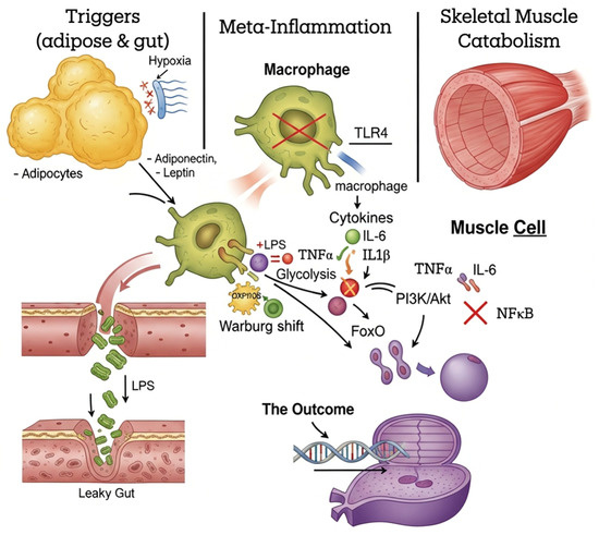
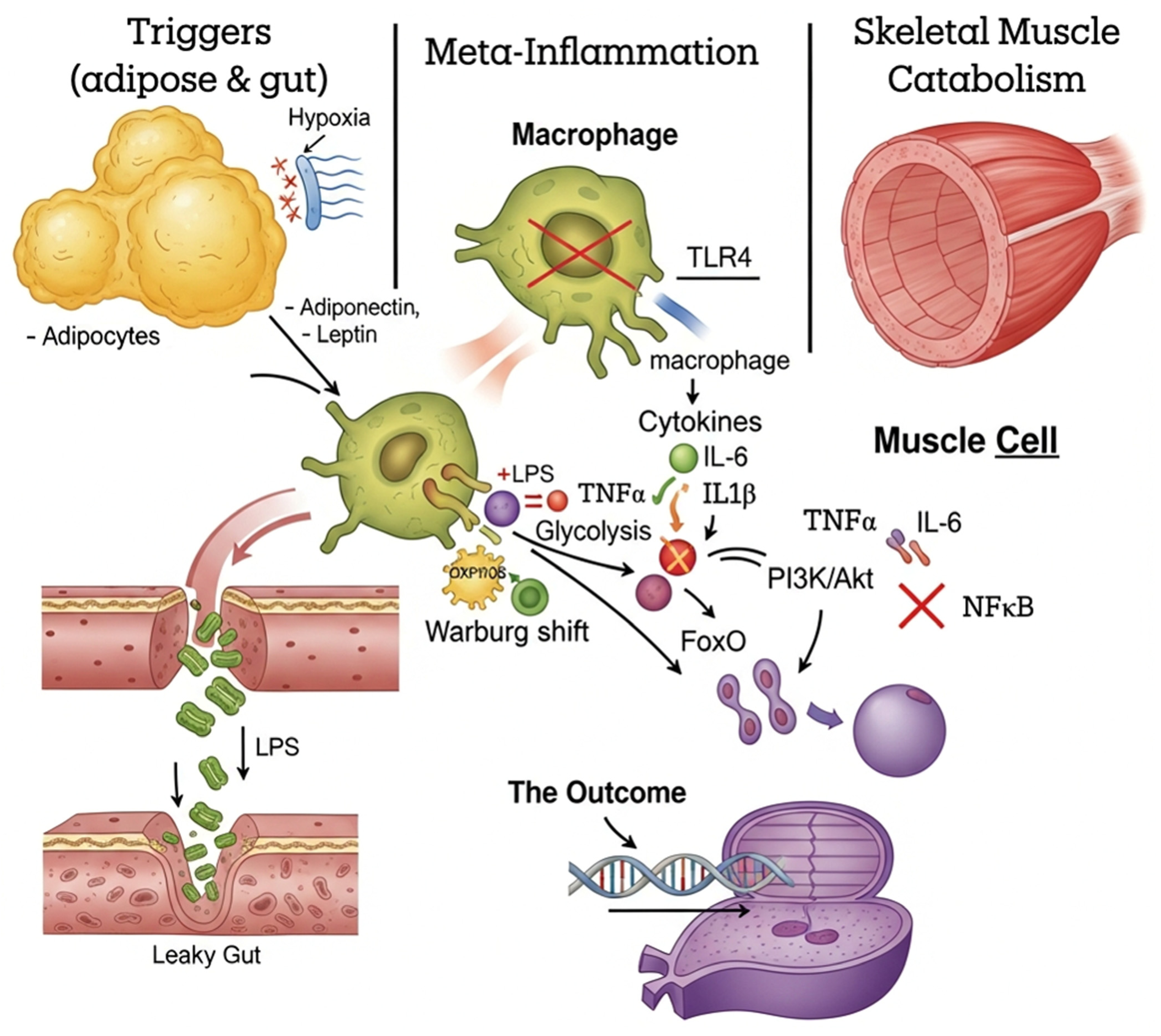
While lipotoxicity and insulin resistance (such as the impairment of the GLUT4 transporter via protein kinase C-) contribute to skeletal muscle metabolic dysfunction in obesity, these mechanisms alone do not fully account for the structural atrophy and impaired muscle accretion that characterize sarcopenia [37,38]. To properly characterize the “sarcopenic” phenotype, it is essential to examine the precise intracellular catabolic pathways activated by systemic meta-inflammation. Skeletal muscle mass is maintained through a dynamic balance between protein synthesis (anabolism) and protein degradation (catabolism) [18]. In PSO, adipose-derived TNF- and IL-6 systemically bombard muscle tissue, tilting this axis heavily toward catabolic signaling and muscle degradation (Figure 1).
Figure 1.
Schematic representation of obesity-induced meta-inflammation and its catabolic effects on skeletal muscle. Excess caloric intake drives visceral adipocyte hypertrophy, leading to local hypoxia and altered adipokine secretion. Concurrently, gut dysbiosis compromises intestinal barrier integrity, allowing for lipopolysaccharide (LPS) translocation (metabolic endotoxemia). LPS activates Toll-like Receptor 4 (TLR4) on infiltrating macrophages, triggering a Warburg-like glycolytic shift (M1 polarization). This results in the systemic release of pro-inflammatory cytokines (TNF-, IL-6). In the developing skeletal muscle, these cytokines impair the anabolic PI3K/Akt pathway, allowing unphosphorylated FoxO to enter the nucleus. Simultaneously, NF-B is activated. Together with upregulated myostatin signaling, these transcription factors drive the expression of the E3 ubiquitin ligases Atrogin-1 and MuRF-1, hyperactivating the ubiquitin–proteasome system (UPS) and leading to severe muscle protein degradation and impaired accretion.
5.1. The Ubiquitin–Proteasome System and E3 Ligases
The chief proteolytic system responsible for the degradation of structural muscle proteins is the ubiquitin–proteasome system (UPS) [18]. Proteins targeted for destruction are tagged with multiple ubiquitin molecules by specific enzymes known as ubiquitin E3 ligases. Once tagged, the polyubiquinated proteins are recognized and degraded into smaller peptides and amino acids by the 26S proteasome [39].
In skeletal muscle, two muscle-specific E3 ubiquitin ligases act as key mediators of atrophy: Muscle Atrophy F-box (MAFbx), commonly known as Atrogin-1 (encoded by the Fbxo32 gene), and Muscle RING-finger protein-1 (MuRF-1, encoded by the Trim63 gene) [18,40]. Under healthy physiological conditions, the expression of Atrogin-1 and MuRF-1 remains tightly regulated and is usually suppressed in growing muscle. However, inflammatory cytokines and oxidative stress markedly upregulate their expression, promoting accelerated muscle protein degradation and catabolic remodeling [18].
5.2. Catabolic Transcription Factors: FoxO, NF-κB, and Myostatin
The expression of the E3 ligases, specifically Fbxo32 (Atrogin-1) and Trim63 (MuRF-1), is tightly regulated by several catabolic transcription factors, primarily the Forkhead box O (FoxO) family, NF-B, and the myostatin/Smad pathway [18,40,41]. Rather than functioning in isolation, these pathways exhibit significant cross-talk; for instance, NF-B activation can further exacerbate insulin resistance, thereby indirectly suppressing Akt and amplifying FoxO signaling.
- The PI3K/Akt/FoxO Axis: Under healthy anabolic conditions, insulin and IGF-1 activate the PI3K/Akt pathway. Activated Akt directly phosphorylates FoxO transcription factors. This phosphorylation traps FoxO in the cell cytoplasm, preventing it from entering the nucleus and transcribing the atrophy-related genes Fbxo32 and Trim63 [18,39,40]. However, systemic TNF- and IL-6, along with oxidative stress, impair PI3K/Akt signaling. Without active Akt, FoxO remains unphosphorylated, freely translocates into the myocyte nucleus, and aggressively drives the transcription of Fbxo32 and Trim63, unleashing the UPS [18].
- The NF-κB Pathway: Systemic TNF- binds to its receptors on the muscle cell membrane, directly activating the IB kinase (IKK) complex. This leads to the phosphorylation and degradation of the NF-B inhibitor IB-, freeing NF-B (a potent inflammatory transcription factor) to enter the nucleus. NF-B acts as a direct inducer of Atrogin-1 and MuRF-1 by binding directly to the Trim63 (MuRF-1) promoter [18,42].
- Myostatin Upregulation: Meta-inflammation also stimulates the expression of myostatin, a profound negative regulator of muscle growth. Myostatin signals through the Smad2/3 transcription factors to independently induce the expression of Fbxo32 and Trim63 while simultaneously suppressing Akt activation, thus establishing a devastating feed-forward loop of muscle catabolism [18,41].
In pediatric patients undergoing active growth and musculoskeletal development, the continuous activation of FoxO and NF-B by an inflamed gut–adipose axis not only accelerates muscle protein degradation but also effectively paralyzes the physiological accretion of new muscle tissue required for normal development.
6. Nutraceutical Interventions: The Role of Dietary Anthocyanins
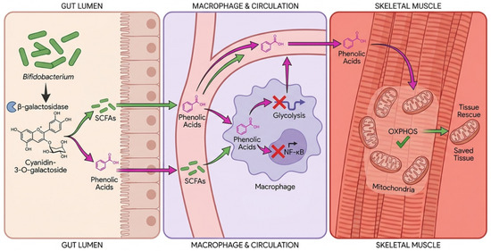
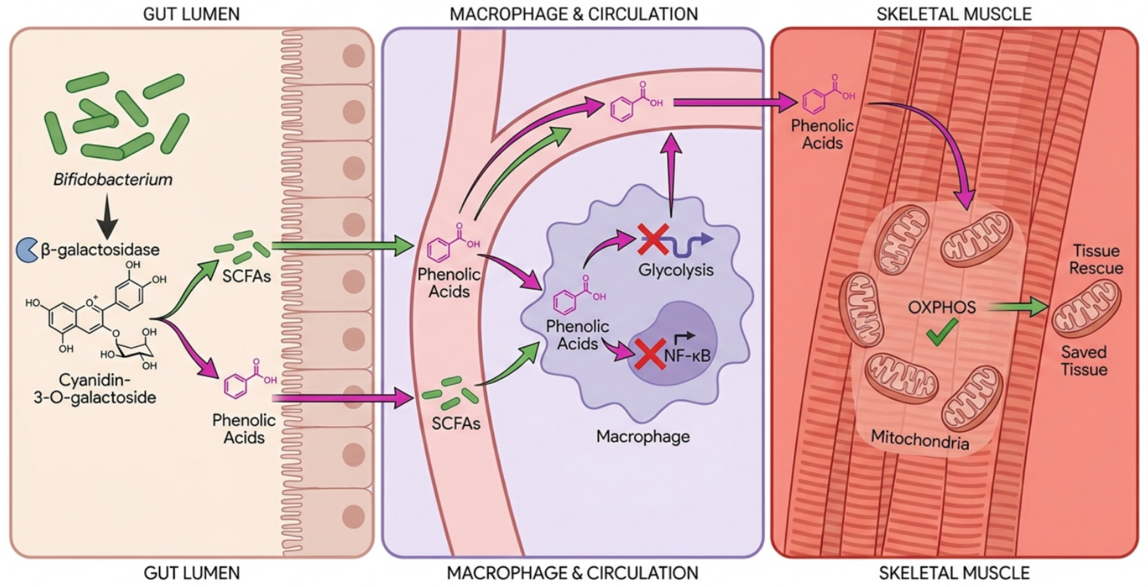
Given the complex pathophysiology linking gut dysbiosis to systemic meta-inflammation and subsequent FoxO-mediated muscle atrophy, single-target pharmacological interventions often fall short. This therapeutic limitation has increased interest in nutritional bioactives capable of simultaneously targeting inflammatory, metabolic, and microbiota-related pathways involved in PSO pathogenesis. Dietary polyphenols, specifically flavonoids such as anthocyanins, possess pleiotropic bioactive properties capable of modulating multiple nodes within this axis simultaneously (Figure 2) [18,40,43]. Anthocyanins are abundant in dark-colored plant foods such as berries, purple corn, and black beans [18].
Figure 2.
Cyanidin-3-O-galactoside gut–muscle axis and dual therapeutic action. The microbial enzyme -galactosidase from commensal Bifidobacterium cleaves cyanidin-3-O-galactoside. The liberated galactose ferments into protective short-chain fatty acids (SCFAs), while the bioactive phenolic acids enter circulation to inhibit macrophage glycolysis (M1 shift) and rescue muscle tissue from catabolism.
The biological efficacy of anthocyanins on skeletal muscle preservation revolves around their ability to interrupt the specific catabolic pathways activated in myocytes.
6.1. Direct Interruption of Myocyte Catabolism
Phenolic aglycones and their derivatives directly intervene in the molecular atrophy cascades. In vitro and in vivo models demonstrate that anthocyanins, particularly cyanidin-3-O-glucoside (C3G) and cyanidin-3-O-galactoside (ideain), act as potent antioxidants that neutralize intracellular reactive oxygen species (ROS) [18,43,44,45,46,47,48]. By mitigating oxidative stress and downregulating pro-inflammatory cytokines, these bioactives contribute to the restoration of the PI3K/Akt pathway activity [18,43].
Upon reaching skeletal muscle tissue, anthocyanin-derived metabolites promote Akt phosphorylation, a critical molecular switch that regulates anabolic signaling. Activated Akt phosphorylates FoxO transcription factors, preventing their nuclear translocation and suppressing the transcription of the atrophy-related genes Fbxo32 and Trim63 [18]. Consequently, this suppression inhibits the ubiquitin–proteasome system’s targeted degradation of muscle proteins [18,40]. Furthermore, anthocyanins have been shown to stabilize IB-, preventing the nuclear translocation of NF-B and thus neutralizing the secondary inflammatory trigger for Trim63 expression [18,42].
Collectively, these mechanisms suggest that anthocyanin-derived phenolic compounds may help preserve skeletal muscle integrity by attenuating inflammatory and proteolytic signaling pathways, directly inhibiting ubiquitin–proteasome-mediated muscle degradation while supporting developmental myogenesis and skeletal muscle accretion during critical stages of pediatric growth.
6.2. Bioavailability, Matrix Effects, and Inter-Individual Variability
While anthocyanins demonstrate potent antioxidant and anti-inflammatory properties in vitro, their clinical efficacy is heavily dictated by their complex pharmacokinetics. Anthocyanins exhibit notoriously poor systemic bioavailability, with typically less than 1–2% of the ingested dose appearing in the systemic circulation unchanged [49]. Furthermore, their absorption is highly susceptible to food matrix effects; for instance, co-ingestion with dairy proteins can bind anthocyanins, further reducing their absorption [50]. Therefore, the biological efficacy of anthocyanins is heavily dependent on inter-individual variability in the gut microbiome. Efficacy relies on the presence of specific microbial enzymes (such as -glucosidases and -galactosidases) capable of cleaving the parent compounds into smaller, more bioavailable phenolic acids (e.g., protocatechuic acid) [6,51]. Importantly, it is critical to emphasize that such nutraceutical interventions are not standalone cures; they must be viewed as adjunctive strategies designed to support foundational dietary interventions and physical activity protocols for obesity management.
7. Pharmacokinetics: Systemic Toxicity vs. Dietary Safety of Galactose Delivery
To maximize the therapeutic potential of anthocyanins, they are often delivered as glycosylated derivatives, such as cyanidin-3-O-galactoside [52,53]. To understand the safety and efficacy of this approach, a critical pharmacokinetic distinction must be made between the toxicological models utilizing injected D-galactose and the safe dietary consumption of complex galactosides.
In pharmacological research, chronic systemic administration of high-dose D-galactose (via subcutaneous or intraperitoneal injections) is used to artificially induce oxidative stress and muscle wasting in rodents. Because these injections bypass the gastrointestinal tract, they overwhelm the hepatic Leloir pathway. The unmetabolized galactose is diverted into producing galactitol and advanced glycation end-products (AGEs), driving severe systemic toxicity.
Conversely, dietary galacto-oligosaccharides (GOS) and anthocyanin–galactosides follow a completely different, safe pharmacokinetic trajectory. Delivered orally, they resist upper GI digestion and transit intact to the colon. Here, they are slowly fermented by commensal Bifidobacterium. This localized, gradual release does not cause a systemic spike in circulating free galactose, entirely avoiding Leloir pathway overload. Safety studies and decades of use of GOS in pediatric infant formulas strongly support the safety and utility of these compounds in children, confirming they do not induce the toxicities associated with systemic D-galactose injections [54,55].
The Systemic Aging Model vs. Dietary Cleavage
In pharmacological research, chronic systemic administration of high-dose D-galactose, most commonly via subcutaneous (s.c.) or intraperitoneal (i.p.) injections ranging from 100 to 800 mg/kg/day (and occasionally up to 1250 mg/kg/day) over a period of 6 to 8 weeks [56,57,58], is widely used to induce accelerated artificial aging, oxidative stress, and muscle wasting in rodent models [18]. At first glance, these findings appear to contradict the proposed therapeutic use of galactosides-bound anthocyanins or galacto-oligosaccharides (GOS) for therapeutic benefit. However, this apparent contradiction is largely explained by fundamental pharmacokinetic differences, specifically the route of administration, the dose, and the site of metabolism.
When high doses of D-galactose are injected systemically, the resulting increase in circulating galactose can exceed the metabolic capacity of the hepatic Leloir pathway, the primary enzymatic route for galactose metabolism [59]. Under these conditions, the liver is unable to convert galactose to glucose-6-phosphate efficiently, leading to the diversion of excess galactose into alternative metabolic pathways, including its reduction to galactitol by aldose reductase. Intracellular accumulation of galactitol has been associated with osmotic stress and cellular dysfunction. Concurrently, the excess systemic galactose reacts non-enzymatically with proteins and lipids to form advanced glycation end-products (AGEs) [18,41]. AGE accumulation enhances reactive oxygen species (ROS) production and systemic inflammation, leading to neurodegeneration and muscle atrophy that perfectly mimics severe sarcopenia.
Conversely, dietary galactosides, including anthocyanin–galactosides and galacto-oligosaccharides (GOS), follow a completely different pharmacokinetic trajectory. These complex molecules are largely resistant to mammalian digestive enzymes in the upper gastrointestinal tract [60]. They transit intact to the colon, where they serve as fermentable substrates for the commensal microbiota.
In the colon, specific beneficial bacteria (such as Bifidobacterium species, which are highly relevant to the pediatric microbiome) express the enzyme -galactosidase. This enzyme locally cleaves the galactose moiety, releasing galactose as a gradual and localized substrate for microbial fermentation. The released galactose can subsequently be utilized by the gut microbiota, contributing to the production of SCFAs like acetate, propionate, and butyrate [61,62,63,64,65,66,67]. This local colonic fermentation is unlikely to generate a systemic spike in circulating free galactose, entirely preventing the overwhelming of the Leloir pathway and the subsequent accumulation of toxic galactitol and AGEs. Thus, the dietary delivery of galactosides acts as a prebiotic trigger for gut barrier restoration without any of the toxicities associated with the systemic D-galactose aging model.
8. Critical Clinical Appraisal: Preclinical and Clinical Evidence
While the molecular targets of bioactives like cyanidin-3-glucoside (C3G), cyanidin-3-galactoside, and galacto-oligosaccharides (GOS) offer a profound mechanistic rationale, translating these findings into pediatric therapies requires a critical appraisal of the current empirical evidence. The literature presents a spectrum of robust preclinical models demonstrating metabolic rescue, alongside emerging clinical trials in human populations that support the safety and efficacy of these interventions.
To bridge the gap between basic in vitro molecular targets and the proposition of human synbiotic therapies, the following section consolidates the current experimental models, dosages, and metabolic outcomes of these critical bioactives.
8.1. Synthesis of Empirical Interventions
The efficacy of targeted bioactives must be evaluated across both highly controlled murine models (to establish mechanistic pathways) and human clinical trials (to establish safety, tolerance, and physiological outcomes). Table 1 summarizes representative studies that anchor the therapeutic claims regarding Cyanidin-3-O-galactoside and galacto-oligosaccharides in mitigating obesity-associated metabolic dysfunction.
Table 1.
Therapeutic interventions using galacto-oligosaccharides in metabolic dysfunctions.
A critical caveat in the current literature is the absence of randomized controlled trials testing these specific targeted synbiotics directly in PSO cohorts. Consequently, the clinical evidence summarized in Table 1 utilizes data from adult and murine models as a necessary translational bridge. While caution is warranted, such extrapolation is supported by the conservation of key biological pathways involved in meta-inflammation, including LPS-mediated TLR4 activation, NF-B signaling, and cytokine-driven muscle catabolism. This extrapolation is scientifically justified by several factors. First, while the macroscopic dynamics of muscle growth differ between adults and children, the fundamental intracellular pathways of meta-inflammation (e.g., LPS-induced TLR4 activation and NF-B-driven catabolism) are biologically conserved across the lifespan. Second, the prebiotic interventions utilized, specifically galacto-oligosaccharides (GOS), have a profound, targeted affinity for Bifidobacterium. Given that Bifidobacterium is the keystone genus of the healthy early-life microbiome, GOS supplementation is highly physiologically relevant to pediatric cohorts. Finally, demonstrating safety, tolerability, and the resolution of metabolic endotoxemia in adult populations provides the ethical and empirical foundation required to design the pediatric-specific clinical trials proposed in this review.
The evidence synthesized in Table 1 highlights several critical findings regarding the potential metabolic effects of anthocyanins and galacto-oligosaccharides. Preclinically, the oral administration of cyanidin derivatives (at physiologically relevant doses of 50–200 mg/kg) consistently demonstrates the capacity to suppress adipogenesis, modulate lipid metabolism, and downregulate the inflammatory cytokines (TNF- and IL-6) responsible for muscle catabolism [52,68]. Crucially, the restoration of AMPK and GLUT4 expression in these models suggests that anthocyanins can actively reverse insulin resistance at the level of the skeletal muscle, providing the energetic substrate required to prevent muscle wasting [43].
Clinically, GOS supplementation in humans (ranging from 5.5 g to 15 g daily) consistently demonstrated its bifidogenic capacity and favorable safety and tolerability profiles [61,66,70]. Given that pediatric obesity is specifically associated with depletion of infant-type Bifidobacterium [5,23,24], these findings support the potential relevance of GOS as a targeted prebiotic mechanism to restore this specific microbial signature. Furthermore, the dose-dependent reduction in plasma LPS (endotoxin) and C-reactive protein (CRP) observed in human trials [61] suggests improvements in gut barrier integrity and systemic inflammatory status. These findings support the hypothesis that microbial fermentation of galactose-containing substrates may contribute to the maintenance of intestinal barrier function and the attenuation of metabolic endotoxemia, a key driver of chronic immunometabolic dysfunction.
However, the available clinical evidence also reveals an important limitation: although prebiotics such as GOS effectively modulate the gut microbiota and improve intestinal barrier function, they may be insufficient on their own to reverse established insulin resistance or advanced muscle atrophy within relatively short intervention periods [66,69]. This limitation highlights the potential value of combining therapeutic strategies targeting both microbial and host metabolic pathways.
8.2. Limitations of Current Evidence and Critical Appraisal
While the theoretical framework supporting synbiotic interventions is robust, a critical appraisal of the literature reveals significant gaps. The vast majority of mechanistic evidence regarding UPS activation, FoxO/NF-B cross-talk, and anthocyanin-mediated muscle rescue is derived from murine models and adult human trials. Direct clinical evidence evaluating these specific targeted synbiotics in pediatric sarcopenic obesity cohorts remains distinctly lacking. Furthermore, while preclinical studies overwhelmingly report positive metabolic outcomes, some human trials utilizing prebiotics in obese populations have yielded contradictory or modest results, often failing to significantly alter deep tissue insulin resistance without concurrent caloric restriction. Therefore, future pediatric randomized controlled trials are urgently needed to validate these mechanistic hypotheses and establish optimal, age-appropriate dosing.
9. Future Directions and Synbiotic Therapies
The complementary mechanisms of action of dietary polyphenols and microbial prebiotics open an innovative frontier in pediatric nutritional therapy: the development of targeted synbiotics. Synbiotics combine prebiotic substrates with specific bioactive compounds or probiotics to synergistically enhance complementary biological effects.
In the context of pediatric sarcopenic obesity, the combination of galacto-oligosaccharides (GOS) with anthocyanin-rich extracts (delivering compounds like cyanidin-3-O-galactoside) offers a comprehensive blockade of the gut–immunometabolism–muscle axis.
- Gut Level Restoration: The GOS component selectively feeds the depleted Bifidobacterium populations characteristic of the pediatric obese microbiome [5,71]. The subsequent proliferation of these beneficial taxa and the concomitant synthesis of short-chain fatty acids (e.g., butyrate and acetate) serve to fortify intestinal tight junction integrity via the modulation of zonulin pathways. This localized barrier restoration effectively limits the paracellular translocation of lipopolysaccharides (LPS), thereby attenuating the primary driver of metabolic endotoxemia [63,64,65,72].
- Systemic and Intracellular Rescue: Simultaneously, the microbial enzyme -galactosidase cleaves the glycosidic bonds of the ingested anthocyanins [6,51]. This releases the highly bioactive phenolic aglycones (cyanidin) into systemic circulation. With the systemic cytokine burden attenuated by upstream prebiotic modulation, these highly bioavailable phenolic aglycones can efficiently partition into skeletal muscle tissue. Within the myocyte, they facilitate the restoration of PI3K/Akt signaling, promoting the cytosolic sequestration of FoxO transcription factors while concurrently suppressing NF-κB nuclear translocation. This coordinated molecular intervention effectively downregulates the ubiquitin–proteasome system (UPS), thereby mitigating proteolytic degradation and preserving skeletal muscle integrity [18].
Future pediatric clinical trials must prioritize investigating these specific synbiotic pairings using standardized diagnostic criteria, such as SMMI z-scores, together with biomarkers of gut barrier function, microbial metabolism, and systemic inflammation (e.g., fecal SCFAs, serum zonulin, and circulating LPS). Such studies will be essential to evaluate the efficacy of non-pharmacological interventions and mechanistic relevance, as well as their potential to support normal skeletal muscle development in pediatric sarcopenic obesity.
10. Conclusions
Pediatric sarcopenic obesity represents a severe, complex metabolic derangement characterized by the coexistence of excess adiposity and impaired skeletal muscle development. Unlike age-related sarcopenia, PSO arises during critical periods of growth and is driven by a combination of energy imbalance, gut dysbiosis, and immunometabolic reprogramming toward a chronic proinflammatory state. This state is characterized by elevated levels of cytokines such as IL-6 and TNF-α, which disrupt normal muscle accretion by activating catabolic pathways, including FoxO- and NF-κB-mediated transcription of Fbxo32 (Atrogin-1) and Trim63 (MuRF-1), ultimately promoting protein degradation through the ubiquitin–proteasome system.
The gut microbiota serves as the crucial central checkpoint in this disease process. Modulating this axis via dietary bioactives offers a highly targeted physiological intervention. Providing structural galactosides, whether as prebiotic galacto-oligosaccharides or bound to anthocyanins like cyanidin-3-O-galactoside, leverages the localized enzymatic power of the colonic microbiota. Through microbial biotransformation, the coordinated release of fermentable galactose and bioactive phenolic acids generates gut-healing SCFAs and systemic antioxidants. By understanding the vital pharmacokinetic distinctions that render colonic galactose safe and highly beneficial, researchers can confidently develop targeted, early-life synbiotic interventions. Although direct clinical evidence remains limited, targeted symbiotic strategies may offer a promising approach to modulating meta-inflammation, improving immunometabolic homeostasis, and supporting health and muscle development in PSO.
Author Contributions
Conceptualization, O.M.-C. and D.C.-E.; methodology, D.C.-E., A.A.R.-E., F.P.-A., P.Z.-D., B.L.-E. and A.M.J.-G.; formal analysis, O.M.-C. and D.C.-E.; investigation, D.C.-E., A.A.R.-E., F.P.-A., P.Z.-D., B.L.-E. and A.M.J.-G.; resources, O.M.-C.; data curation, O.M.-C., D.C.-E. and A.A.R.-E.; writing—original draft preparation, A.A.R.-E., F.P.-A., P.Z.-D., B.L.-E., A.M.J.-G., D.C.-E. and O.M.-C.; supervision, O.M.-C. and D.C.-E.; project administration, O.M.-C. All authors have read and agreed to the published version of the manuscript.
Funding
This research received no external funding.
Data Availability Statement
No new data were created or analyzed in this study. Data sharing is not applicable to this article.
Acknowledgments
During the preparation of this manuscript, the authors used Google’s Imagen 4 for the generation of the figures. The authors have reviewed and edited the output and take full responsibility for the content of this publication.
Conflicts of Interest
The authors declare no conflicts of interest.
Abbreviations
The following abbreviations are used in this manuscript:
| AGEs | Advanced glycation end-products |
| AMPK/pAMPK | AMP-activated protein kinase/phosphorylated AMPK |
| BIA | Bioelectrical impedance analysis |
| C3G | Cyanidin-3-O-glucoside |
| CRP | C-reactive protein |
| DXA | Dual-energy X-ray absorptiometry |
| FoxO | Forkhead box O |
| GLUT4 | Glucose transporter type 4 |
| GOS | Galacto-oligosaccharides |
| HIF-1α | Hypoxia-inducible factor 1-alpha |
| IGF-1 | Insulin-like growth factor-1 |
| IKK | IκB kinase complex |
| IL-1β | Interleukin-1 beta |
| IL-6 | Interleukin-6 |
| i.p. | Intraperitoneal |
| LPS | Lipopolysaccharide |
| MAFbx | Muscle atrophy F-box (also known as Atrogin-1) |
| mTOR | Mammalian target of rapamycin |
| MuRF-1 | Muscle RING-finger protein-1 |
| NF-κB | Nuclear factor-kappa B |
| OXPHOS | Oxidative Phosphorylation |
| PI3K | Phosphatidylinositol 3-kinase |
| PSO | Pediatric sarcopenic obesity |
| ROS | Reactive oxygen species |
| s.c. | Subcutaneous |
| SCFAs | Short-chain fatty acids |
| SMMI | Skeletal Muscle Mass Index |
| STZ | Streptozotocin |
| TLR4 | Toll-Like Receptor 4 |
| TNF-α | Tumor necrosis factor-alpha |
| UPS | Ubiquitin–proteasome system |
References
- Zembura, M.; Matusik, P. Sarcopenic Obesity in Children and Adolescents: A Systematic Review. Front. Endocrinol. 2023, 14, 1269546. [Google Scholar] [CrossRef] [Scilit] [PubMed]
- World Health Organization (WHO). Obesity. World Health Organization (WHO): Geneva, Switzerland, 2024. Available online: https://www.who.int/health-topics/obesity (accessed on 19 February 2026).
- Zhao, J.; Huang, Y.; Yu, X. A Narrative Review of Gut-Muscle Axis and Sarcopenia: The Potential Role of Gut Microbiota. Int. J. Gen. Med. 2021, 14, 1263–1273. [Google Scholar] [CrossRef] [Scilit] [PubMed]
- O’Neill, L.A.J.; Kishton, R.J.; Rathmell, J. A Guide to Immunometabolism for Immunologists. Nat. Rev. Immunol. 2016, 16, 553–565. [Google Scholar] [CrossRef] [Scilit] [PubMed]
- Saeed, N.; Al-Beltagi, M.; Bediwy, A.; El-Sawaf, Y.; Toema, O. Gut Microbiota in Various Childhood Disorders: Implication and Indications. World J. Gastroenterol. 2022, 28, 1875–1901. [Google Scholar] [CrossRef] [Scilit] [PubMed]
- Cassidy, A.; Minihane, A.-M. The Role of Metabolism (and the Microbiome) in Defining the Clinical Efficacy of Dietary Flavonoids. Am. J. Clin. Nutr. 2017, 105, 10–20. [Google Scholar] [CrossRef] [Scilit] [PubMed]
- Cruz-Jentoft, A.J.; Bahat, G.; Bauer, J.; Boirie, Y.; Bruyère, O.; Cederholm, T.; Cooper, C.; Landi, F.; Rolland, Y.; Sayer, A.A.; et al. Sarcopenia: Revised European Consensus on Definition and Diagnosis. Age Ageing 2019, 48, 16–31. [Google Scholar] [CrossRef] [Scilit] [PubMed]
- Marques, M.; Baptista, F. Pediatric Sarcopenia: What Do We Know? Acta Médica Port. 2025, 38, 800–807. [Google Scholar] [CrossRef] [Scilit] [PubMed]
- Flores-Opazo, M.; Monsalves-Álvarez, M.; Sepúlveda-Guzmán, C.; Jannas-Vela, S.; Valero, P.F.; Burrows, R.; Correa-Burrows, P.; Troncoso, R. Perspective Review: Advancing toward the Diagnosis of Sarcopenia in the Pediatric Population. Pediatr. Res. 2025, 1–7. [Google Scholar] [CrossRef] [Scilit] [PubMed]
- Kiosis, G.; Ioannou, D.; Skourtsidis, K.; Fouskas, V.; Stergiou, K.; Kavvadas, D.; Papamitsou, T.; Karachrysafi, S.; Kourti, M. Newer Insights on the Occurrence of Sarcopenia in Pediatric Patients with Cancer: A Systematic Review of the Past 5 Years of Literature. Cancers 2025, 17, 3188. [Google Scholar] [CrossRef] [Scilit] [PubMed]
- Wang, S.; Zhang, W.; Qin, Z.; Zhou, X.; Xue, C.; Wang, D.; Liang, X.; Zhang, Z.; Zhan, S.; Wang, S.; et al. Sarcopenic Obesity in Children: An Emerging Complication Evidenced by Clinical Data and a Juvenile Mouse Model. J. Cachexia Sarcopenia Muscle 2026, 17, e70278. [Google Scholar] [CrossRef] [Scilit] [PubMed]
- Shamah-Levy, T.; Gaona-Pineda, E.B.; Cuevas-Nasu, L.; Valenzuela-Bravo, D.G.; Morales-Ruan, C.; Rodríguez-Ramírez, S.; Méndez-Gómez-Humarán, I.; Ávila-Arcos, M.A.; Álvarez-Sánchez, C.; Ávila-Curiel, A.; et al. Overweight and Obesity in Mexican School-Aged and Adolescents. Salud Pública De México 2024, 66, 404–413. [Google Scholar] [CrossRef] [Scilit] [PubMed]
- Centers for Disease Control and Prevention (CDC) Overweight and Obesity 2024. Available online: https://www.cdc.gov/obesity/ (accessed on 19 February 2026).
- Weihrauch-Blüher, S.; Wiegand, S. Risk Factors and Implications of Childhood Obesity. Curr. Obes. Rep. 2018, 7, 254–259. [Google Scholar] [CrossRef] [Scilit] [PubMed]
- Zhang, X.; Zhou, Y.; Ji, Y.; Zhao, L.; Gong, J.; Chen, Y.; Shen, L.; Cai, S.; Li, X. Comparison of Body Composition Between Thin and Normal-Weight Sarcopenic Children: A Single-Center, Propensity Score Matching Analysis of a Large-Scale Study. BMC Pediatr. 2026, 26, 353. [Google Scholar] [CrossRef] [Scilit] [PubMed]
- Bodine, S.C.; Baehr, L.M. Skeletal Muscle Atrophy and the E3 Ubiquitin Ligases MuRF1 and MAFbx/Atrogin-1. Am. J. Physiol.-Endocrinol. Metab. 2014, 307, E469–E484. [Google Scholar] [CrossRef] [Scilit] [PubMed]
- Thoma, A.; Lightfoot, A.P. NF-kB and Inflammatory Cytokine Signalling: Role in Skeletal Muscle Atrophy. Adv. Exp. Med. Biol. 2018, 1088, 267–279. [Google Scholar] [CrossRef] [Scilit] [PubMed]
- Nikawa, T.; Ulla, A.; Sakakibara, I. Polyphenols and Their Effects on Muscle Atrophy and Muscle Health. Molecules 2021, 26, 4887. [Google Scholar] [CrossRef] [Scilit] [PubMed]
- Vlachopapadopoulou, E.; Mazur, A.; Gawlik, A.; Telega, G.; Wojcik, M.; Molnár, D. Editorial: Endocrine and Metabolic Consequences of Childhood Obesity, Volume III. Front. Endocrinol. 2025, 16, 1546470. [Google Scholar] [CrossRef] [Scilit] [PubMed]
- Raksadawan, N.; Sermswan, A.; Ukkrit, J. Monitoring Skeletal Muscle Growth in Children. Asia Pac. J. Paediatr. Child Health 2022, 5, 45–56. [Google Scholar]
- Zhu, J.; He, M.; Li, S.; Lei, Y.; Xiang, X.; Guo, Z.; Wang, Q. Shaping Oral and Intestinal Microbiota and the Immune System during the First 1,000 Days of Life. Front. Pediatr. 2025, 13, 1471743. [Google Scholar] [CrossRef] [Scilit] [PubMed]
- Tassoni, D.S.; Macedo, R.C.O.; Delpino, F.M.; Santos, H.O. Gut Microbiota and Obesity: The Chicken or the Egg? Obesities 2023, 3, 296–321. [Google Scholar] [CrossRef] [Scilit]
- Riva, A.; Borgo, F.; Lassandro, C.; Verduci, E.; Morace, G.; Borghi, E.; Berry, D. Pediatric obesity is associated with an altered gut microbiota and discordant shifts in Firmicutes populations. Environ. Microbiol. 2017, 19, 95–105. [Google Scholar] [CrossRef] [Scilit] [PubMed]
- dos Santos Pereira Indiani, C.M.; Rizzardi, K.F.; Castelo, P.M.; Ferraz, L.F.C.; Darrieux, M.; Parisotto, T.M. Childhood Obesity and Firmicutes/Bacteroidetes Ratio in the Gut Microbiota: A Systematic Review. Child. Obes. 2018, 14, 501–509. [Google Scholar] [CrossRef] [Scilit] [PubMed]
- Cani, P.D.; Amar, J.; Iglesias, M.A.; Poggi, M.; Knauf, C.; Bastelica, D.; Neyrinck, A.M.; Fava, F.; Tuohy, K.M.; Chabo, C.; et al. Metabolic Endotoxemia Initiates Obesity and Insulin Resistance. Diabetes 2007, 56, 1761–1772. [Google Scholar] [CrossRef] [Scilit] [PubMed]
- Gupta, V.K.; Paul, S.; Dutta, C. Geography, Ethnicity or Subsistence-Specific Variations in Human Microbiome Composition and Diversity. Front. Microbiol. 2017, 8, 1162. [Google Scholar] [CrossRef] [Scilit] [PubMed]
- Rothschild, D.; Weissbrod, O.; Barkan, E.; Kurilshikov, A.; Korem, T.; Zeevi, D.; Costea, P.I.; Godneva, A.; Kalka, I.N.; Bar, N.; et al. Environment Dominates over Host Genetics in Shaping Human Gut Microbiota. Nature 2018, 555, 210–215. [Google Scholar] [CrossRef] [Scilit] [PubMed]
- Ajamian, M.; Steer, D.; Rosella, G.; Gibson, P.R. Serum Zonulin as a Marker of Intestinal Mucosal Barrier Function: May Not Be What It Seems. PLoS ONE 2019, 14, e0210728. [Google Scholar] [CrossRef] [Scilit] [PubMed]
- Scheffler, L.; Crane, A.; Heyne, H.; Tönjes, A.; Schleinitz, D.; Ihling, C.H.; Stumvoll, M.; Freire, R.; Fiorentino, M.; Fasano, A.; et al. Widely Used Commercial ELISA Does Not Detect Precursor of Haptoglobin2, but Recognizes Properdin as a Potential Second Member of the Zonulin Family. Front. Endocrinol. 2018, 9, 22. [Google Scholar] [CrossRef] [Scilit] [PubMed]
- Manco, M.; Putignani, L.; Bottazzo, G.F. Gut Microbiota, Lipopolysaccharides, and Innate Immunity in the Pathogenesis of Obesity and Cardiovascular Risk. Endocr. Rev. 2010, 31, 817–844. [Google Scholar] [CrossRef] [Scilit] [PubMed]
- Saltiel, A.R.; Olefsky, J.M. Inflammatory Mechanisms Linking Obesity and Metabolic Disease. J. Clin. Investig. 2017, 127, 1–4. [Google Scholar] [CrossRef] [Scilit] [PubMed]
- Weisberg, S.P.; McCann, D.; Desai, M.; Rosenbaum, M.; Leibel, R.L.; Ferrante, A.W. Obesity Is Associated with Macrophage Accumulation in Adipose Tissue. J. Clin. Investig. 2003, 112, 1796–1808. [Google Scholar] [CrossRef] [Scilit] [PubMed]
- Hotamisligil, G.S.; Shargill, N.S.; Spiegelman, B.M. Adipose Expression of Tumor Necrosis Factor-Alpha: Direct Role in Obesity-Linked Insulin Resistance. Science 1993, 259, 87–91. [Google Scholar] [CrossRef] [Scilit] [PubMed]
- Kosteli, A.; Sugaru, E.; Haemmerle, G.; Martin, J.F.; Lei, J.; Zechner, R.; Ferrante, A.W. Weight Loss and Lipolysis Promote a Dynamic Immune Response in Murine Adipose Tissue. J. Clin. Investig. 2010, 120, 3466–3479. [Google Scholar] [CrossRef] [Scilit] [PubMed]
- Boutens, L.; Stienstra, R. Adipose Tissue Macrophages: Going off Track during Obesity. Diabetologia 2016, 59, 879–894. [Google Scholar] [CrossRef] [Scilit] [PubMed]
- Makowski, L.; Chaib, M.; Rathmell, J.C. Immunometabolism: From Basic Mechanisms to Translation. Immunol. Rev. 2020, 295, 5–14. [Google Scholar] [CrossRef] [Scilit] [PubMed]
- Xu, H.; Barnes, G.T.; Yang, Q.; Tan, G.; Yang, D.; Chou, C.J.; Sole, J.; Nichols, A.; Ross, J.S.; Tartaglia, L.A.; et al. Chronic Inflammation in Fat Plays a Crucial Role in the Development of Obesity-Related Insulin Resistance. J. Clin. Investig. 2003, 112, 1821–1830. [Google Scholar] [CrossRef] [Scilit] [PubMed]
- Gil-Iturbe, E.; Arbones-Mainar, J.M.; Moreno-Aliaga, M.J.; Lostao, M.P. GLUT12 and Adipose Tissue: Expression, Regulation and Its Relation with Obesity in Mice. Acta Physiol. 2019, 226, e13283. [Google Scholar] [CrossRef] [Scilit] [PubMed]
- Kim, C.; Hwang, J.-K. Flavonoids: Nutraceutical Potential for Counteracting Muscle Atrophy. Food Sci. Biotechnol. 2020, 29, 1619–1640. [Google Scholar] [CrossRef] [Scilit] [PubMed]
- Yun, C.E.; Vuong, T.A.; So, H.-K.; Lee, S.J.; Na, M.W.; Anh, S.; Lee, H.-K.; Kim, K.H.; Kang, J.-S.; Bae, G.-U. Aronia Upregulates Myogenic Differentiation and Augments Muscle Mass and Function Through Muscle Metabolism. Front. Nutr. 2021, 8, 753643. [Google Scholar] [CrossRef] [Scilit] [PubMed]
- Li, M.; Qin, Y.; Geng, R.; Fang, J.; Kang, S.-G.; Huang, K.; Tong, T. Effects and Mechanisms of Phytochemicals on Skeletal Muscle Atrophy in Glucolipid Metabolic Disorders: Current Evidence and Future Perspectives. Food Innov. Adv. 2025, 4, 83–98. [Google Scholar] [CrossRef] [Scilit]
- Verdi, A.M.O.H.; Rizzardi, M.L.; Soares, J.M.; Schiessel, D.L.; Coletti, D.; Seelaender, M.C.L.; Gonçalves, D.C. Nutraceutical Interception of Cachexia: Grape-Derived Compounds as Pathophysiological Network Modulators. Biology 2025, 14, 1159. [Google Scholar] [CrossRef] [Scilit] [PubMed]
- Randeni, N.; Luo, J.; Xu, B. Critical Review on Anti-Obesity Effects of Anthocyanins Through PI3K/Akt Signaling Pathways. Nutrients 2025, 17, 1126. [Google Scholar] [CrossRef] [Scilit] [PubMed]
- Deepa, P.; Sowndhararajan, K.; Hong, M. A Review of the Role of an Anthocyanin, Cyanidin-3-o-$\beta$-Glucoside in Obesity-Related Complications. Plants 2023, 12, 3889. [Google Scholar] [CrossRef] [Scilit] [PubMed]
- Mattioli, R.; Francioso, A.; Mosca, L.; Silva, P. Anthocyanins: A Comprehensive Review of Their Chemical Properties and Health Effects on Cardiovascular and Neurodegenerative Diseases. Molecules 2020, 25, 3809. [Google Scholar] [CrossRef] [Scilit] [PubMed]
- Molonia, M.S.; Occhiuto, C.; Muscarà, C.; Speciale, A.; Bashllari, R.; Villarroya, F.; Saija, A.; Cimino, F.; Cristani, M. Cyanidin-3-o-Glucoside Restores Insulin Signaling and Reduces Inflammation in Hypertrophic Adipocytes. Arch. Biochem. Biophys. 2020, 691, 108488. [Google Scholar] [CrossRef] [Scilit] [PubMed]
- Huang, F.; Zhao, R.; Xia, M.; Shen, G.X. Impact of Cyanidin-3-Glucoside on Gut Microbiota and Relationship with Metabolism and Inflammation in High Fat-High Sucrose Diet-Induced Insulin Resistant Mice. Microorganisms 2020, 8, 1238. [Google Scholar] [CrossRef] [Scilit] [PubMed]
- Tu, P.; Zheng, X.; Niu, H.; Chen, Z.; Wang, X.; Wu, L.; Tang, Q. Characterizing the Gut Microbial Metabolic Profile of Mice with the Administration of Berry-Derived Cyanidin-3-Glucoside. Metabolites 2023, 13, 818. [Google Scholar] [CrossRef] [Scilit] [PubMed]
- Czank, C.; Cassidy, A.; Zhang, Q.; Morrison, D.J.; Preston, T.; Kroon, P.A.; Botting, N.P.; Kay, C.D. Human Metabolism and Elimination of the Anthocyanin, Cyanidin-3-Glucoside: A 13C-Tracer Study. Am. J. Clin. Nutr. 2013, 97, 995–1003. [Google Scholar] [CrossRef] [Scilit] [PubMed]
- Bohn, T. Dietary Factors Affecting Polyphenol Bioavailability. Nutr. Rev. 2014, 72, 429–452. [Google Scholar] [CrossRef] [Scilit] [PubMed]
- Faria, A.; Fernandes, I.; Norberto, S.; Mateus, N.; Calhau, C. Interplay between Anthocyanins and Gut Microbiota. J. Agric. Food Chem. 2014, 62, 6898–6902. [Google Scholar] [CrossRef] [Scilit] [PubMed]
- Lim, S.M.; Jung, J.I.; Lee, H.S.; Kim, S.M.; Kim, N.Y.; Seo, T.S.; Bae, J.S.; Kim, E.J. Cyanidin-3-o-Galactoside-Enriched Aronia Melanocarpa Extract Attenuates Weight Gain and Adipogenic Pathways in High-Fat Diet-Induced Obese C57BL/6 Mice. Nutrients 2019, 11, 1190. [Google Scholar] [CrossRef] [Scilit] [PubMed]
- Wu, P.; Li, F.; Zhang, J.; Yang, B.; Ji, Z.; Chen, W. Phytochemical Compositions of Extract from Peel of Hawthorn Fruit, and Its Antioxidant Capacity, Cell Growth Inhibition, and Acetylcholinesterase Inhibitory Activity. BMC Complement. Altern. Med. 2017, 17, 151. [Google Scholar] [CrossRef] [Scilit] [PubMed]
- Vandenplas, Y.; Greef, E.D.; Veereman, G. Prebiotics in Infant Formula. Gut Microbes 2014, 5, 681–687. [Google Scholar] [CrossRef] [Scilit] [PubMed]
- Braegger, C.; Chmielewska, A.; Decsi, T.; Kolacek, S.; Mihatsch, W.; Moreno, L.; Pieścik, M.; Puntis, J.; Shamir, R.; Szajewska, H.; et al. Supplementation of Infant Formula with Probiotics and/or Prebiotics: A Systematic Review and Comment by the ESPGHAN Committee on Nutrition. J. Pediatr. Gastroenterol. Nutr. 2011, 52, 238–250. [Google Scholar] [CrossRef] [Scilit] [PubMed]
- Liu, T.; Liu, X.; Chen, Q.; Ren, J.; Li, Z.; Qiu, X.; Wang, X.; Wu, L.; Hu, M.; Chen, D.; et al. Systematic Evaluation of a Mouse Model of Aging-Associated Parkinson’s Disease Induced with MPTP and D-Galactose. Biology 2026, 15, 169. [Google Scholar] [CrossRef] [Scilit] [PubMed]
- Niu, S.; Azzouz, R.S.; Yan, L.-J. Rodent Models of D-Galactose Induction of Accelerated Aging: A Platform for Exploring Kidney Aging Mechanisms and Anti-Kidney Aging Strategies. Cells 2026, 15, 766. [Google Scholar] [CrossRef] [Scilit] [PubMed]
- Sadigh-Eteghad, S.; McCann, S.K.; Majdi, A.; Mahmoudi, J.; Vafaee, M.S.; Macleod, M.R. D-Galactose-Induced Brain Ageing Model: A Systematic Review and Meta-Analysis on Cognitive Outcomes and Oxidative Stress Indices. PLoS ONE 2017, 12, e0184122. [Google Scholar] [CrossRef] [Scilit] [PubMed]
- Azman, K.F.; Zakaria, R. D-Galactose-Induced Accelerated Aging Model: An Overview. Biogerontology 2019, 20, 763–782. [Google Scholar] [CrossRef] [Scilit] [PubMed]
- Davani-Davari, D.; Negahdaripour, M.; Karimzadeh, I.; Seifan, M.; Mohkam, M.; Masoumi, S.J.; Berenjian, A.; Ghasemi, Y. Prebiotics: Definition, Types, Sources, Mechanisms, and Clinical Applications. Foods 2019, 8, 92. [Google Scholar] [CrossRef] [Scilit] [PubMed]
- Vulevic, J.; Juric, A.; Tzortzis, G.; Gibson, G.R. A Mixture of Trans-Galactooligosaccharides Reduces Markers of Metabolic Syndrome and Modulates the Fecal Microbiota and Immune Function of Overweight Adults. J. Nutr. 2013, 143, 324–331. [Google Scholar] [CrossRef] [Scilit] [PubMed]
- Umasankar, P.; K., N.; Govindarajalu, S.; Sundar, J.S.; Srinivasan, V.; Ramachandran, K.; Chandran, N.; Prem, D. Healthier Gut, Stronger Kids: Harnessing Prebiotics for Childhood Obesity-a Narrative Review. Int. J. Community Med. Public Health 2025, 12, 3820–3826. [Google Scholar] [CrossRef] [Scilit]
- Hamer, H.M.; Jonkers, D.; Venema, K.; Vanhoutvin, S.; Troost, F.J.; Brummer, R.J. The Role of Butyrate on Colonic Function. Aliment. Pharmacol. Ther. 2008, 27, 104–119. [Google Scholar] [CrossRef] [Scilit] [PubMed]
- Bäckhed, F.; Ding, H.; Wang, T.; Hooper, L.V.; Koh, G.Y.; Nagy, A.; Semenkovich, C.F.; Gordon, J.I. The Gut Microbiota as an Environmental Factor That Regulates Fat Storage. Proc. Natl. Acad. Sci. USA 2004, 101, 15718–15723. [Google Scholar] [CrossRef] [Scilit] [PubMed]
- Kelly, C.J.; Zheng, L.; Campbell, E.L.; Saeedi, B.; Scholz, C.C.; Bayless, A.J.; Wilson, K.E.; Glover, L.E.; Kominsky, D.J.; Magnuson, A.; et al. Crosstalk Between Microbiota-Derived Short-Chain Fatty Acids and Intestinal Epithelial HIF Augments Tissue Barrier Function. Cell Host Microbe 2015, 17, 662–671. [Google Scholar] [CrossRef] [Scilit] [PubMed]
- Canfora, E.E.; van der Beek, C.M.; Hermes, G.D.A.; Goossens, G.H.; Jocken, J.W.E.; Holst, J.J.; van Eijk, H.M.; Venema, K.; Smidt, H.; Zoetendal, E.G.; et al. Supplementation of Diet With Galacto-Oligosaccharides Increases Bifidobacteria, but Not Insulin Sensitivity, in Obese Prediabetic Individuals. Gastroenterology 2017, 153, 87–97.e3. [Google Scholar] [CrossRef] [Scilit] [PubMed]
- Rivière, A.; Selak, M.; Lantin, D.; Leroy, F.; Vuyst, L.D. Bifidobacteria and Butyrate-Producing Colon Bacteria: Importance and Strategies for Their Stimulation in the Human Gut. Front. Microbiol. 2016, 7, 979. [Google Scholar] [CrossRef] [Scilit] [PubMed]
- Sivamaruthi, B.S.; Kesika, P.; Chaiyasut, C. The Influence of Supplementation of Anthocyanins on Obesity-Associated Comorbidities: A Concise Review. Foods 2020, 9, 687. [Google Scholar] [CrossRef] [Scilit] [PubMed]
- Beteri, B.; Barone, M.; Turroni, S.; Brigidi, P.; Tzortzis, G.; Vulevic, J.; Sekulic, K.; Motei, D.-E.; Costabile, A. Impact of Combined Prebiotic Galacto-Oligosaccharides and Bifidobacterium Breve-Derived Postbiotic on Gut Microbiota and HbA1c in Prediabetic Adults: A Double-Blind, Randomized, Placebo-Controlled Study. Nutrients 2024, 16, 2205. [Google Scholar] [CrossRef] [Scilit] [PubMed]
- Johnstone, N.; Dart, S.; Knytl, P.; Nauta, A.; Hart, K.; Kadosh, K.C. Nutrient Intake and Gut Microbial Genera Changes After a 4-Week Placebo Controlled Galacto-Oligosaccharides Intervention in Young Females. Nutrients 2021, 13, 4384. [Google Scholar] [CrossRef] [Scilit] [PubMed]
- Koller, A.; Săsăran, M.; Mărginean, C. The Role of Gut Microbiota in Pediatric Obesity and Metabolic Disorders: Insights from a Comprehensive Review. Nutrients 2025, 17, 1883. [Google Scholar] [CrossRef] [Scilit] [PubMed]
- Fasano, A. Zonulin and Its Regulation of Intestinal Barrier Function: The Biological Door to Inflammation, Autoimmunity, and Cancer. Physiol. Rev. 2011, 91, 151–175. [Google Scholar] [CrossRef] [Scilit] [PubMed]

Disclaimer/Publisher’s Note: The statements, opinions and data contained in all publications are solely those of the individual author(s) and contributor(s) and not of MDPI and/or the editor(s). MDPI and/or the editor(s) disclaim responsibility for any injury to people or property resulting from any ideas, methods, instructions or products referred to in the content. |
© 2026 by the authors. Licensee MDPI, Basel, Switzerland. This article is an open access article distributed under the terms and conditions of the Creative Commons Attribution (CC BY) license.