


Highlights
Public health relevance—How does this work relate to a public health issue?
- Melioidosis frequently involves the liver and spleen, but the prevalence of hepatosplenic involvement has not previously been quantitatively synthesized.
- This systematic review and meta-analysis provides pooled estimates of hepatic, splenic, and concomitant hepatosplenic involvement among patients with melioidosis.
Public health significance—Why is this work of significance to public health?
- Hepatic and splenic involvement affects approximately one in nine and one in seven patients with melioidosis, respectively, highlighting a substantial burden of visceral disease.
- Significant geographic variation was identified, with higher prevalence observed in Southeast Asia than in Oceania.
Public health implications—What are the key implications or messages for practitioners, policy makers and/or researchers in public health?
- These findings support consideration of abdominal imaging in patients with suspected or confirmed melioidosis, particularly in endemic settings, to facilitate detection of visceral involvement.
- Prospective multicenter studies using standardized imaging protocols are needed to improve prevalence estimates and optimize clinical management.
Abstract
Background: Melioidosis is a potentially life-threatening infection caused by Burkholderia pseudomallei. Hepatic and splenic involvement are recognized manifestations of disseminated melioidosis; however, reported prevalence varies considerably across studies, and no quantitative synthesis has been available. This systematic review and meta-analysis aimed to estimate the pooled prevalence of hepatic, splenic, and concomitant hepatosplenic involvement in patients with melioidosis. Methods: A systematic search of PubMed, Embase, and Scopus was conducted from database inception to May 2026, following the PRISMA 2020 guidelines. Observational studies reporting hepatic and/or splenic involvement among patients with confirmed melioidosis were eligible. Random-effects meta-analyses were performed to estimate pooled prevalence with 95% confidence intervals (CIs). Subgroup analyses were conducted according to geographic region and age group. Methodological quality was assessed using the Joanna Briggs Institute critical appraisal checklist. Results: Fourteen eligible studies involving 1844 patients with melioidosis were included. The pooled prevalence of hepatic involvement was 11% (95% CI, 6–18%; I2 = 84.8%), splenic involvement was 15% (95% CI, 7–27%; I2 = 95.8%), and concomitant hepatosplenic involvement was 13% (95% CI, 4–34%; I2 = 89.5%). Subgroup analyses demonstrated significantly higher pooled prevalence of hepatic and splenic involvement in Southeast Asia than in Oceania (p = 0.0164 and p = 0.0003, respectively). No significant differences were observed between adult and pediatric populations for hepatic (p = 0.1607) or splenic involvement (p = 0.8243). Most included studies were judged to have a moderate risk of bias, and no evidence of small-study effects was identified for analyses of hepatic or splenic involvement. Conclusions: Hepatic and splenic involvement are common manifestations of melioidosis, affecting approximately one in nine and one in seven patients, respectively. Significant geographic variation suggests that disease burden is greater in Southeast Asia than in Oceania. These findings support the routine consideration of abdominal imaging in patients with suspected or confirmed melioidosis, particularly in endemic regions, to facilitate early recognition of visceral involvement. Further prospective multicenter studies using standardized imaging protocols are needed to refine prevalence estimates and improve understanding of hepatosplenic melioidosis.
1. Introduction
Melioidosis is a potentially life-threatening infectious disease caused by Burkholderia pseudomallei, a Gram-negative saprophytic bacterium endemic to Southeast Asia and northern Australia, with an increasing number of reported cases from South Asia, China, Africa, and the Americas [1]. Human infection is typically acquired through direct inoculation, inhalation, or ingestion of contaminated soil or water. Clinical manifestations range from localized infection to fulminant septicemia with multiple organ involvement, particularly among individuals with diabetes mellitus, chronic kidney disease, excessive alcohol use, or other immunocompromising conditions [1,2].
Visceral organ involvement is a characteristic feature of disseminated melioidosis, with the liver and spleen representing the most commonly affected abdominal organs. Hepatic and splenic abscesses may occur independently or simultaneously and are frequently detected during abdominal ultrasonography or computed tomography (CT). Multiple small abscesses involving both organs have been regarded as a characteristic radiological feature of melioidosis in endemic regions and may provide an important diagnostic clue in patients presenting with prolonged fever or sepsis [1,2]. Nevertheless, hepatosplenic lesions are not pathognomonic and often resemble pyogenic abscesses, tuberculosis, fungal infections, or metastatic malignancies, making accurate diagnosis challenging.
Several observational studies have investigated the frequency of hepatic and splenic involvement among patients with melioidosis. For example, Currie et al. reported that visceral abscesses occurred in only a small proportion of Australian patients [3]. In contrast, studies from Thailand and Malaysia described substantially higher frequencies of hepatic and splenic abscesses [4,5,6,7,8]. Likewise, Anunnatsiri et al. demonstrated that routine abdominal imaging detected asymptomatic visceral abscesses in a considerable number of patients, suggesting that organ involvement may be underrecognized without systematic imaging [4]. These findings indicate marked geographic variation in the reported prevalence of hepatosplenic melioidosis.
Although previous studies have improved the understanding of visceral melioidosis, important knowledge gaps remain. First, most published studies are single-center observational cohorts conducted in endemic regions with heterogeneous patient populations, diagnostic protocols, and imaging practices, resulting in considerable variation in reported prevalence estimates [3,4,5,6,7,8]. Second, existing reviews, including the landmark review by White (2003) [1] and the comprehensive review by Cheng and Currie (2005) [2], primarily focused on the overall epidemiology, pathogenesis, diagnosis, and management of melioidosis rather than specifically evaluating hepatic and splenic involvement. Third, despite numerous cohort studies published over the past two decades, no systematic review and meta-analysis has quantitatively synthesized the prevalence of hepatic, splenic, and concomitant hepatosplenic involvement or explored sources of heterogeneity such as geographic region and patient age.
Birnie et al. (2019) [9] conducted a systematic review and data synthesis of the global burden of melioidosis and reported intra-abdominal abscesses in 18.3% of patients. However, these were analyzed as a composite outcome encompassing the liver, spleen, pancreas, and other abdominal sites rather than as organ-specific manifestations. Thus, separate pooled estimates of hepatic, splenic, and concomitant hepatosplenic involvement, including potential variations by geographic region and age, remain insufficiently characterized.
To address these gaps, we conducted a systematic review and meta-analysis of observational studies reporting hepatic and splenic involvement in patients with melioidosis. Specifically, this study aimed to estimate the pooled prevalence of hepatic, splenic, and concomitant hepatosplenic involvement and to explore variations by geographic region and age group. By providing organ-specific quantitative estimates, this study may improve understanding of the epidemiological burden of hepatosplenic melioidosis and inform future clinical and epidemiological research.
2. Materials and Methods
2.1. Study Design and Registration
This systematic review and meta-analysis was conducted according to the Preferred Reporting Items for Systematic Reviews and Meta-Analyses (PRISMA 2020) guidelines [10] (Supplementary File S3). The review protocol was prospectively registered in the International Prospective Register of Systematic Reviews (PROSPERO; registration number CRD420261405642).
2.2. Eligibility Criteria
Studies were eligible if they enrolled human participants of any age with confirmed melioidosis and reported the number or prevalence of hepatic involvement, splenic involvement, or concomitant hepatosplenic involvement. Hepatic and splenic involvement included liver abscesses, splenic abscesses, or concurrent hepatic and splenic abscesses identified by imaging or surgical findings.
Eligible study designs included observational studies, namely cohort, case–control, and cross-sectional studies, that provided sufficient quantitative data to calculate the prevalence of hepatic and/or splenic involvement. Case series were considered eligible when they included a defined group of patients with melioidosis and provided an appropriate denominator from which prevalence could be calculated. Mixed-method studies were eligible only when quantitative data relevant to the prevalence outcomes were separately extractable; however, no mixed-method studies met the eligibility criteria for inclusion in the final meta-analysis. Reviews, editorials, and letters without original quantitative data, conference abstracts with insufficient information for data extraction and methodological appraisal, and animal or in vitro studies were excluded. No restrictions were imposed on publication year, language, or geographic region.
2.3. Information Sources and Search Strategy
A comprehensive literature search was performed in PubMed, Embase, and Scopus from database inception to May 2026. Additional studies were identified through manual screening of reference lists and forward citation searching of eligible articles. No language or publication date restrictions were applied.
The search strategy combined controlled vocabulary terms and free-text keywords related to melioidosis, B. pseudomallei, hepatic abscess, splenic abscess, hepatosplenic abscess, and visceral melioidosis. The complete search strategies for each database are provided in Supplementary File S1.
2.4. Study Selection
All records identified through the database searches were imported into reference management software, and duplicate records were removed. Two reviewers independently screened the titles and abstracts of retrieved records according to the predefined eligibility criteria. Articles considered potentially relevant underwent full-text assessment for inclusion. Full texts of potentially eligible articles were sought through the sources available during the review. Reports for which the full text could not be obtained were classified as not retrieved. The full texts were independently evaluated by the same reviewers to determine eligibility. Any disagreements regarding study selection were resolved through discussion and consensus.
2.5. Data Extraction
Two reviewers independently extracted data using a standardized data collection form. The extracted information included study characteristics (author, year, country, study design), patient demographics, underlying medical conditions, clinical manifestations, laboratory findings, imaging characteristics, microbiological diagnostic methods, treatment regimens, complications, mortality, recurrence, and clinical recovery. Any disagreements during data extraction were resolved by consensus between the reviewers.
2.6. Risk-of-Bias Assessment
The methodological quality of the included studies was assessed independently by two reviewers using the Joanna Briggs Institute critical appraisal checklist for studies reporting prevalence data [11]. The checklist evaluates the appropriateness of the sampling frame and recruitment process, adequacy of sample size, description and coverage of the study population, validity and consistency of case and outcome measurement, appropriateness of statistical analysis, and management of incomplete data. Each domain was rated as “yes,” “no,” or “unclear,” with disagreements resolved through discussion and consensus. For descriptive presentation, studies satisfying 8–9 domains were considered to have low risk of bias, those satisfying 5–7 domains were considered to have moderate risk, and those satisfying four or fewer domains were considered to have high risk. No study was excluded solely based on its methodological-quality assessment.
2.7. Outcomes
The primary outcomes were the pooled prevalence of hepatic involvement, splenic involvement, and concomitant hepatosplenic involvement in patients with melioidosis. Secondary outcomes included demographic characteristics, geographic distribution, underlying comorbidities, clinical manifestations, and diagnostic imaging findings. Subgroup analyses were performed according to age group and geographic region.
2.8. Statistical Analysis
Meta-analyses of proportions were performed using R version 4.4.3 (R Foundation for Statistical Computing, Vienna, Austria) with the meta package (version 8.5.0). For each outcome, study-specific prevalence was calculated as the number of patients with hepatic, splenic, or concomitant hepatosplenic involvement divided by the total number of confirmed melioidosis cases. Pooled prevalence estimates and 95% confidence intervals (CIs) were calculated using random-effects models with logit-transformed proportions and inverse-variance weighting. Between-study variance (τ2) was estimated using the DerSimonian–Laird method. Statistical heterogeneity was assessed using Cochran’s Q test and the I2 statistic. Subgroup analyses were performed according to geographic region and age group. Small-study effects were assessed by visual inspection of funnel plots and Egger’s regression test when at least 10 studies were available. Leave-one-out sensitivity analyses were performed by sequentially excluding each study and recalculating the pooled prevalence and heterogeneity statistics to assess the influence of individual studies on the overall estimates. A two-sided p < 0.05 was considered statistically significant.
3. Results
3.1. Included Study
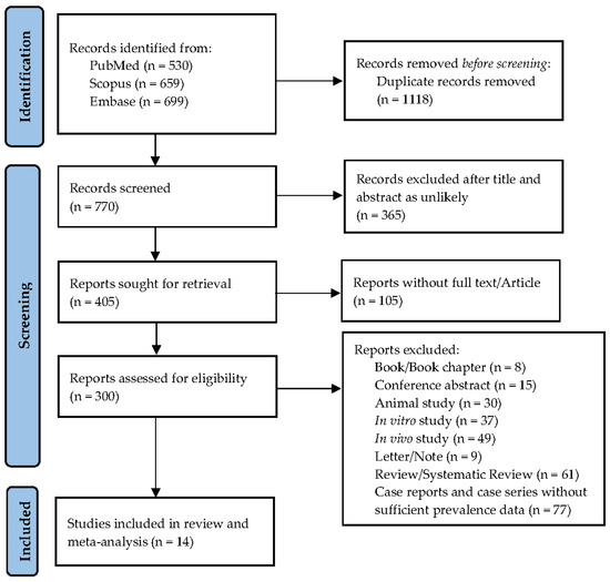
The literature search identified 1888 records from PubMed (n = 530), Scopus (n = 659), and Embase (n = 699). After removing 1118 duplicate records, 770 studies underwent title and abstract screening. Of these, 365 records were excluded. Full-text reports were sought for 405 records; however, 105 (25.9%) were unavailable through the sources used during the review and therefore could not be assessed for eligibility. Consequently, 300 full-text articles were assessed for eligibility. Following full-text assessment, 286 articles were excluded according to the predefined eligibility criteria, including case reports and case series without an appropriate denominator or sufficient prevalence data, reviews, conference abstracts, in vitro or animal studies, and studies without extractable data on hepatic or splenic involvement. The numbers excluded for each reason are presented in the PRISMA flow diagram (Figure 1). Ultimately, 14 studies met the inclusion criteria. No mixed-method studies met the eligibility criteria for inclusion in the final meta-analysis. Of these, 10 reported hepatic involvement, 10 reported splenic involvement, and 6 reported concomitant hepatic and splenic involvement and were included in the corresponding meta-analyses.
Figure 1.
PRISMA flow diagram.
3.2. Study Characteristics
The characteristics of the included studies are summarized in Table 1. The 14 eligible studies were published between 2004 and 2025 and included a total of 1844 patients with confirmed melioidosis [3,4,5,6,7,8,9,12,13,14,15,16,17,18]. The studies were conducted predominantly in Southeast Asia, including Thailand (n = 5), Malaysia (n = 3), Laos (n = 1), China (n = 1), and India (n = 2), with two additional studies from Australia and one from the Netherlands involving imported melioidosis cases.
Table 1.
Characteristics of included studies.
Most studies employed retrospective observational cohort designs (n = 10), while three were prospective cohorts or prospective observational studies, and one combined retrospective and prospective data collection. Study sample sizes ranged from 7 to 540 patients, with the largest cohorts reported by Currie et al. (n = 540) [3], Anunnatsiri et al. (n = 356) [4], and Prideaux et al. (n = 321) [15].
The study populations consisted primarily of culture-confirmed melioidosis, although one study included patients with diabetes and Burkholderia pseudomallei septicemia, and another evaluated imported melioidosis among returned travelers. Three studies exclusively enrolled pediatric patients, whereas the remaining studies included predominantly adult populations. The median or mean age ranged from 5 to 57 years, and males accounted for the majority of participants in most studies, with reported proportions ranging from 50.0% to 85.7%.
Diabetes mellitus was the most frequently reported underlying comorbidity, with prevalence ranging from 3.3% to 100% among the included cohorts. Ten studies reported data on hepatic involvement, ten on splenic involvement, and six on concomitant hepatic and splenic involvement, enabling quantitative synthesis of these outcomes. Reported mortality varied substantially across studies, ranging from 3.7% to 63.0%, reflecting differences in study populations, disease severity, and clinical settings.
3.3. Pooled Prevalence of Hepatic and Splenic Involvement
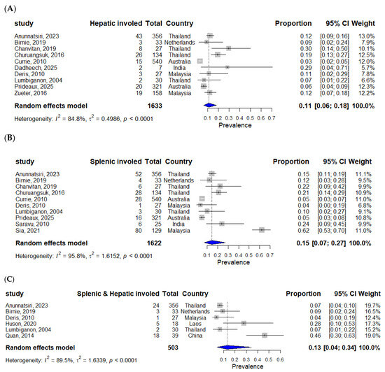
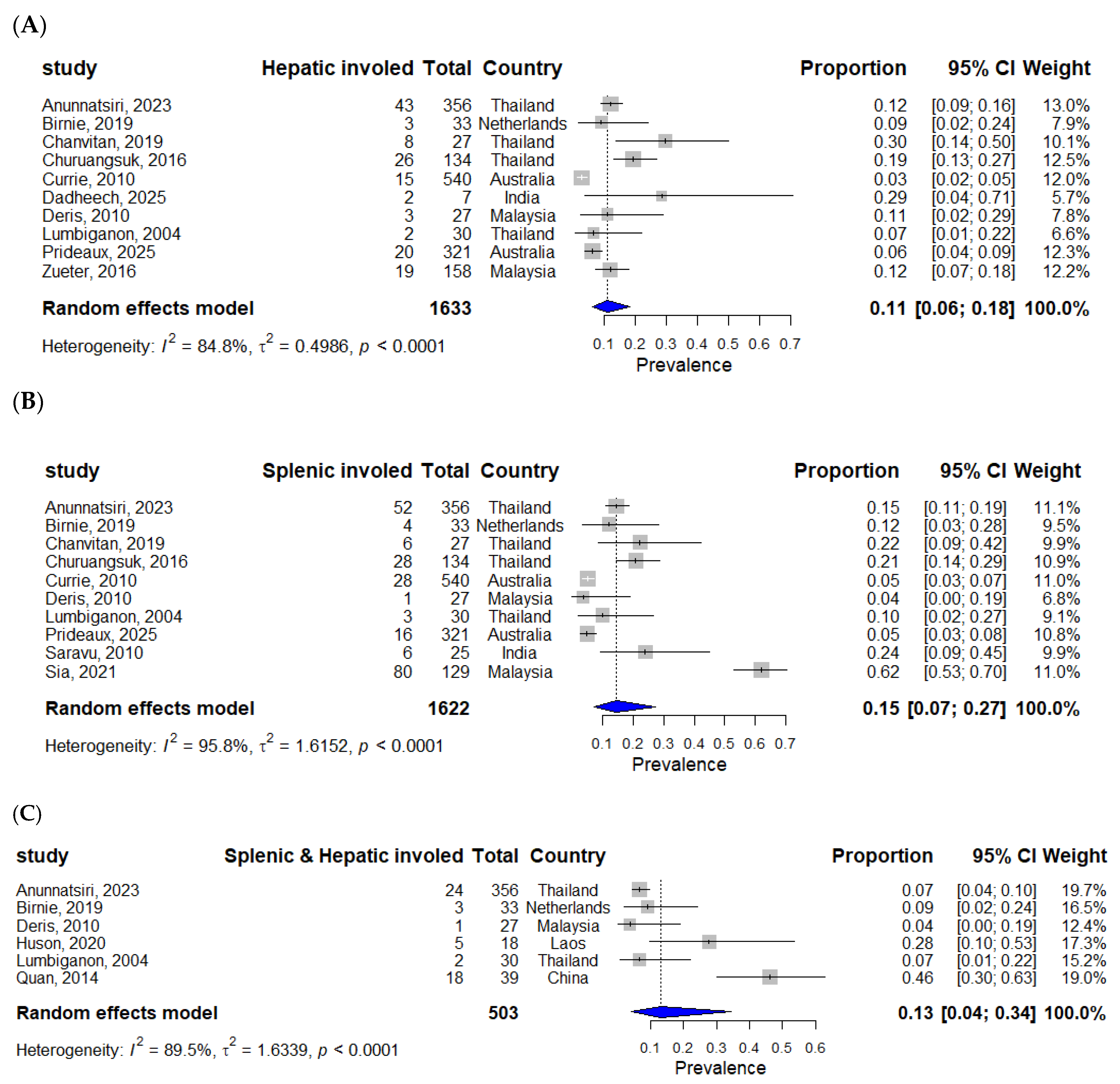
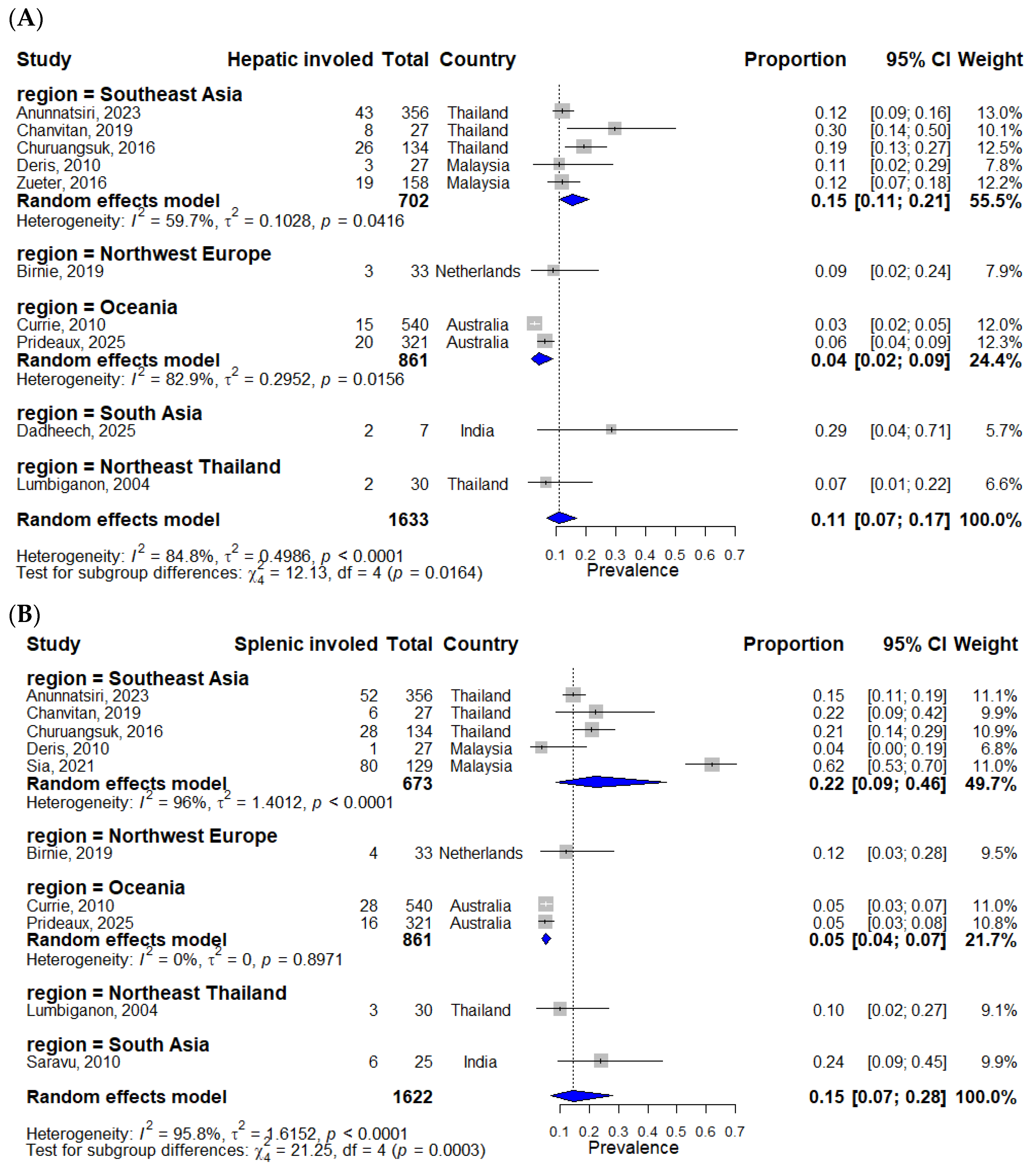
Ten studies involving 1633 patients with confirmed melioidosis reported hepatic involvement (Figure 2A). Individual-study prevalence ranged from 3% in the Australian cohort reported by Currie et al. [3] to 30% in the pediatric cohort reported by Chanvitan et al. [5]. The random-effects pooled prevalence of hepatic involvement was 11% (95% CI, 6–18%), with substantial between-study heterogeneity (I2 = 84.8%, p < 0.001).
Figure 2.
Forest plots of the prevalence of hepatic (A) [3,4,5,6,7,8,9,12,14,15], splenic (B) [3,4,5,6,7,9,14,15,17,18], and concomitant hepatic-splenic involvement (C) [4,7,9,13,14,16]. Gray squares represent individual study effect sizes, with horizontal lines indicating 95% confidence intervals. The size of each square reflects the study weight, and the blue diamond represents the pooled estimate from the random-effects model.
Ten studies comprising 1622 patients reported splenic involvement (Figure 2B). Individual-study prevalence ranged from 4% in the Malaysian cohort reported by Deris et al. [7] to 62% in the Malaysian cohort reported by Sia et al. [18]. The random-effects pooled prevalence of splenic involvement was 15% (95% CI, 7–27%), with considerable between-study heterogeneity (I2 = 95.8%, p < 0.001).
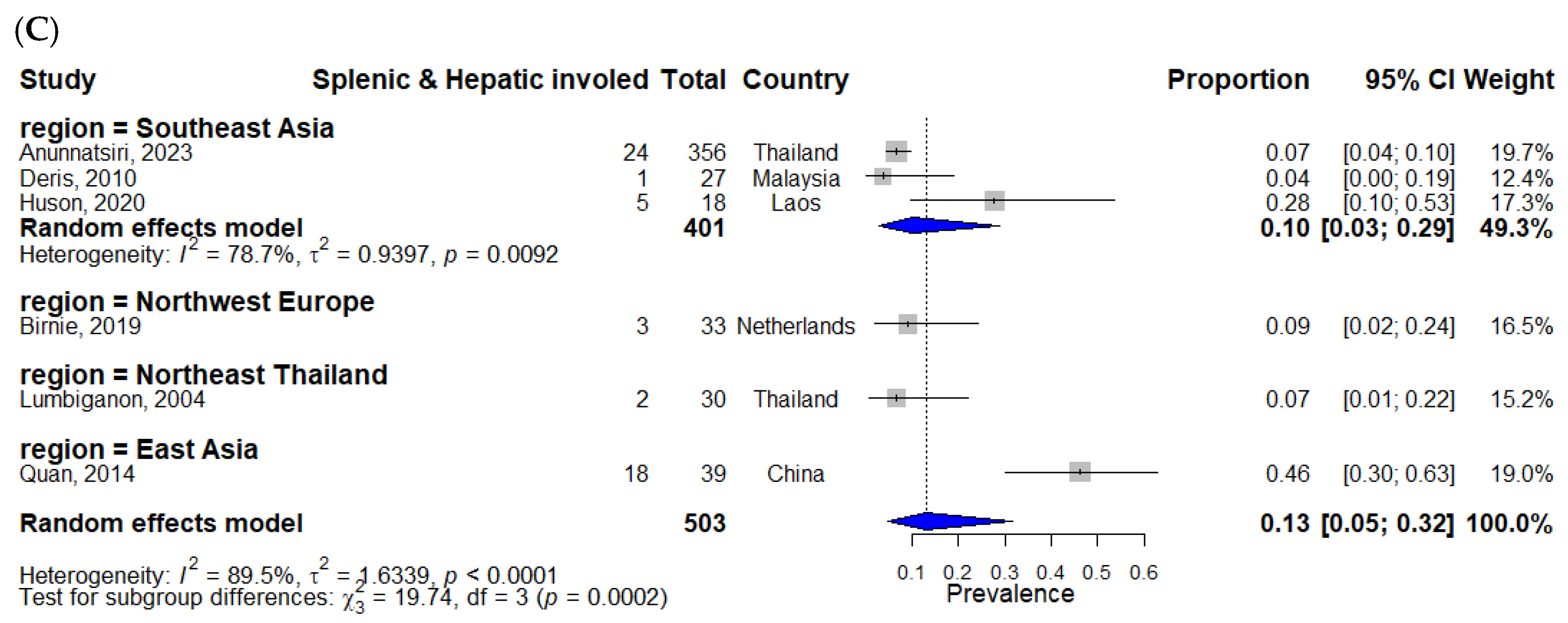
Six studies involving 503 patients reported concomitant hepatic and splenic involvement (Figure 2C). Individual-study prevalence ranged from 4% in the Malaysian study by Deris et al. [7] to 46% in the Chinese cohort reported by Quan et al. [16]. The random-effects pooled prevalence of concomitant hepatosplenic involvement was 13% (95% CI, 4–34%), with substantial between-study heterogeneity (I2 = 89.5%, p < 0.001).
3.4. Subgroup Analysis by Geographic Region
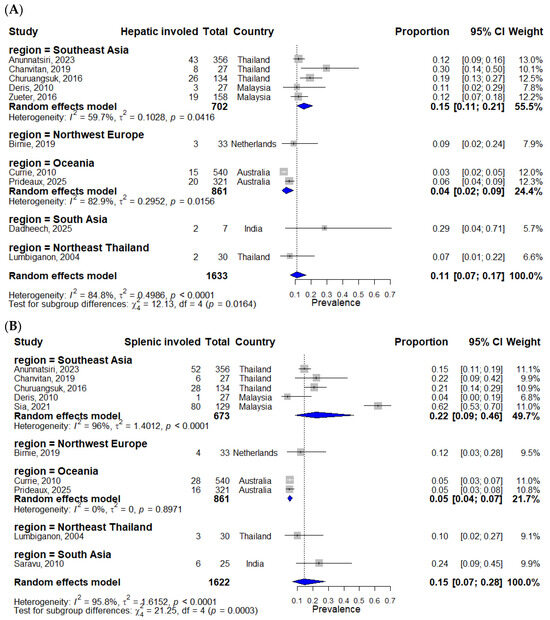
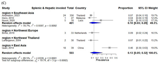
The prevalence of visceral organ involvement varied across geographic regions (Figure 3). The pooled prevalence of hepatic involvement was 15% (95% CI 11–21%) in Southeast Asia and 4% (95% CI 2–9%) in Oceania, with a statistically significant subgroup difference (p = 0.0164; Figure 3A). Similarly, splenic involvement was more prevalent in Southeast Asia (22%; 95% CI 9–46%) than in Oceania (5%; 95% CI 4–7%), with a significant subgroup difference (p = 0.0003; Figure 3B). For concomitant hepatic and splenic involvement, estimates varied across geographic subgroups (test for subgroup differences, p = 0.0002); however, this finding should be interpreted cautiously because several geographic subgroups were represented by a single study (Figure 3C).
Figure 3.
Forest plots of subgroup analysis by geographic region. Hepatic (A) [3,4,5,6,7,8,9,12,14,15], splenic (B) [3,4,5,6,7,9,14,15,17,18], and concomitant hepatic-splenic involvement (C) [4,7,9,13,14,16]. Gray squares represent individual study effect sizes, with horizontal lines indicating 95% confidence intervals. The size of each square reflects the study weight, and the blue diamond represents the pooled estimate from the random-effects model.
3.5. Subgroup Analysis by Age Group
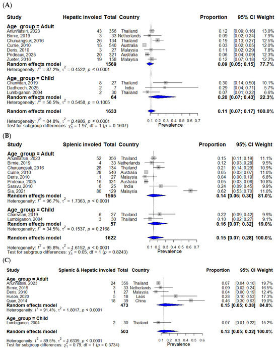
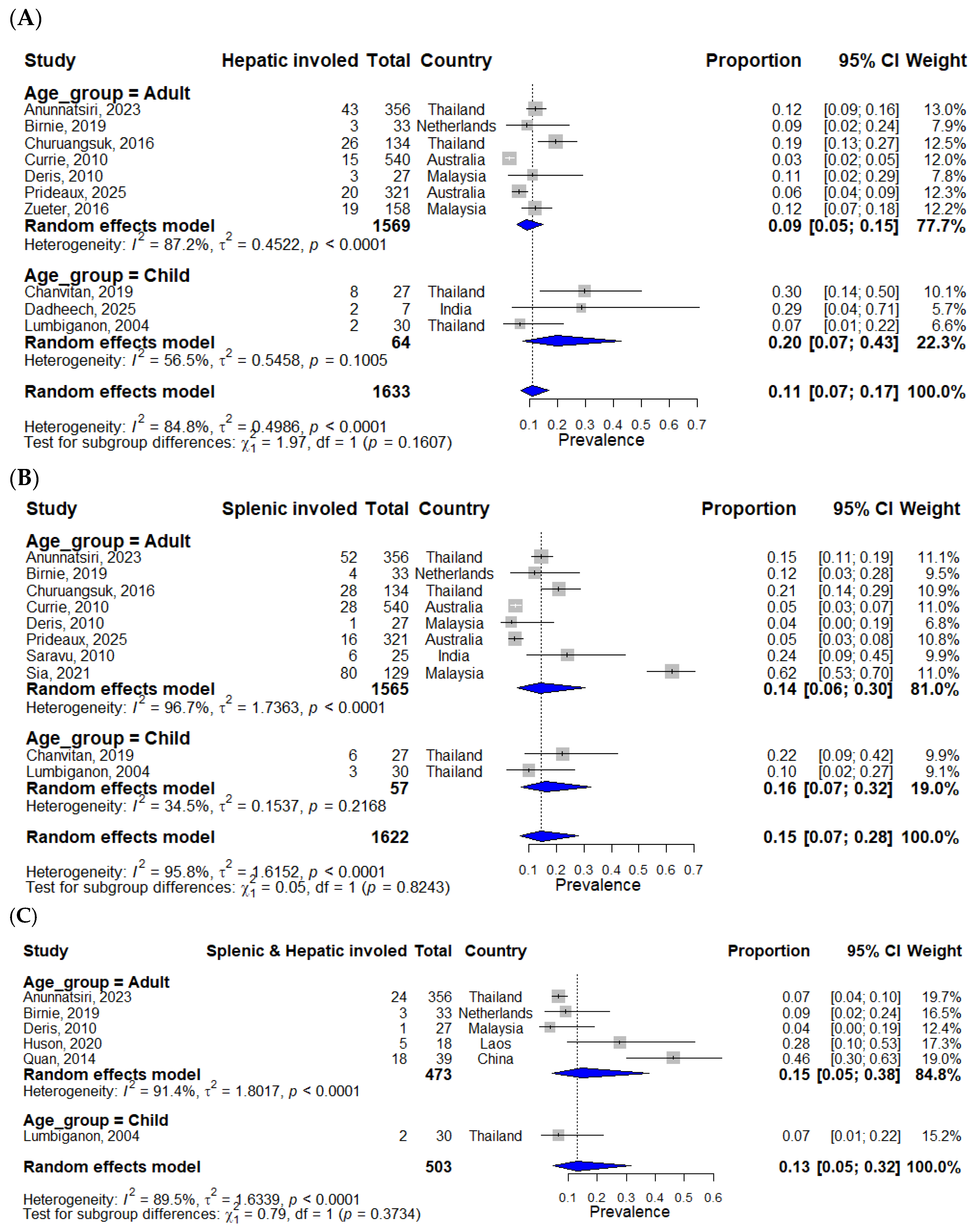
Age subgroup analyses were conducted for studies exclusively enrolling adult or pediatric populations (Figure 4). The pooled prevalence of hepatic involvement was 9% (95% CI, 5–15%) among adults and 20% (95% CI, 7–43%) among children, with no significant difference between age groups (p = 0.1607), Figure 4A. Similarly, the pooled prevalence of splenic involvement was 14% (95% CI, 6–30%) in adults and 16% (95% CI, 7–32%) in children (p = 0.8243), Figure 4B. For concomitant hepatic and splenic involvement, the pooled prevalence among adults was 15% (95% CI, 5–38%), Figure 4C. Because only one pediatric study contributed data, a pooled pediatric estimate was not calculated, and the subgroup comparison should be interpreted cautiously.
Figure 4.
Forest plots of subgroup analysis by age. Hepatic (A) [3,4,5,6,7,8,9,12,14,15], splenic (B) [3,4,5,6,7,9,14,15,17,18], and concomitant hepatic-splenic involvement (C) [4,7,9,13,14,16]. Gray squares represent individual study effect sizes, with horizontal lines indicating 95% confidence intervals. The size of each square reflects the study weight, and the blue diamond represents the pooled estimate from the random-effects model.
3.6. Risk of Bias in Included Studies
The methodological quality of the 14 included studies was assessed using the Joanna Briggs Institute (JBI) critical appraisal checklist for prevalence studies. Overall, three studies were judged to have a low risk of bias and 11 to have a moderate risk of bias; no study was classified as having a high risk of bias. Most studies clearly defined the study population, used microbiologically confirmed diagnoses of melioidosis, and applied appropriate statistical analyses. The main methodological limitations were retrospective study designs, inconsistent use of abdominal imaging for detecting hepatic and splenic involvement, small sample sizes, and single-center recruitment, which may have contributed to the substantial heterogeneity observed across studies. Detailed domain-level assessments are presented in Supplementary Table S1.
3.7. Small-Study Effects
Visual inspection of the funnel plots showed no marked asymmetry for the analyses of splenic or hepatic involvement. Egger’s regression test did not indicate evidence of small-study effects for splenic involvement (p = 0.679) or hepatic involvement (p = 0.869). For the analysis of concomitant hepatic and splenic involvement, formal assessment of small-study effects was not performed because fewer than ten studies were available. The details are in Supplementary File S2.
3.8. Leave-One-Out Sensitivity Analysis
Leave-one-out sensitivity analyses showed that the pooled prevalence estimates were generally robust to the sequential exclusion of individual studies (Supplementary Figure S1). For hepatic involvement, the pooled prevalence ranged from 10% to 13%, compared with the overall estimate of 11% (95% CI: 6–18%); excluding Currie et al. (2010) [3] reduced I2 from 84.8% to 69.4%. For splenic involvement, the pooled prevalence ranged from 12% to 16%, compared with an overall estimate of 15% (95% CI: 7–27%); excluding Sia et al. (2021) [18] reduced I2 from 95.8% to 85.4%. For concomitant hepatosplenic involvement, the pooled prevalence ranged from 9% to 15%, compared with the overall estimate of 13% (95% CI: 4–34%); excluding Quan et al. (2014) [16] reduced I2 from 89.5% to 57.9%. Overall, no single study materially altered the pooled estimates, supporting the robustness of the primary findings.
4. Discussion
This systematic review and meta-analysis synthesized evidence from 14 observational studies involving 1844 patients with confirmed melioidosis and provides the first quantitative synthesis of the prevalence of hepatic, splenic, and concomitant hepatosplenic involvement. The pooled prevalence of hepatic involvement was 11%, splenic involvement was 15%, and concomitant hepatic and splenic involvement was 13%. These findings confirm that visceral organ involvement is a common manifestation of disseminated melioidosis and support previous observations that the liver and spleen are among the most frequently affected organs following hematogenous dissemination of Burkholderia pseudomallei [1,2]. Large cohort studies from Australia, Thailand, and Malaysia have similarly demonstrated that abdominal abscesses represent an important component of the clinical spectrum of melioidosis, although the reported prevalence has varied considerably across settings [3,6,8].
Our pooled estimates are generally consistent with previous observational studies but provide a more comprehensive summary of disease burden than individual reports. For example, Currie et al. (2010) reported relatively low frequencies of hepatic and splenic abscesses in the Darwin prospective cohort [3], whereas studies from Thailand and Malaysia reported substantially higher proportions of visceral involvement [4,8,18]. Similarly, Chanvitan et al. (2019) found that hepatic and splenic abscesses were important predictors of pediatric melioidosis [5], while Mohan et al. (2020) demonstrated that systematic identification of splenic abscesses substantially improved the diagnosis of melioidosis in Malaysian children [19]. By pooling data across diverse populations, our study provides more stable prevalence estimates that may better reflect the overall burden of hepatosplenic melioidosis.
A major finding of this meta-analysis was the considerable between-study heterogeneity observed for all pooled estimates. Several methodological and clinical factors likely contributed to this variability. First, the included studies differed in study design, patient selection, and disease severity. Some cohorts enrolled only patients with culture-confirmed bacteremic melioidosis or severe disease treated at tertiary referral centers, whereas others included broader populations of hospitalized patients or imported cases [7,9]. Second, the prevalence of important risk factors, particularly diabetes mellitus, varied substantially between studies. Diabetes is the most common underlying disease associated with melioidosis and has consistently been linked to increased susceptibility to disseminated infection [2,15,16]. Third, differences in diagnostic imaging protocols probably influenced case ascertainment. Studies that routinely performed abdominal ultrasonography or computed tomography were more likely to detect asymptomatic visceral abscesses than studies in which imaging was reserved for patients with localized abdominal symptoms [4,13].
The subgroup analyses further demonstrated significant geographic variation in the prevalence of hepatic and splenic involvement, with higher estimates observed in Southeast Asia than in Oceania. These findings are consistent with previous epidemiological studies reporting regional differences in the clinical presentation of melioidosis [3,8,18]. Several explanations have been proposed, including differences in host characteristics, environmental exposure to B. pseudomallei, bacterial genetic diversity, healthcare accessibility, and diagnostic practices [1,2]. In addition, routine abdominal ultrasonography is commonly incorporated into the evaluation of suspected melioidosis in several Southeast Asian centers, facilitating the detection of clinically silent hepatic and splenic abscesses that might otherwise remain undiagnosed [4,13]. Although our subgroup analyses cannot establish causality, they suggest that regional variation should be considered when interpreting the epidemiology of visceral melioidosis.
No statistically significant differences were identified between adult and pediatric populations; however, pediatric studies tended to report higher prevalence estimates for hepatic involvement. This finding should be interpreted cautiously because relatively few pediatric studies were available, and only one study contributed data for concomitant hepatosplenic involvement. Previous pediatric studies have shown that hepatic and splenic abscesses may be particularly useful diagnostic markers in children presenting with fever of unknown origin in endemic regions [5,14,19]. Nevertheless, larger multicenter pediatric cohorts are required to determine whether age independently influences the risk of visceral organ involvement.
The present findings have important clinical implications. Approximately one in seven patients with melioidosis had splenic involvement, one in nine had hepatic involvement, and one in eight had concomitant hepatosplenic involvement. These results support existing recommendations that abdominal imaging should be considered in patients with suspected or confirmed melioidosis, particularly those with prolonged fever, bacteremia, or evidence of disseminated infection [1,3]. Ultrasonography provides a rapid and accessible screening tool in resource-limited settings, whereas computed tomography offers greater sensitivity for detecting small or multiple abscesses [13,20]. Characteristic radiological findings, including multiple small hypodense lesions and the “honeycomb” appearance of hepatic abscesses, may further increase diagnostic suspicion, although these findings should always be interpreted alongside microbiological confirmation because they are not entirely specific for melioidosis [21,22].
Several limitations should also be acknowledged. Most included studies were retrospective observational cohorts, making them susceptible to selection bias, incomplete clinical data, and inconsistent reporting. Considerable statistical heterogeneity remained despite subgroup analyses, indicating that additional unmeasured factors likely contributed to the variability of prevalence estimates. Diagnostic imaging protocols were not standardized across studies, potentially leading to underestimation of asymptomatic hepatic or splenic abscesses in cohorts where routine abdominal imaging was not performed [13,20]. Additionally, 105 reports (25.9% of those sought for retrieval) could not be obtained and therefore could not be assessed for eligibility. This may have introduced selection bias if the unavailable reports differed systematically from those that were successfully retrieved. Most included studies originated from Southeast Asia and northern Australia, limiting the generalizability of the findings to emerging endemic regions. Finally, relatively few studies reported concomitant hepatosplenic involvement, reducing the precision of pooled estimates and preventing more detailed analyses of prognostic factors.
Future research should prioritize prospective multicenter cohort studies using standardized imaging protocols and uniform diagnostic criteria to improve the comparability of prevalence estimates across different geographic settings. Individual patient-data meta-analyses could further clarify the influence of age, diabetes mellitus, bacteremia, and other host factors on visceral organ involvement. Additional studies evaluating the diagnostic performance and cost-effectiveness of routine abdominal ultrasonography or computed tomography in patients with suspected melioidosis are also warranted, particularly in resource-limited endemic settings [12,19]. Such evidence would strengthen clinical guidelines for the early recognition and management of hepatosplenic melioidosis.
5. Conclusions
This systematic review and meta-analysis demonstrate that hepatic and splenic involvement are common manifestations of melioidosis, with pooled prevalence estimates of 11%, 15%, and 13% for hepatic, splenic, and concomitant hepatosplenic involvement, respectively. Significant geographic variation was observed, with higher prevalence reported in Southeast Asia than in Oceania, whereas age-related differences were not statistically significant. Despite substantial heterogeneity among studies, these findings provide the first quantitative synthesis of the burden of hepatosplenic melioidosis and highlight the importance of maintaining a high index of suspicion for visceral organ involvement in patients with confirmed or suspected melioidosis. Routine abdominal imaging should be considered, particularly in endemic settings, to facilitate early detection and appropriate management. Future well-designed prospective studies using standardized diagnostic protocols are needed to refine prevalence estimates and improve understanding of the epidemiology of hepatosplenic melioidosis.
Supplementary Materials
The following supporting information can be downloaded at https://www.mdpi.com/article/10.3390/ijerph23091127/s1. File S1: Search Strategies; File S2: Funnel plot and linear regression; File S3: PRISMA 2020 Checklist; Table S1: Quality of studies by Joanna Briggs Institute (JBI) assessment. Figure S1: Leave-one-out sensitivity analyses of hepatosplenic involvement in melioidosis.
Author Contributions
Conceptualization, W.K.K.; Methodology, W.K.K. and J.T. (Jongkonnee Thanasai); Software, W.K.K.; Validation, W.K.K. and J.T. (Jongkonnee Thanasai); Formal analysis, W.K.K., M.C., S.-n.L., A.P., A.C., J.T. (Jitbanjong Tangpong), J.T. (Jongkonnee Thanasai) and J.S.; Investigation, W.K.K., M.C., S.-n.L., J.S., J.T. (Jitbanjong Tangpong), J.T. (Jongkonnee Thanasai), S.K., A.C. and A.P.; Resources, W.K.K. and J.T. (Jongkonnee Thanasai); Data curation, W.K.K. and J.T. (Jongkonnee Thanasai); Writing—original draft, W.K.K. and J.T. (Jongkonnee Thanasai); Writing—review and editing, W.K.K. and J.T. (Jongkonnee Thanasai); Visualization, W.K.K. and J.T. (Jongkonnee Thanasai); Supervision, W.K.K. and J.T. (Jongkonnee Thanasai); Project administration, W.K.K. All authors have read and agreed to the published version of the manuscript.
Funding
This work was supported by Mahasarakham University.
Institutional Review Board Statement
Not applicable.
Informed Consent Statement
Not applicable.
Data Availability Statement
No new data were created or analyzed in this study. Data sharing is not applicable to this article.
Conflicts of Interest
The authors declare no conflicts of interest.
Abbreviations
The following abbreviations are used in this manuscript:
| CI | confidence interval |
| CT | computed tomography |
| PRISMA | preferred reporting items for systematic reviews and meta-analyses |
References
- White, N.J. Melioidosis. Lancet 2003, 361, 1715–1722. [Google Scholar] [CrossRef] [Scilit] [PubMed]
- Cheng, A.C.; Currie, B.J. Melioidosis: Epidemiology, pathophysiology, and management. Clin. Microbiol. Rev. 2005, 18, 383–416. [Google Scholar] [CrossRef] [Scilit] [PubMed]
- Currie, B.J.; Ward, L.; Cheng, A.C. The epidemiology and clinical spectrum of melioidosis: 540 cases from the 20 year Darwin prospective study. PLoS Negl. Trop. Dis. 2010, 4, e900. [Google Scholar] [CrossRef] [Scilit] [PubMed]
- Anunnatsiri, S.; Chetchotisakd, P. Intra-abdominal Abscesses due to Melioidosis: Clinical Course and Outcomes. J. Med. Assoc. Thai. 2023, 106, S12–S18. [Google Scholar]
- Chanvitan, S.; Geater, A.; Laoprasopwattana, K. Hepatic/splenic abscess and/or skin and soft tissue infection as predictors of melioidosis in children. J. Infect. Dev. Ctries. 2019, 13, 149–153. [Google Scholar] [CrossRef] [Scilit] [PubMed]
- Churuangsuk, C.; Chusri, S.; Hortiwakul, T.; Charernmak, B.; Silpapojakul, K. Characteristics, clinical outcomes and factors influencing mortality of patients with melioidosis in southern Thailand: A 10-year retrospective study. Asian Pac. J. Trop. Med. 2016, 9, 256–260. [Google Scholar] [CrossRef] [Scilit] [PubMed]
- Deris, Z.Z.; Hasan, H.; Siti Suraiya, M.N. Clinical characteristics and outcomes of bacteraemic melioidosis in a teaching hospital in a northeastern state of Malaysia: A five-year review. J. Infect. Dev. Ctries. 2010, 4, 430–435. [Google Scholar] [CrossRef] [Scilit] [PubMed]
- Zueter, A.; Yean, C.Y.; Abumarzouq, M.; Rahman, Z.A.; Deris, Z.Z.; Harun, A. The epidemiology and clinical spectrum of melioidosis in a teaching hospital in a North-Eastern state of Malaysia: A fifteen-year review. BMC Infect. Dis. 2016, 16, 333. [Google Scholar] [CrossRef] [Scilit] [PubMed]
- Birnie, E.; Savelkoel, J.; Reubsaet, F.; Roelofs, J.J.T.H.; Soetekouw, R.; Kolkman, S.; Cremers, A.L.; Grobusch, M.P.; Notermans, D.W.; Wiersinga, W.J. Dutch Melioidosis Study Group Melioidosis in travelers: An analysis of Dutch melioidosis registry data 1985–2018. Travel Med. Infect. Dis. 2019, 32, 101461. [Google Scholar] [CrossRef] [Scilit] [PubMed]
- Page, M.J.; McKenzie, J.E.; Bossuyt, P.M.; Boutron, I.; Hoffmann, T.C.; Mulrow, C.D.; Shamseer, L.; Tetzlaff, J.M.; Akl, E.A.; Brennan, S.E.; et al. The PRISMA 2020 statement: An updated guideline for reporting systematic reviews. BMJ Clin. Res. Ed. 2021, 372, n71. [Google Scholar] [CrossRef] [Scilit] [PubMed]
- Munn, Z.; Moola, S.; Riitano, D.; Lisy, K. The development of a critical appraisal tool for use in systematic reviews addressing questions of prevalence. Int. J. Health Policy Manag. 2014, 3, 123–128. [Google Scholar] [CrossRef] [Scilit] [PubMed]
- Dadheech, M.; Gupta, A.; Richariya, D.; Yadav, Y.S. Pediatric Melioidosis in Central India: A Clinico-Epidemiological Study from a Landlocked Region. Cureus 2025, 17, e98488. [Google Scholar] [CrossRef] [Scilit] [PubMed]
- Huson, M.A.M.; Kling, K.; Chankongsin, S.; Phongluxa, K.; Keoluangkhot, V.; Newton, P.N.; Dance, D.; Heller, T.; Neumayr, A. Point-of-Care Ultrasound in the Diagnosis of Melioidosis in Laos. Am. J. Trop. Med. Hyg. 2020, 103, 675–678. [Google Scholar] [CrossRef] [Scilit] [PubMed]
- Lumbiganon, P.; Chotechuangnirun, N.; Kosalaraksa, P. Clinical experience with treatment of melioidosis in children. Pediatr. Infect. Dis. J. 2004, 23, 1165–1166. [Google Scholar] [CrossRef] [Scilit]
- Prideaux, L.; Sandeman, M.; Stratton, H.; Kelleher, A.D.; Smith, S.; Hanson, J. Melioidosis in people living with diabetes; clinical presentation, clinical course and implications for patient management. Acta Trop. 2025, 263, 107559. [Google Scholar] [CrossRef] [Scilit] [PubMed]
- Quan, H.B.; Li, T.Y.; Gao, Y.Y.; Chen, D.X. Clinical features of diabetes mellitus cases complicated by Burkholderia pseudomallei septicemia. Genet. Mol. Res. GMR 2014, 13, 3108–3116. [Google Scholar] [CrossRef] [Scilit] [PubMed]
- Saravu, K.; Mukhopadhyay, C.; Vishwanath, S.; Valsalan, R.; Docherla, M.; Vandana, K.E.; Shastry, B.A.; Bairy, I.; Rao, S.P. Melioidosis in southern India: Epidemiological and clinical profile. Southeast. Asian J. Trop. Med. Public Health 2010, 41, 401–409. [Google Scholar] [PubMed]
- Sia, T.L.L.; Mohan, A.; Ooi, M.H.; Chien, S.L.; Tan, L.S.; Goh, C.; Pang, D.C.L.; Currie, B.J.; Wong, J.S.; Podin, Y. Epidemiological and Clinical Characteristics of Melioidosis Caused by Gentamicin-Susceptible Burkholderia pseudomallei in Sarawak, Malaysia. Open Forum Infect. Dis. 2021, 8, ofab460. [Google Scholar] [CrossRef] [Scilit] [PubMed]
- Mohan, A.; Manan, K.; Tan, L.S.; Tan, Y.C.; Chin, S.T.; Ahmad, R.; Hamli, Q.I.; Chuah, T.B.; Chien, S.L.; Lau, P.S.; et al. Detection of spleen abscesses facilitates diagnosis of melioidosis in Malaysian children. Int. J. Infect. Dis. IJID Off. Publ. Int. Soc. Infect. Dis. 2020, 98, 59–66. [Google Scholar] [CrossRef] [Scilit] [PubMed]
- Muttarak, M.; Peh, W.C.; Euathrongchit, J.; Lin, S.E.; Tan, A.G.; Lerttumnongtum, P.; Sivasomboon, C. Spectrum of imaging findings in melioidosis. Br. J. Radiol. 2009, 82, 514–521. [Google Scholar] [CrossRef] [Scilit] [PubMed]
- Chong, V.H. Is a honeycomb appearance on computer tomography characteristic for Burkholderia pseudomallei liver abscess? Clin. Infect. Dis. 2006, 43, 265–266. [Google Scholar] [CrossRef] [Scilit] [PubMed][Green Version]
- Khiangte, H.L.; Vimala, L.R.; Eapen, A.; Veeraraghavan, B.; Karuppusami, R.; Gibikote, S. A Retrospective case–control study to evaluate the Diagnostic Accuracy of Honeycomb sign in melioid liver abscess. Am. J. Trop. Med. Hyg. 2018, 99, 852–857. [Google Scholar] [CrossRef] [Scilit] [PubMed]

Disclaimer/Publisher’s Note: The statements, opinions and data contained in all publications are solely those of the individual author(s) and contributor(s) and not of MDPI and/or the editor(s). MDPI and/or the editor(s) disclaim responsibility for any injury to people or property resulting from any ideas, methods, instructions or products referred to in the content. |
© 2026 by the authors. Licensee MDPI, Basel, Switzerland. This article is an open access article distributed under the terms and conditions of the Creative Commons Attribution (CC BY) license.