


Highlights
Public health relevance—How does this work relate to a public health issue?
- Temporomandibular disorders are an important source of musculoskeletal pain and may negatively affect patients’ function and quality of life.
- This study evaluates a clinically applicable non-pharmacological intervention that may broaden conservative rehabilitation options for myofascial TMD.
Public health significance—Why is this work of significance to public health?
- The addition of dry needling to standard rehabilitation was associated with a statistically significant between-group difference in early pressure-evoked pain response and with differences in quality-of-life changes.
- No statistically significant between-group difference was demonstrated in the overall change in pain intensity from baseline to the end of treatment.
Public health implications—What are the key implications or messages for practitioners, policy makers and/or researchers in public health?
- The use of dry needling as an adjunct to multimodal rehabilitation in patients with myofascial temporomandibular disorders (TMD) warrants further investigation, particularly regarding early pain response and quality-of-life outcomes.
Abstract
Background: Temporomandibular disorders (TMD) represent a significant source of musculoskeletal pain in the head and orofacial region. Myofascial TMD is characterised by pain, the presence of myofascial trigger points, and functional limitations. Methods: Forty patients (30 women and 10 men; mean age 32.4 ± 8.6 years) were enrolled in this pilot randomised controlled trial. The experimental group (n = 20) received a standard rehabilitation programme supplemented by a single dry needling session targeting the masseter and temporalis muscles, whereas the control group (n = 20) underwent standard rehabilitation alone. Pressure-evoked pain intensity (VAS) was assessed using a 0–10 visual analogue scale following the application of standardised algometric pressure to clinically identified myofascial trigger points. Quality of life was assessed using the WHOQOL-BREF questionnaire. Both outcomes were evaluated at baseline (T0), after the first treatment session (T1), and at the end of the rehabilitation programme (T2). The trial was registered in the ISRCTN registry (ISRCTN17648638). Results: After the first treatment session, both groups demonstrated a one-point reduction in pressure-evoked pain intensity (VAS). Quality-of-life scores increased by 10 points in the experimental group and by 8 points in the control group. At the end of the rehabilitation programme (T2), median pressure-evoked pain intensity (VAS) decreased to 2 (IQR 1) in the experimental group and to 3 (IQR 2) in the control group. WHOQOL-BREF scores increased by 36 points in the experimental group (50 [IQR 10] to 86 [IQR 7]) and by 30 points in the control group (54 [IQR 12] to 84 [IQR 8]). The Mann–Whitney U test revealed a statistically significant between-group difference in the early change in pressure-evoked pain intensity (VAS) from T0 to T1 (p = 0.023) and in WHOQOL-BREF score changes from T0 to T1 (p = 0.007) and from T0 to T2 (p = 0.035). However, the between-group difference in the overall change in pressure-evoked pain intensity (VAS) from T0 to T2 was not statistically significant (p = 0.585). Conclusions: The addition of a single dry needling session to standard rehabilitation was associated with a potentially more favourable early response in pressure-evoked pain intensity (VAS) and greater improvements in quality-of-life scores. However, no statistically significant between-group difference was observed in the overall change in pressure-evoked pain intensity (VAS) at the end of treatment. Given the pilot design and small sample size, these findings should be considered preliminary and interpreted with caution.
1. Introduction
Temporomandibular disorders (TMD) comprise a heterogeneous group of musculoskeletal and neuromuscular conditions affecting the temporomandibular joint, the masticatory muscles, and associated craniofacial structures. They affect approximately 5–12% of the general population and are considered the second most common cause of musculoskeletal pain after low back pain [1]. TMD may significantly affect oral function and activities of daily living, including chewing, speaking, yawning, and other mandibular movements. In addition to local symptoms, persistent temporomandibular pain may negatively affect quality of life, sleep, psychosocial well-being, and participation in routine occupational and social activities. The multifactorial nature of TMD requires comprehensive clinical assessment and, in many patients, a multimodal therapeutic approach.
The myofascial form of TMD is particularly associated with painful dysfunction of the masticatory muscles. Typical clinical manifestations include local or referred pain in the jaw, face, temporal region, and preauricular area, together with restricted mandibular movement and impaired functional performance. Myofascial TMD is also frequently associated with the presence of hypersensitive myofascial trigger points within the masticatory muscles, particularly the masseter and temporalis muscles [2]. These trigger points may contribute to spontaneous pain, pressure-evoked pain, pain during mandibular movement, increased muscle tension, and altered motor control. Persistent nociceptive input from the affected muscles may also contribute to peripheral and central sensitisation, potentially facilitating the persistence or chronification of symptoms.
The primary therapeutic goals in patients with myofascial TMD are therefore to reduce pain, restore mandibular mobility, improve masticatory muscle function, and support a return to normal daily activities. Conservative treatment is generally recommended as the first-line therapeutic strategy. Standard management may include soft tissue techniques, manual therapy, joint mobilisation, targeted therapeutic exercise, patient education, behavioural interventions, and pharmacotherapy [3]. Rehabilitation interventions are commonly directed towards reducing excessive muscle tension, improving joint mobility, restoring coordinated mandibular movement, and addressing functional impairments that may contribute to the condition. As TMD is often influenced by multiple biological, mechanical, and psychosocial factors, combining different conservative therapeutic modalities may provide additional clinical benefits compared with a single intervention. Dry needling (DN) is increasingly used as a minimally invasive therapeutic technique in the management of myofascial pain. The procedure involves the insertion of a thin, solid filiform needle into myofascial trigger points or other clinically relevant soft-tissue structures without the injection of any pharmacological substance. DN is considered a therapeutic technique aimed at modulating nociceptive input, reducing local muscle sensitivity, and potentially influencing neuromuscular activity associated with myofascial trigger points [4].
The proposed mechanisms underlying the effects of DN include mechanical stimulation of myofascial trigger points, local biochemical responses, modulation of nociceptive signalling, and changes in the local metabolic and microcirculatory environment. These mechanisms may contribute to reduced peripheral nociceptive activity and muscle sensitivity. Experimental studies have suggested that DN, particularly when associated with a local twitch response, may alter the concentrations of selected nociceptive mediators, including substance P and calcitonin gene-related peptide (CGRP). Changes in other pain- and inflammation-related mediators have also been reported [5,6,7]. Although these findings provide potential biological explanations for the analgesic effects of DN, the clinical significance and relative contribution of these mechanisms remain incompletely understood.
In clinical practice, DN is frequently incorporated into comprehensive rehabilitation programmes rather than used as an isolated therapeutic intervention. In patients with myofascial TMD, targeted treatment of trigger points in the masticatory muscles may reduce local tenderness and pressure-evoked pain, potentially facilitating subsequent active rehabilitation. An early reduction in pressure-evoked pain intensity (VAS) following DN may indicate decreased mechanical sensitivity of the treated muscles and could support improved tolerance of therapeutic exercise and functional mandibular movements. Accordingly, the addition of DN to standard rehabilitation may provide an early analgesic benefit compared with rehabilitation alone. However, whether this early response translates into sustained clinical improvements or additional benefits in quality of life requires further investigation.
Current evidence regarding the effectiveness of DN in myofascial TMD remains inconclusive. A recent sham-controlled randomised controlled trial demonstrated a significant immediate analgesic effect of a single DN intervention applied to the masseter and temporalis muscles, although no consistent changes in electromyographic activity were observed, suggesting that the observed effects may be predominantly short-term [8]. In contrast, studies using active comparators and systematic reviews have not demonstrated clear superiority of DN over manual therapy and have highlighted substantial heterogeneity in treatment protocols and limited certainty of the available evidence [9]. A multicentre randomised controlled trial by Dunning et al. reported more favourable outcomes for a combined intervention consisting of DN and upper cervical spinal manipulation; however, because the treatment was multimodal, the observed effects cannot be attributed to DN alone [10].
Accordingly, an important gap in current knowledge concerns the limited evidence regarding the additional effect of a single DN session incorporated into a standardised multimodal rehabilitation programme. Previous studies have frequently evaluated DN as a stand-alone intervention, compared it with another active treatment, or included it within combined therapeutic protocols, thereby limiting the ability to determine its specific incremental contribution. Moreover, relatively limited attention has been paid to the temporal pattern of changes in pressure-evoked pain intensity (VAS) and quality of life following a single DN session combined with rehabilitation.
Therefore, the aim of this pilot randomised controlled trial was to evaluate the additional effect of a single DN session targeting the masseter and temporalis muscles when combined with standardised rehabilitation in patients with myofascial TMD. The study specifically focused on changes in pressure-evoked pain intensity (VAS), assessed using standardised algometric stimulation of clinically identified myofascial trigger points, and on quality-of-life outcomes measured using the WHOQOL-BREF questionnaire. Particular attention was given to the early treatment response (T0–T1) and changes observed at the end of the rehabilitation programme (T0–T2).
2. Materials and Methods
2.1. Study Design
This prospective, randomised, controlled pilot study was conducted between November 2025 and June 2026 at two clinical and academic sites in Olomouc, Czech Republic: the Rehabilitation Outpatient Clinic (FBLR Practice) and the Department of Clinical Rehabilitation, Faculty of Health Sciences, Palacký University Olomouc. The study was designed to compare the clinical effectiveness of standard rehabilitation alone with standard rehabilitation supplemented by dry needling in patients with myofascial temporomandibular disorders.
The study followed a parallel-group design with two treatment arms and a 1:1 allocation ratio. The pilot character of the study was chosen to evaluate the feasibility of the proposed therapeutic protocol and to obtain preliminary data regarding treatment effects on pressure-evoked pain intensity (VAS) and quality of life. The study protocol included baseline clinical assessment, allocation to the experimental or control group, completion of the assigned rehabilitation programme, and repeated assessment of selected clinical outcomes.
The study was conducted in accordance with the ethical principles of biomedical research involving human participants. The study protocol was approved by the Ethics Committee of the Faculty of Health Sciences, Palacký University Olomouc, on 7 January 2025 (protocol no. UPOL-3853/1030S-2025).
Prior to enrolment, all participants received information regarding the purpose and course of the study, the proposed therapeutic procedures, and their participation in the research. Written informed consent was obtained from every participant before inclusion in the study.
2.2. Participants
A total of 40 patients with clinically diagnosed myofascial temporomandibular disorders were enrolled in the study. The sample comprised 30 women and 10 men aged between 18 and 65 years. The diagnosis of myofascial TMD was established prior to study inclusion according to the current version of the internationally recognised Diagnostic Criteria for Temporomandibular Disorders (DC/TMD).
Patients were recruited from individuals presenting for rehabilitation assessment and treatment of temporomandibular dysfunction.
Eligible participants were required to have clinical findings compatible with myofascial TMD and to be able to participate actively in the planned rehabilitation programme and repeated outcome assessment. The inclusion of patients with predominantly myofascial involvement was intended to create a relatively homogeneous study population and to focus specifically on a subgroup in which dysfunction of the masticatory muscles and myofascial trigger points may play an important role.
Exclusion criteria were defined to minimise the influence of conditions that could substantially affect pain perception, muscle tone, temporomandibular joint function, or response to therapy. Patients with structural TMJ disorders, particularly disc displacement, osteoarthritis, or arthritis, as well as those with a history of trauma or surgery involving the TMJ or cervical spine, were excluded. Additional exclusion criteria included severe neurological disorders, including trigeminal neuralgia, primary headache disorders, fibromyalgia and other chronic pain syndromes, autoimmune and systemic rheumatic diseases, uncontrolled diabetes, and clinically significant endocrine or metabolic disorders. Patients with severe cardiovascular or cerebrovascular disease, active infection, malignancy, severe psychiatric or cognitive disorders, and pregnant women were also excluded. Contraindications to dry needling were also considered, particularly coagulation disorders, severe needle phobia, or local infection at the treatment site. Patients who had undergone physiotherapy, acupuncture, or occlusal splint therapy within the previous 3 months, or who regularly used analgesics or NSAIDs, were not included in the study.
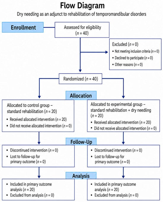
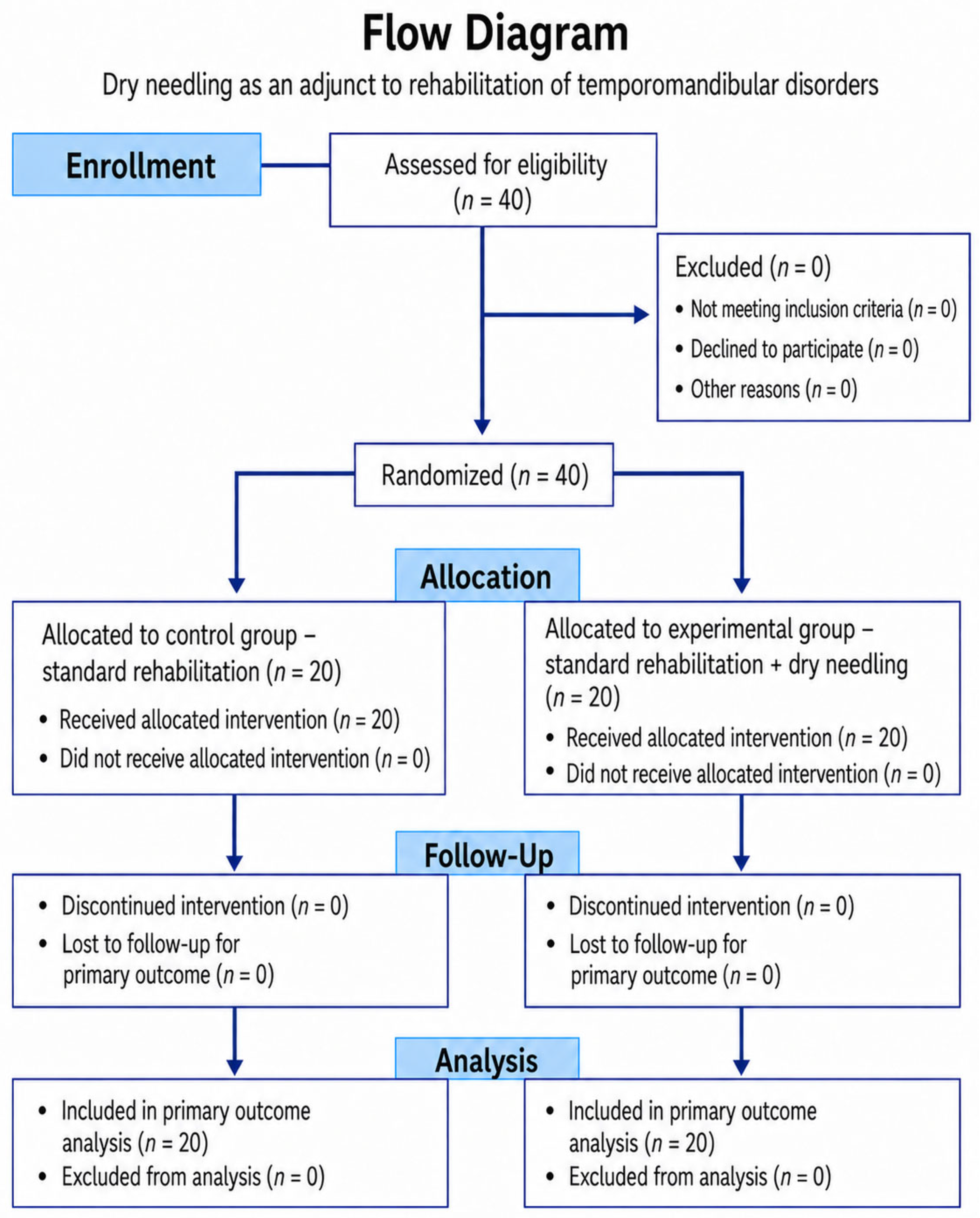
All 40 screened participants met the eligibility criteria and were included in the study. As shown in the study flow diagram, no participant was excluded prior to randomisation, and all randomised patients proceeded to their allocated treatment group.
2.3. Randomisation and Group Allocation
Following baseline assessment and confirmation of eligibility, participants were randomly allocated to one of two treatment groups in a 1:1 ratio. Random allocation was performed using sealed envelopes. This procedure was used to distribute participants between the experimental and control groups while reducing the possibility of systematic allocation according to clinical characteristics or investigator preference.
Twenty patients were allocated to the control group and received standard rehabilitation only, whereas 20 patients were allocated to the experimental group and received the same standard rehabilitation programme supplemented by dry needling. The study flow diagram demonstrates that all 40 enrolled participants underwent randomisation and that each treatment arm consisted of 20 participants. No participant failed to receive the allocated intervention, discontinued treatment, or was excluded from the primary analysis. The complete participant flow is presented in Figure 1 of the manuscript. The reporting of this pilot randomised controlled trial was guided by the CONSORT 2025 statement to ensure transparent and comprehensive reporting of the study design, methodology, and results [11] (File S1).
Figure 1.
Study flow diagram. The diagram illustrates participant enrolment, randomisation, allocation to the control and experimental groups, follow-up, and inclusion in the final analysis.
The use of equal allocation was intended to ensure comparable group sizes and to facilitate between-group comparison of clinical outcomes. Because this was a pilot study with a relatively small sample, randomisation was particularly important for limiting systematic differences between treatment groups at baseline. Subsequent analysis confirmed that the groups were comparable with respect to age, sex, and baseline values of pressure-evoked pain intensity (VAS) and quality of life.
2.4. Intervention
The control group (n = 20) underwent a standardised multimodal rehabilitation programme consisting of 10 treatment sessions, delivered twice weekly for 30 min per session. The therapy included soft tissue techniques aimed at reducing increased muscle tension and improving the mobility of myofascial structures of the masticatory and related cervical muscles, treatment of myofascial trigger points using post-isometric relaxation, and mobilisation of hypomobile segments of the temporomandibular joint and cervical spine, followed by targeted stabilisation and sensorimotor exercises focused on retraining coordinated and optimal mouth opening. Symptomatic pharmacotherapy was used only when clinically indicated as part of standard care. The rehabilitation protocol was based on the previously published methodology of Konečný et al. [12].
The experimental group (n = 20) underwent an identical rehabilitation programme with the same number, frequency, and duration of treatment sessions. In addition, a single dry needling intervention was performed during the first treatment session in accordance with standard therapeutic and safety procedures. Sterile filiform needles measuring 0.25 × 25 mm (Seirin, Japan) were inserted into clinically identified active myofascial trigger points in the masseter and temporalis muscles. The dry needling procedure itself lasted approximately 1 min.
Apart from the single dry needling intervention, all other therapeutic components, including their frequency, duration, and total number of sessions, were identical in both groups. This approach was intended to minimise between-group differences and to enable assessment of the additional effect of dry needling when added to standard rehabilitation therapy.
2.5. Outcome Measures
The primary clinical outcomes were pressure-evoked pain intensity (VAS) and quality of life. Pressure-evoked pain intensity (VAS) was assessed using a 0–10 visual analogue scale following the application of standardised algometric pressure to clinically identified active myofascial trigger points [13]. Lower scores indicated lower pressure-evoked pain intensity, whereas higher scores reflected greater subjectively perceived pain intensity in response to the applied pressure. The VAS was selected as a simple, widely used tool suitable for repeated assessment of pain intensity throughout the course of treatment.
The assessment did not focus on spontaneous pain but exclusively on pressure-evoked pain intensity (VAS). Active myofascial trigger points were clinically identified by palpation based on the presence of a palpable taut band, a hypersensitive spot, and reproduction of the patient’s characteristic local or referred pain.
Following identification of an active trigger point, a pressure algometer was positioned perpendicular to the examined site, and a standardised pressure of 1 kg/cm2 (approximately 98 kPa) was applied. The patient subsequently rated the pressure-evoked pain intensity using the 0–10 visual analogue scale (VAS).
Quality of life was assessed using the validated Czech version of the WHOQOL-BREF questionnaire [14]. This instrument was used to assess the patient-reported impact of the condition and treatment on overall quality of life. Including a quality-of-life measure enabled the study to assess not only changes in pressure-evoked pain intensity (VAS) but also broader changes from the patient’s perspective.
Both outcome measures were assessed at three predefined time points. Baseline assessment was performed before the start of treatment (T0), the second assessment 24 h after the first therapeutic intervention (T1), and the final assessment after completion of the entire rehabilitation programme (T2). The T1 time point was defined identically for both groups and allowed assessment of the early treatment response following the first intervention, including the potential additional effect of dry needling in the experimental group. This repeated-measures design enabled evaluation of both early changes in pressure-evoked pain intensity (VAS) and quality of life after the initial treatment session and the overall course of these outcomes following completion of the 10-session rehabilitation programme.
All therapeutic interventions were performed by a single therapist with 15 years of clinical experience in the treatment of temporomandibular disorders (TMD), thereby ensuring consistency and standardisation of therapeutic procedures. Data on pressure-evoked pain intensity (VAS) and quality of life (WHOQOL-BREF) were collected by the treating therapist. The subsequent statistical analysis was performed by an independent assessor who was blinded to treatment group allocation and was unaware of whether the analysed data originated from the experimental or control group.
2.6. Statistical Analysis
Statistical analysis was performed using SPSS version 30. Before comparative testing, the distribution of the collected data was assessed using the Shapiro–Wilk test to evaluate normality. The choice of subsequent statistical tests was based on the measurement level and distributional characteristics of the variables.
Interval data meeting the assumptions required for parametric analysis were evaluated using t-tests. Ordinal data and variables for which parametric assumptions were not considered appropriate were analysed using non-parametric statistical methods. Within-group changes over time were assessed using the Wilcoxon signed-rank test, while between-group comparisons were performed using the Mann–Whitney U test.
Results were primarily expressed as medians accompanied by interquartile ranges (IQR), providing information on both the central tendency and variability of the observed values. This method of presentation was particularly appropriate for ordinal and non-normally distributed data. Statistical significance was defined as a two-sided p value of <0.05.
The statistical analysis was designed to compare changes in pressure-evoked pain intensity (VAS) and quality of life within each treatment group over time and to explore whether the addition of dry needling resulted in a different treatment response compared with standard rehabilitation alone. No formal adjustment for multiple comparisons was applied because of the exploratory pilot nature of the study. Accordingly, all inferential analyses should be interpreted as exploratory.
3. Results
At baseline (T0), the control and experimental groups were comparable with respect to age, sex distribution, and the initial values of the main outcome measures. No statistically significant baseline differences were observed between the groups in pressure-evoked pain intensity (VAS) or quality of life assessed using the WHOQOL-BREF questionnaire (p = n.s.). The baseline median pressure-evoked pain intensity (VAS) score was 6 (IQR 2) in the control group and 5 (IQR 2) in the experimental group. Baseline WHOQOL-BREF scores were 54 (IQR 12) in the control group and 50 (IQR 10) in the experimental group. After the first therapeutic session (T1), an early reduction in pressure-evoked pain intensity (VAS) was observed in both groups. The median pressure-evoked pain intensity (VAS) score decreased by one point, from 6 to 5 in the control group and from 5 to 4 in the experimental group. In the experimental group, a descriptively narrower interquartile range was observed compared with the control group (IQR 1 vs. 2). At the final assessment (T2), the median pressure-evoked pain intensity (VAS) score decreased to 3 (IQR 2) in the control group and to 2 (IQR 1) in the experimental group (Table 1). Quality of life also improved during the treatment programme. In the control group, the WHOQOL-BREF score increased from a median of 54 (IQR 12) at baseline to 62 (IQR 16) after the first intervention and subsequently to 84 (IQR 8) at the end of therapy. In the experimental group, the corresponding values increased from 50 (IQR 10) at baseline to 60 (IQR 9) after the first intervention and to 86 (IQR 7) at the end of the programme. Thus, the total improvement in WHOQOL-BREF score from baseline to the end of therapy was 30 points in the control group and 36 points in the experimental group (Table 1).
Table 1.
Medians with interquartile ranges (IQR) for pain intensity: pressure-evoked pain (VAS) and quality of life (WHOQOL-BREF) in the control and experimental groups at baseline (T0), after the first therapy session (T1), and at the end of therapy (T2).
An additional analysis of between-group variability was performed using Levene’s test. No statistically significant difference in variability was demonstrated for VAS at T0 or for WHOQOL-BREF at T0 and T1 (p > 0.05). In contrast, statistically significant heterogeneity of variances was found for VAS at T1 (F = 4.11; p = 0.049), VAS at T2 (F = 5.11; p = 0.047), and WHOQOL-BREF at T2 (F = 4.66; p = 0.012) (Table 2).
Table 2.
Homogeneity of Variance Test.
Subsequently, further inferential statistical analyses were performed to evaluate changes over time and between-group differences in the main outcome measures.
The Mann–Whitney U test demonstrated a statistically significant difference between the experimental and control groups in the early change in pain intensity between T0 and T1 (ΔVAS T0–T1: U = 275.0; Z = 2.268; p = 0.023). In contrast, the overall change in pain intensity between T0 and T2 did not differ significantly between the groups (U = 219.0; Z = 0.546; p = 0.585). For quality of life, statistically significant between-group differences were observed both in the early change between T0 and T1 (ΔWHOQOL-BREF: U = 101.0; Z = −2.682; p = 0.007) and in the overall change between T0 and T2 (U = 122.5; Z = −2.099; p = 0.035) (Table 3).
Table 3.
Mann–Whitney U Test for Independent Samples.
The Wilcoxon signed-rank test demonstrated statistically significant within-group changes across all assessed intervals in both treatment groups. Pressure-evoked pain intensity (VAS) decreased significantly from T0 to T1 and from T1 to T2 in both the control group (Z = −3.557 and −3.880, respectively; both p < 0.001) and the experimental group (Z = −3.828 and −4.134, respectively; both p < 0.001). Similarly, WHOQOL-BREF scores increased significantly from T0 to T1 and from T1 to T2 in both groups (all p < 0.001). These findings indicate statistically significant within-group reductions in pressure-evoked pain intensity (VAS) and improvements in quality of life throughout the rehabilitation programme in both treatment groups (Table 4).
Table 4.
Related-Samples Wilcoxon Signed Rank Test.
Additional exploratory effect-size analysis demonstrated a more pronounced between-group effect on the early change in pressure-evoked pain intensity from T0 to T1 (η2 = 0.148; 95% CI: 0.006–0.346). In contrast, the effect size for the overall change in pain intensity from T0 to T2 was negligible (η2 = 0.008; 95% CI: 0.000–0.136). For quality of life assessed using the WHOQOL-BREF questionnaire, the between-group effect size for the T0–T1 change was η2 = 0.191 (95% CI: 0.020–0.389), whereas the effect size for the T0–T2 change was η2 = 0.122 (95% CI: 0.000–0.317). Given the pilot nature of the study, the limited sample size, and the absence of adjustment for multiple comparisons, these estimates should be interpreted with caution. The findings are primarily exploratory and require confirmation in larger controlled studies. During the study, three participants used a single rescue dose of diclofenac 50 mg: one participant in the experimental group and two participants in the control group. No repeated or regular analgesic use was reported.
4. Discussion
This pilot randomised controlled trial evaluated the additional effect of a single dry needling (DN) session incorporated into a standardised rehabilitation programme in patients with myofascial temporomandibular disorders (TMD). Both treatment groups demonstrated statistically significant within-group reductions in pressure-evoked pain intensity (VAS) and improvements in quality of life throughout the rehabilitation programme. Between-group analysis revealed a statistically significant difference in the early change in pressure-evoked pain intensity (VAS) from T0 to T1 and in WHOQOL-BREF score changes from T0 to T1 and from T0 to T2. However, the overall change in pressure-evoked pain intensity (VAS) from T0 to T2 did not differ significantly between groups. These findings suggest a potential additional effect of DN during the early phase of treatment but do not demonstrate superior overall analgesic efficacy at the end of the rehabilitation programme.
From a clinical perspective, the median pressure-evoked pain intensity (VAS) score decreased by approximately three points from baseline to the end of treatment in both groups. Although both groups demonstrated substantial within-group reductions in pressure-evoked pain intensity (VAS), the between-group difference in overall pain reduction was small and statistically non-significant. Therefore, the principal potential contribution of DN observed in this study appears to be associated with the early treatment response rather than with a superior overall reduction in pressure-evoked pain intensity (VAS) at the end of rehabilitation.
Pressure-evoked pain intensity (VAS) decreased in both groups after the first therapeutic intervention and continued to decline throughout the rehabilitation programme. At certain assessment points, the experimental group demonstrated descriptively lower pressure-evoked pain intensity (VAS) scores and narrower interquartile ranges. However, these descriptive differences should be interpreted cautiously and cannot independently establish greater clinical effectiveness or a more consistent treatment response. Between-group analysis demonstrated a statistically significant difference only in the early change in pressure-evoked pain intensity (VAS) from T0 to T1, whereas the overall change from T0 to T2 did not differ significantly between groups.
A possible explanation for the early treatment response may involve the targeted stimulation of active myofascial trigger points in the masseter and temporalis muscles and subsequent modulation of nociceptive input. DN may influence local muscle sensitivity, peripheral nociceptive processing, and the conditions facilitating subsequent active rehabilitation. However, because the present study did not directly investigate physiological mechanisms, these explanations remain hypothetical and cannot be established from the available data.
Quality of life improved significantly in both treatment groups. The experimental group demonstrated a numerically greater increase in WHOQOL-BREF scores, and between-group analysis identified statistically significant differences in score changes from T0 to T1 and from T0 to T2. Nevertheless, given the pilot study design, small sample size, and absence of a formal assessment of the minimal clinically important difference (MCID), the clinical relevance of these differences remains uncertain. Furthermore, clinically meaningful thresholds for WHOQOL-BREF scores are not uniformly established across patient populations and clinical conditions. Therefore, although the findings suggest a potentially favourable additional effect of DN on quality-of-life outcomes, definitive conclusions regarding its clinical importance cannot be drawn.
The findings should be interpreted in the context of the current international literature, which suggests that the effects of DN in myofascial TMD may be predominantly short-term and influenced by the type of comparator, treatment protocol, and duration of follow-up. A recent sham-controlled randomised controlled trial by Durmaz et al. demonstrated a greater immediate reduction in pain following a single DN intervention targeting the masseter and temporalis muscles compared with sham treatment, although no consistent changes in electromyographic activity were observed [8]. These findings support the possibility that the effects of a single DN session may be most evident during the early post-intervention period, which is consistent with the statistically significant early between-group difference in pressure-evoked pain intensity (VAS) observed in the present study.
In contrast, a randomised controlled trial comparing DN with manual therapy demonstrated significant improvements in pain, maximum mouth opening, cervical disability, and pressure pain threshold in both groups, without statistically significant differences between the therapeutic approaches [15]. Similarly, the systematic review and network meta-analysis by Menéndez-Torre et al. did not demonstrate clear superiority of DN over manual therapy and highlighted considerable heterogeneity in treatment protocols and limited certainty of the available evidence [9]. A systematic review published in 2025 reported predominantly favourable outcomes associated with DN regarding pain and selected functional parameters, while emphasising substantial methodological heterogeneity among the included studies [16]. Differences in study design, treatment frequency, target muscles, comparator interventions, and follow-up duration considerably limit direct comparisons between studies.
In this context, DN may be more appropriately considered a potential adjunct to multimodal rehabilitation rather than a superior stand-alone therapeutic intervention. Importantly, the present study evaluated only a single DN session added to an otherwise identical standardised rehabilitation programme. The observed early between-group difference in pressure-evoked pain intensity (VAS) suggests that even a single DN intervention may influence the initial treatment response. However, the present pilot study does not provide sufficient evidence to determine the optimal number, frequency, or timing of DN sessions. These questions should be addressed in future randomised controlled trials with larger samples, longer follow-up periods, and adequate statistical power.
Study Limitations
Several methodological limitations should be considered when interpreting the findings of this pilot study. The relatively small sample size of 40 participants limits the generalisability of the results and reduces the precision of the estimated between-group effects. Follow-up was restricted to the completion of the 10-session rehabilitation programme; therefore, the persistence of the observed changes in pressure-evoked pain intensity (VAS) and quality of life over the medium and long term remains unknown.
Furthermore, DN was administered only once, preventing conclusions regarding the optimal number, frequency, or timing of treatment sessions. The principal outcome measures were patient-reported pressure-evoked pain intensity (VAS) and WHOQOL-BREF scores. Consequently, the absence of participant blinding and a sham control represents an important limitation, as the specific effects of DN cannot be reliably distinguished from patient expectations, placebo effects, or the additional attention associated with the adjunct intervention.
Outcome assessment was not blinded because pressure-evoked pain intensity (VAS) and WHOQOL-BREF data were collected by the treating therapist. This introduces a potential risk of measurement and observer bias. Future clinical trials should therefore employ an independent, blinded outcome assessor whenever feasible.
Another potential confounding factor was the limited use of symptomatic analgesic medication. During the study, diclofenac 50 mg was taken once by three participants, including one patient in the experimental group and two patients in the control group. Although analgesic exposure was minimal, its potential influence on pressure-evoked pain intensity (VAS) and quality-of-life outcomes cannot be completely excluded.
Additionally, the study was not designed to formally assess the minimal clinically important difference (MCID). Therefore, the clinical relevance of the observed changes in pressure-evoked pain intensity (VAS) and WHOQOL-BREF scores cannot be conclusively determined from the available data.
A further limitation was the absence of adjustment for multiple statistical comparisons. Given the number of within-group and between-group analyses, the possibility of type I error cannot be excluded. Accordingly, the reported p-values should be considered exploratory and hypothesis-generating rather than confirmatory.
Despite these limitations, the study demonstrated good feasibility of the proposed therapeutic and research protocol. All randomised participants completed their allocated treatment programmes, no participants were lost to follow-up, and no serious treatment-related adverse events were reported. These findings support the feasibility of conducting a subsequent adequately powered randomised controlled trial using a similar study design.
Future research should include larger, adequately powered, preferably multicentre randomised controlled trials with longer follow-up periods and appropriate control conditions. In addition to pressure-evoked pain intensity (VAS) and quality-of-life assessment, future studies should incorporate objective functional outcome measures, including mandibular range of motion, masticatory function, pressure pain thresholds, muscle tenderness, and other clinically relevant functional parameters. Such measures could provide a more comprehensive understanding of treatment effects and their clinical relevance.
Future investigations should also determine the optimal dosage, frequency, and timing of DN interventions and identify patient subgroups with myofascial TMD that may derive the greatest benefit from this therapeutic approach.
Overall, the findings of this pilot study support further investigation of DN as an adjunct to standard rehabilitation in patients with myofascial TMD. The results suggest a potential early benefit regarding pressure-evoked pain intensity (VAS) and additional improvements in quality-of-life outcomes. However, the absence of a statistically significant between-group difference in the overall change in pressure-evoked pain intensity (VAS), together with the exploratory design and limited sample size, requires cautious interpretation. Confirmation in larger, methodologically rigorous randomised controlled trials is warranted.
5. Conclusions
The findings of this pilot randomised controlled trial demonstrated a statistically significant between-group difference in the early change in pressure-evoked pain intensity (VAS) from T0 to T1 and in quality-of-life changes from T0 to T1 and from T0 to T2. However, the overall change in pressure-evoked pain intensity (VAS) from T0 to T2 did not differ significantly between the groups.
The addition of a single dry needling session to standard rehabilitation may therefore be associated with a more favourable early response in pressure-evoked pain intensity (VAS) and differences in quality-of-life outcomes. Nevertheless, no statistically significant additional reduction in pressure-evoked pain intensity (VAS) was demonstrated at the end of the rehabilitation programme. Given the exploratory pilot design, small sample size, and limited follow-up period, these findings should be interpreted with caution.
The study also demonstrated the feasibility of the proposed therapeutic and research protocol, providing a foundation for future adequately powered randomised controlled trials with larger samples and longer follow-up periods to confirm the clinical relevance and potential additional benefits of dry needling in patients with myofascial temporomandibular disorders.
Supplementary Materials
The following supporting information can be downloaded at: https://www.mdpi.com/article/10.3390/ijerph23111392/s1, File S1: CONSORT checklist.
Author Contributions
Conceptualisation: D.D. and P.K.; Methodology: D.D. and P.K.; Formal Analysis: D.D. and P.K.; Investigation: D.D. and P.K.; Data Curation: D.D. and P.K.; Writing—Original Draft Preparation: D.D., P.K. and P.G.A.; Writing—Review and Editing: J.V., A.M., P.K. and P.G.A. All authors have read and agreed to the published version of the manuscript.
Funding
This study was supported by the Internal Grant 2025, Faculty of Health Sciences, Palacký University Olomouc, Czech Republic (project: IGA_FZV_2025_015). The trial was registered at ISRCTN (ISRCTN17648638).
Institutional Review Board Statement
The study was conducted in accordance with the Declaration of Helsinki and approved by the Ethics Committee of the Faculty of Health Sciences, Palacký University Olomouc (protocol code UPOL-3853/1030S-2025, approved on 7 January 2025).
Informed Consent Statement
All participants signed informed consent prior to inclusion in the study.
Data Availability Statement
The data presented in this study are available on request from the corresponding author.
Conflicts of Interest
The authors declare no conflicts of interest.
References
- De Leeuw, R.; Klasser, G.D. Orofacial Pain: Guidelines for Assessment, Diagnosis, and Management, 5th ed.; Quintessence Publishing: Hanover Park, IL, USA, 2013. [Google Scholar]
- Okeson, J.P. Management of Temporomandibular Disorders and Occlusion, 8th ed.; Elsevier: St. Louis, MO, USA, 2019. [Google Scholar]
- Wright, E.F. Manual of Temporomandibular Disorders, 4th ed.; Wiley-Blackwell: Hoboken, NJ, USA, 2019. [Google Scholar]
- Dommerholt, J.; Fernández-de-Las-Peñas, C. Trigger Point Dry Needling, 2nd ed.; Elsevier Books: London, UK, 2018. [Google Scholar]
- Shah, J.P.; Danoff, J.V.; Desai, M.J.; Parikh, S.; Nakamura, L.Y.; Phillips, T.M.; Gerber, L.H. Biochemicals associated with pain and inflammation are elevated in sites near to and remote from active myofascial trigger points. Arch. Phys. Med. Rehabil. 2008, 89, 16–23. [Google Scholar] [CrossRef] [Scilit] [PubMed]
- Cagnie, B.; Dewitte, V.; Barbe, T.; Timmermans, F.; Delrue, N.; Meeus, M. Physiologic effects of dry needling. Curr. Pain Headache Rep. 2013, 17, 348. [Google Scholar] [CrossRef] [Scilit] [PubMed]
- Fernández-de-Las-Peñas, C.; Nijs, J. Trigger point dry needling for the treatment of myofascial pain syndrome: Current perspectives within a pain neuroscience paradigm. J. Pain Res. 2019, 12, 1899–1911. [Google Scholar] [CrossRef] [Scilit] [PubMed]
- Durmaz, N.; Gözler, S.; Demircioğlu, G.; Burma, K. Immediate Effects of Dry Needling in Patients with Myofascial Temporomandibular Disorders: A Randomized Controlled Trial. Healthcare 2026, 14, 2189. [Google Scholar] [CrossRef] [Scilit] [PubMed]
- Menéndez-Torre, Á.; Martín Pintado-Zugasti, A.; Cuenca Zaldivar, J.N.; García-Bermejo, P.; Gómez-Costa, D.; Molina-Álvarez, M.; Arribas-Romano, A.; Fernández-Carnero, J. Effectiveness of deep dry needling versus manual therapy in the treatment of myofascial temporomandibular disorders: A systematic review and network meta-analysis. Chiropr. Man. Therap. 2023, 31, 46. [Google Scholar] [CrossRef] [Scilit] [PubMed]
- Dunning, J.; Butts, R.; Bliton, P.; Vathrakokoilis, K.; Smith, G.; Lineberger, C.; Eshleman, N.; Fernández-De-Las-Peñas, C.; Young, I.A. Dry needling and upper cervical spinal manipulation in patients with temporomandibular disorder: A multi-center randomized clinical trial. Cranio 2024, 42, 809–822. [Google Scholar] [CrossRef] [Scilit] [PubMed]
- Hopewell, S.; Chan, A.-W.; Collins, G.S.; Hróbjartsson, A.; Moher, D.; Schulz, K.F.; Tunn, R.; Aggarwal, R.; Berkwits, M.; Berlin, J.A.; et al. CONSORT 2025 Statement: Updated Guideline for Reporting Randomised Trials. BMJ 2025, 389, e081123. [Google Scholar] [CrossRef] [Scilit]
- Konečný, P.; Havlíčková, J.; Elfmark, M.; Tvrdý, P.; Hanáková, D.; Jureček, M. Effects of Rehabilitation in Patients with Temporomandibular Joint Disorders. Rehabil. Fyz. Lek. 2007, 14, 95–100. [Google Scholar]
- Huskisson, E.C. Measurement of Pain. Lancet 1974, 304, 1127–1131. [Google Scholar] [CrossRef] [Scilit] [PubMed]
- The WHOQOL Group. Development of the World Health Organization WHOQOL-BREF quality of life assessment. Psychol. Med. 1998, 28, 551–558. [Google Scholar] [CrossRef] [Scilit] [PubMed]
- García-de la-Banda-García, R.; Cortés-Pérez, I.; Ibancos-Losada, M.D.R.; López-Ruiz, M.D.C.; Obrero-Gaitán, E.; Osuna-Pérez, M.C. Effectiveness of Dry Needling versus Manual Therapy in Myofascial Temporomandibular Disorders: A Single-Blind Randomized Controlled Trial. J. Pers. Med. 2023, 13, 1415. [Google Scholar] [CrossRef] [Scilit] [PubMed]
- Khayamzadeh, M.; Razmara, F.; Tavassoli, A. Dry Needling in Treatment of Temporomandibular Joint Disorders: A Systematic Review. Clin. Exp. Dent. Res. 2025, 11, e70214. [Google Scholar] [CrossRef] [Scilit] [PubMed]

Disclaimer/Publisher’s Note: The statements, opinions and data contained in all publications are solely those of the individual author(s) and contributor(s) and not of MDPI and/or the editor(s). MDPI and/or the editor(s) disclaim responsibility for any injury to people or property resulting from any ideas, methods, instructions or products referred to in the content. |
© 2026 by the authors. Licensee MDPI, Basel, Switzerland. This article is an open access article distributed under the terms and conditions of the Creative Commons Attribution (CC BY) license.