


Abstract
Ferroptosis is a critical driver of tubular necrosis, and its effective inhibition holds significant potential for mitigating renal injury. In this study, chemical investigation of the strain Penicillium rubens MABC05 yielded nine drimane-pyrone-type meroterpenoids (1–9), including six previously undescribed analogues, a previously undescribed tetrahydroxanthone-ergosterol hybrid (10), four steroids (11–14), and three xanthone derivatives (15–17). Their structures were elucidated via comprehensive spectroscopic analysis, supported by single-crystal X-ray diffraction (Mo Kα for 1 and 3, and Cu Kα for 6) and 13C NMR calculations (2 and 10). The absolute configurations of 1–5 and 10 were assigned by ECD calculation. Compounds 1–6 represent a rare group of meroterpenoids containing a fused drimane-type sesquiterpene and pyrone skeleton. Additionally, two storage-induced oxidative artifacts of 10 were also characterized. These compounds were evaluated for anti-ferroptotic and cytotoxic activities. Compound 15 exhibited inhibition against RSL3-induced ferroptosis in human renal proximal tubular epithelial cells with an EC50 value of 12.8 μM; it effectively reduced MDA levels, elevated GSH content, and suppressed lipid radical generation. Compounds 11 and 16 exhibited cytotoxicity against MCF-7 cells with IC50 values of 4.3 and 8.4 μM, respectively. Compound 15 represents a promising chemotype for ferroptosis inhibitors with potential for mitigating renal tubular injury.
1. Introduction
The talarolutins and penicillipyrones constitute a small group of meroterpenoids (drimane-pyrone-type meroterpenoids), featuring a drimane-type sesquiterpene fused with a 2-methyl-pyrone (or its hydrogenated derivative) moiety [1,2]. The proposed biosynthesis of these meroterpenoids is initiated by the ionization of farnesyl diphosphate (FPP), generating an allylic carbocation that attacks the electron-rich pyrone ring to afford the C-farnesylated intermediate. Subsequent epoxidation, ring-opening, and cyclization (forming either 6/6 or 6/5 bicyclic systems) proceed on the sesquiterpenoid framework. Notably, a key carbocation rearrangement occurs during the cyclization stage for the 6/5 bicyclic systems, leading to a distinct carbon-carbon connectivity between the pyrone ring and the sesquiterpene unit (penicillipyrones). Moreover, the enol tautomerism of the 4-hydroxy-2-pyrone core and the subsequent intramolecular nucleophilic addition of the enolic hydroxyl to the sesquiterpene carbocation dictate the final carbonyl orientation in the pyrone moiety. The structural variety of drimane-pyrone-type meroterpenoids arises from oxidation of the ring bearing the gem-dimethyl group to generate alcohol, ketone, or alkene functionalities, or from the saturation of the methyl-substituted double bond in the pyrone ring. To date, only six members, talarolutins A–D and penicillipyrones A–B, have been reported from the endophytic Talaromyces minioluteus and a marine Penicillium sp., respectively, and penicillipyrone B elicited significant induction of quinone reductase.
Although Penicillium notatum (a historical synonym of P. rubens) has long been famous for penicillin production, the systematic chemical investigation of P. rubens as a source of diverse metabolites has only begun in recent years [3,4,5,6,7,8,9,10,11], leading to the identification of the common polyketide citrinin [4], new meroterpenoids with an oxygenated seudenone core and isoprenyl unit containing side chain [3,6,9], new drimane-seudenone-type meroterpenoids [3,7], new amino-bis-tetrahydrofuran derivatives [6], rare steroids bearing a 6/6/6/6/5 pentacyclic system [10], a new linear sesquiterpenoid [9], and a novel polyketide with bicyclo[2.2.2]undecane skeleton [7].
As part of our ongoing program to discover bioactive molecules from marine-derived fungi [12,13,14,15], 1H NMR analysis of the EtOAc extract of Penicillium rubens MABC05 revealed talarolutin D (7) as its major constituent (Figure S1). Despite the fact that drimane-pyrone meroterpenoids possess novel scaffolds and promising pharmacological potential, the limited number of congeners and the lack of comprehensive bioactivity evaluations have impeded the assessment of their therapeutic prospects. Therefore, we conducted a chemical investigation of the metabolites of this strain using a 1H NMR-guided isolation strategy, with a focus on the drimane-pyrone meroterpenoids. Specifically, the isolation was directed by monitoring the characteristic 1H NMR signals of the drimane-pyrone skeleton, including the four tertiary methyl singlets (δH 0.80–1.50) and the distinctive pyrone methyl doublet at ca. δH 1.45 or olefinic methyl singlet at ca. δH 2.20 in the subfractions. As a result, 17 compounds, including six previously undescribed (1–6) and three known (7–9) drimane-pyrone meroterpenoids, a previously undescribed tetrahydroxanthone–ergosterol hybrid (10), four known sterols, and three xanthone derivatives, were obtained (Figure 1). It should be stated that a SciFinder search indicated that compounds sharing the same planar structures as 1 and 4–6 are commercially available. However, the complete absence of any literature reports (and thus any physical or spectroscopic data) and unassigned relative and absolute configurations precluded their unambiguous identification. Therefore, compounds 1 and 4–6 are reported as new compounds in this study, with their relative and absolute configurations established here for the first time. Herein, the isolation, structural elucidation, and bioactivity evaluation of these compounds were described.
2. Results and Discussion
2.1. Structural Elucidation
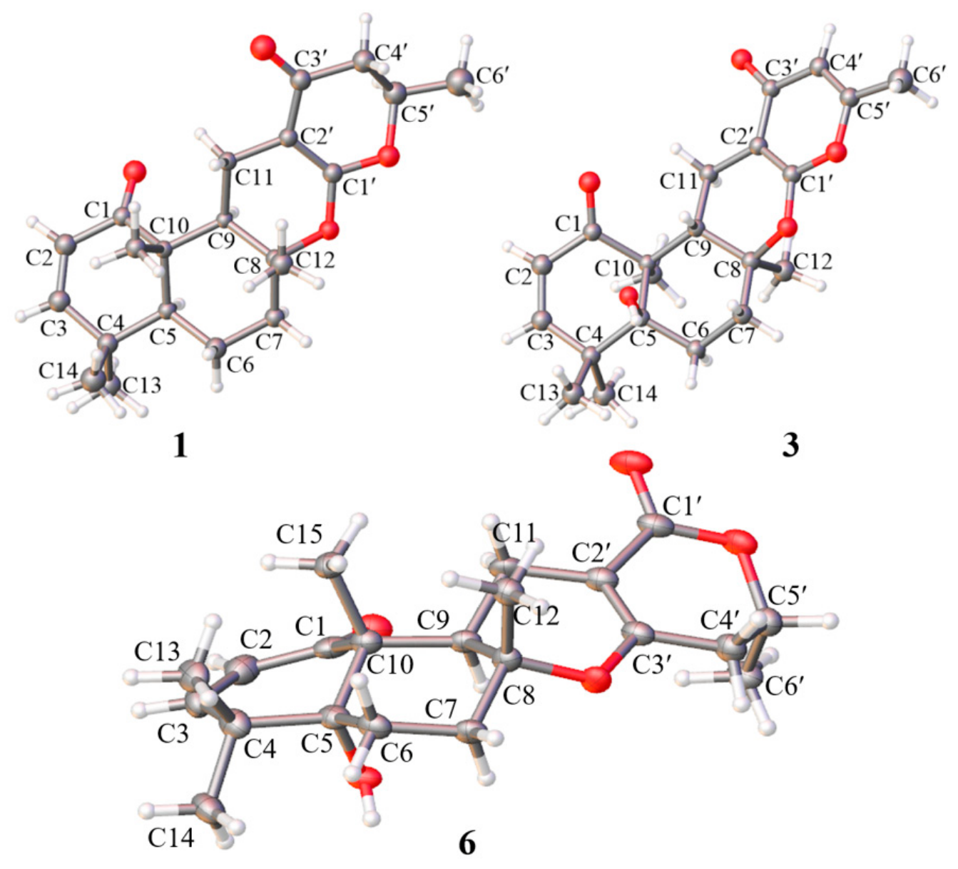
The molecular formula of penirubenoid A (1) was determined to be C21H28O4 by HRESIMS ion at m/z 345.2054 [M + H]+ (calcd. 345.2066), accounting for eight degrees of unsaturation. The 1H NMR spectrum exhibited resonances attributable to four methyl singlets (δH 1.28, 1.18, 1.082, 1.079), one secondary methyl doublet (δH 1.43, d, J = 6.3 Hz), an oxymethine proton (δH 4.57, ddq, J = 13.9, 6.3, 3.2 Hz), and a cis-disubstituted double bond [δH 6.33 (d, J = 10.2 Hz) and 5.70 (d, J = 10.2 Hz)], together with multiple aliphatic signals (Table 1). The 13C NMR data, assisted by HSQC, resolved 21 carbon signals, which were classified as five methyls (δC 31.0, 20.4, 21.7, 20.3, 14.9), four sp3 methylenes (δC 42.6, 38.9, 19.8, 17.6), three sp3 methines (δC 75.5, 50.8, 43.7) including one oxygenated methine, three sp3 quaternary carbons (δC 83.1, 47.3, 36.2), two carbonyls (δC 204.3, 191.0), and two olefinic moieties [δC 154.7/123.9 and 168.1/91.1]. Since the two carbonyls and two double bonds accounted for four of the eight degrees of unsaturation, the remaining four required a tetracyclic framework for compound 1. These data revealed the structural features for a talarolutin-type meroterpenoid, closely related to the co-isolated known analogue talarolutin D (7). The key structural difference was the presence of an additional methine (δH 1.66; δC 50.8) in 1 instead of the oxygenated carbon at δC 78.4 (C-5) observed in 7, implying replacement of the hydroxy group by a hydrogen atom. This deduction was supported by the COSY correlation between H-5 (δH 1.66) and H2-6 (δH 1.79, 1.49), and further corroborated by HMBC cross-peaks from the gem-dimethyl protons (δH 1.079 and 1.082) to C-5 (δC 50.8) (Figure 2). The gross structure was determined as depicted. The NOESY correlations (Figure 3) from H-11β (δH 2.00) to H3-15 (δH 1.18) and H3-12 (δH 1.28) and from H-5 (δH 1.66) to H-9 (δH 1.90) indicated H3-15 and H3-12 were in the same orientation, while H-5 and H-9 were in the opposite direction. Thus, the relative configurations at C-5, C-8, C-9, and C-10 were successfully assigned, while the configuration at C-5′ could not be established from NOESY data, as this stereocenter was too remote from the others to yield diagnostic correlations. Nevertheless, the NMR data of the pyrone moiety were almost identical to those of talarolutins A–D (the structure of talarolutin A was confirmed by single-crystal X-ray diffraction analysis) [1], suggesting that C-5′ possessed the same relative configuration as that of talarolutins A–D. This assignment was ultimately confirmed by single-crystal X-ray diffraction analysis of 1 using Mo Kα radiation (Figure 4).
Table 1.
1H and 13C NMR Data of 1–6 (J in Hz, δ in ppm).
Figure 1.
Structures of compounds 1–17 from the fungus Penicillium rubens MABC05.
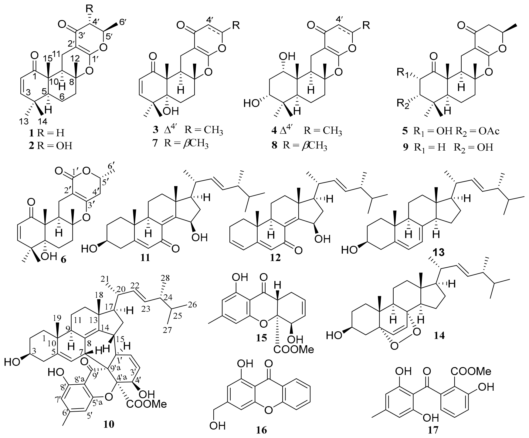
Figure 2.
Key COSY (▬) and HMBC (→) correlations of 1–6.
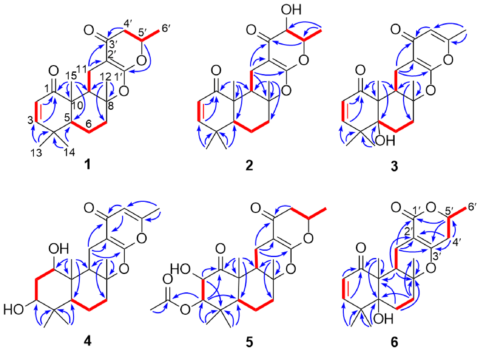
Figure 3.
Key NOESY correlations ( ) of 1–6.
) of 1–6.
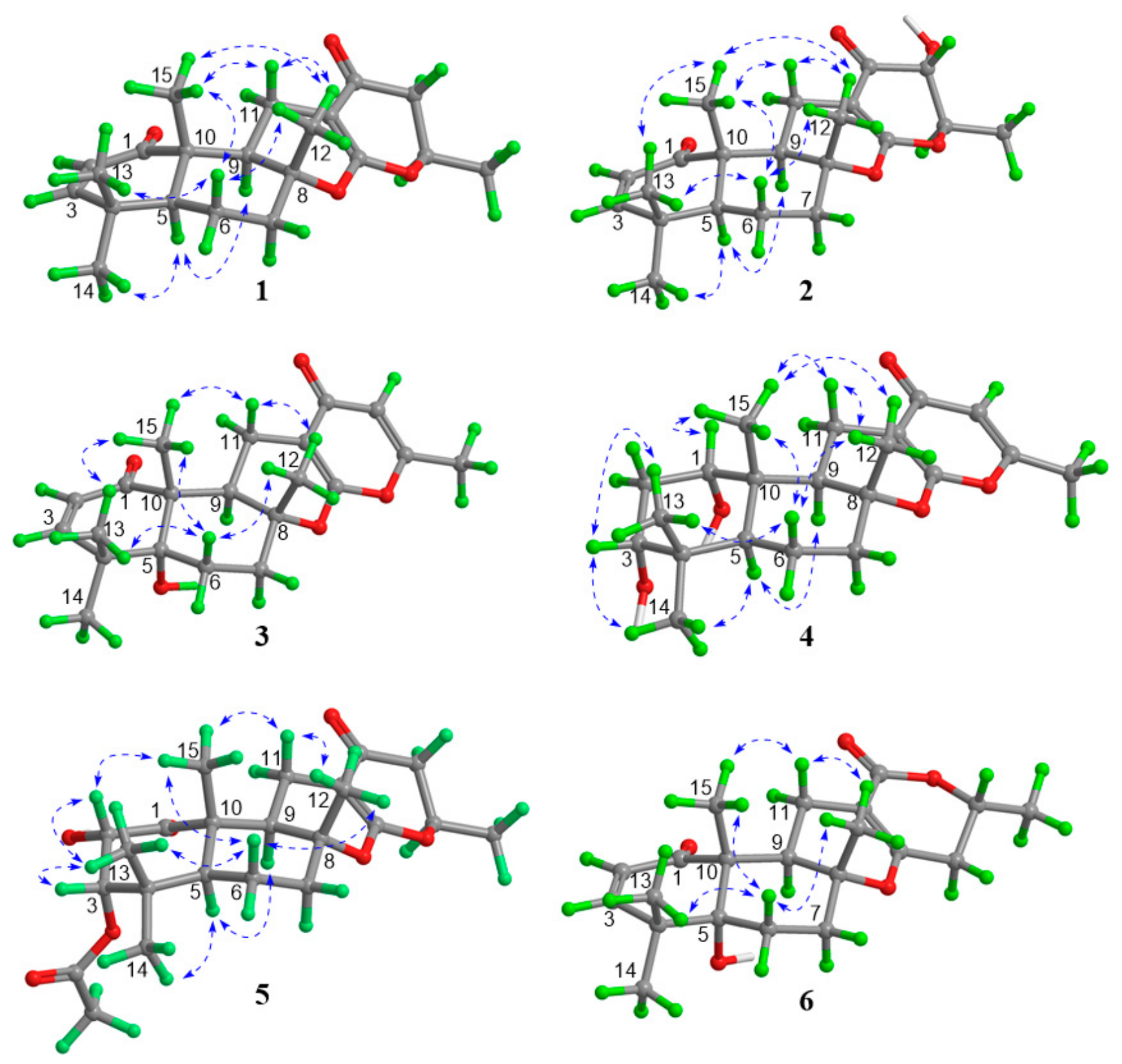
Figure 4.
X-ray crystal structures of compounds 1 and 3 determined using Mo Kα radiation, and compound 6 determined using Cu Kα radiation.
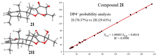
The molecular formula of compound 2 was established as C21H28O5 by HRESIMS. Its NMR data were very similar to those of 1, with obvious differences being the presence of an additional methine (δH 3.86, d, J = 12.2 Hz; δC 71.6) and the upfield-shifted chemical shifts in H-5′. These spectral distinctions suggested that 2 is a hydroxylated derivative of 1, with the hydroxy group most plausibly located at C-4′. This structural assignment was corroborated by the HMBC correlations (Figure 2) from H3-6′ to the methine carbons C-5′ and C-4′, and was further substantiated by the H-4′/H-5′/H3-6′ spin system established by the COSY spectrum. The relative configuration of the drimane moiety was determined to be the same as that of 1 by comparison of the NMR data, and was confirmed by NOESY correlations (Figure 3). The coupling constant JH-4′/H-5′ (12.2 Hz) was indicative of the trans-relationship of H-4′ and H-5′ in an axial orientation. Similar to 1, the relative configuration of C-4′ and C-5′ relative to the drimane moiety could not be assigned from the NOESY data, leaving two plausible structural candidates (Figure 5 and Table S1). Subsequent DP4+ probability analysis of the 13C NMR data supported 2I as the correct isomer (Figure 5 and Table S2), a structural assignment also consistent with biogenetic considerations.
Figure 5.
Two plausible structural candidates (2I and 2II) and regression analysis of experimental and calculated 13C NMR chemical shifts (ppm) of 2I.
Penirubenoid C (3) had the molecular formula of C21H26O5 as established by HRESIMS, indicating one more degree of unsaturation than 7. Its NMR data were closely related to those of 7, with a key difference being the replacement of the original methyl doublet (δH 1.44; δC 20.4) and the CH2-4′–CH-5′ (δH 4.60, 2.49, 2.36; δC 75.6, 42.6) by one olefinic methyl (δH 2.20; δC 19.2) and one additional double bond (δH 6.0; δC 112.0, 160.4). This suggested that the 2,3-dihydro-2-methyl-4-pyrone unit in 7 was converted into a 2-methyl-γ-pyrone moiety. The deduction was evidenced by the HMBC correlation from the olefinic methyl protons (δH 2.20) to the new olefinic carbons (δC 112.0, 160.4). The relative configuration of 3 was determined to be the same as that of 7 by NOESY analysis (Figure 3) and confirmed by single-crystal X-ray crystallography (Figure 4).
Penirubenoid D (4) was assigned the molecular formula C21H30O5 by HRESIMS. Comparison of the 1D NMR data showed that the differences between 4 and 8 mirrored those between 3 and 7, indicating a 2-methyl-γ-pyrone moiety, which was confirmed by HMBC correlations from the olefinic methyl (δH 2.21) to C-4′ (δC 111.4) and C-5′ (δC 160.7). The relative configuration of 4 was determined by NOESY analysis (Figure 3).
The molecular formula of 5 was determined to be C23H32O7 by HRESIMS, accounting for seven degrees of unsaturation. The 1H and 13C NMR data of 5 closely resembled those of 1, with the most notable differences being the presence of two additional oxygenated methines (δH 5.31, 4.96; δC 83.4, 68.8) and an acetoxy group (δH 2.02; δC 20.7, 170.0) instead of the double bond Δ2 in 1. The structure of 5 was subsequently established by detailed 2D NMR analysis (Figure 2 and Figure 3). In particular, the 1H-1H COSY cross-peaks between the two oxymethine protons H-2 (δH 4.96) and H-3 (δH 5.31), together with the HMBC correlations from both H-2 and H-3 to the carbonyl carbon C-1 (δC 210.3) and from H-3 to the acetoxy carbonyl carbon (δC 170.0), assigned the hydroxy group to C-2 and the acetoxy group to C-3. The NOESY correlations from H-2 to H3-15 (δH 1.30) and H3-13 (δH 1.24) and from H-5 (δH 1.73) to H-9 (δH 2.15) and from H-11β (δH 1.97) to H3-12 (δH 1.298) and H3-15 (δH 1.305) revealed that the two cyclohexane rings were trans-fused and adopted chair-chair conformations. These data also indicated that H-2 and the methyl groups CH3-12, CH3-13 and CH3-15 were β-axial oriented, whereas H-5 and H-9 were in α-axial orientation. The coupling constant of JH-2–H-3 (4.0 Hz) reflected an axial–equatorial relationship of H-2 and H-3. A comparison of the NMR data of the dihydropyran-4-one moiety in compounds 1 and 5 indicated that C-5′ in 5 possessed the same relative configuration as that in 1. Thus, the structure of 5 was determined as depicted.
Compound 6 had a molecular formula of C21H28O5 as determined by the HRESIMS, the same as that of 7. The 1H NMR data of 6 were nearly identical to 7, except for the splitting pattern of the methylene protons H2-11 and H2-4′. In 6, the protons of both methylene groups (H2-11 and H2-4′) each displayed additional small allylic couplings (ca. 1 Hz) compared with those in 7. This observation suggested the presence of a (CH2-11)–C=C–(CH2-4′) fragment in 6. Further comparison of their 13C NMR data revealed that the carbonyl carbon of the pyrone moiety was significantly upfield shifted from 191.0 in 7 to 165.7 ppm in 6, indicating the presence of an α-pyrone moiety in 6 (the α-pyrone moiety did not undergo the keto-enol tautomerization process to give the γ-pyrone unit) instead of the γ-pyrone unit in 7. The HMBC correlations from H2-11 to C-1′, C-2′, C-3′, from H2-4′ to C-3′ and C-5′, and from H-5′ to C-1′ and C-3′, along with the COSY relationship of H2-4′/H-5′/H3-6′ confirmed the presence of the α-pyrone moiety and its connection with the drimane unit via C-2′ and C-3′. The relative configuration of the drimane moiety was assigned by NOESY analysis (Figure 3), while the relative configuration at C-5′ and the absolute configuration of 6 were unambiguously established by single-crystal X-ray diffraction using Cu Kα radiation [Flack parameter = 0.1(3)], which also revealed the α-axial orientation of the 6′-CH3 group in the solid state.
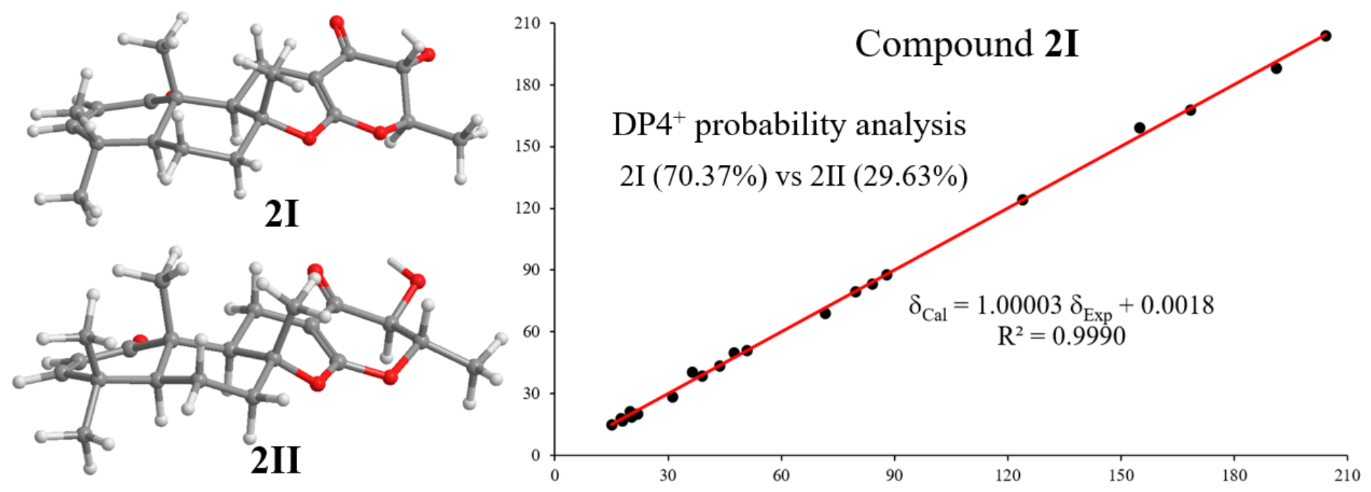
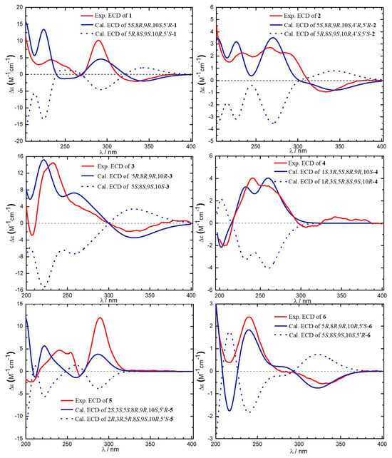
The absolute configurations of compounds 1–5 were assigned by comparison of their experimental and calculated ECD spectra (Figure 6). For compound 6, the ECD calculation was also performed and the result was fully consistent with that determined by single-crystal X-ray diffraction.
Figure 6.
Experimental and calculated ECD spectra of 1–6.
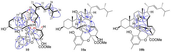
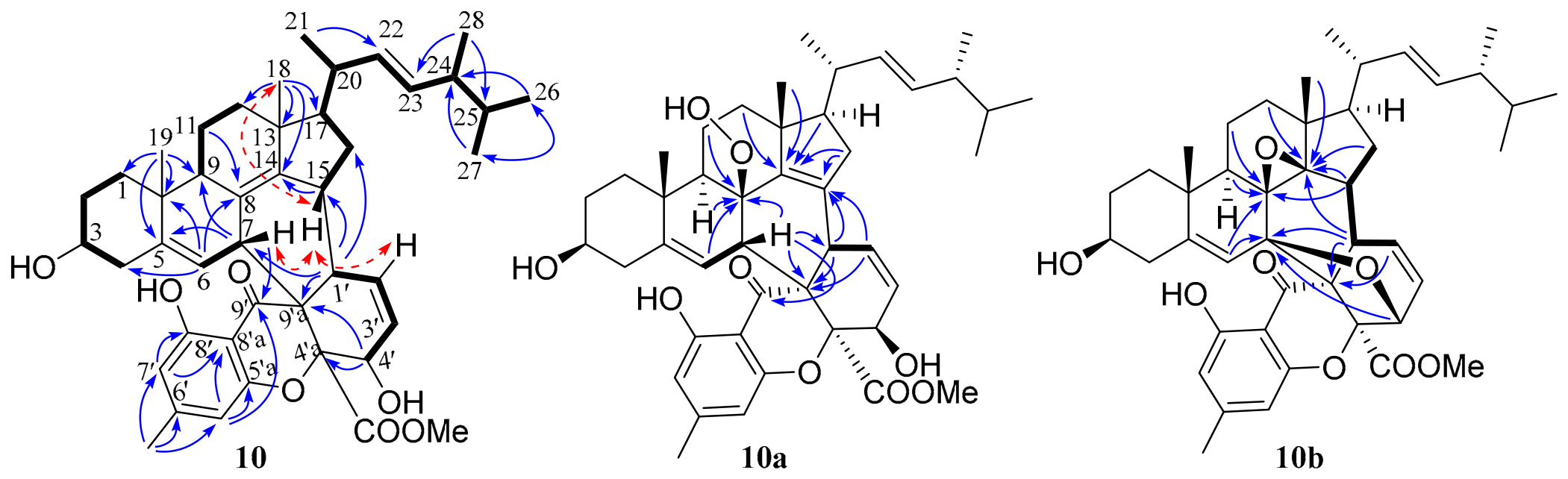
The molecular formula of peniruxanthosterol (10) was assigned as C44H56O7 by the HRESIMS. Its structure was established as a tetrahydroxanthone–ergosterol hybrid by analysis of the NMR data (Table 2), comprising subunits related to compounds 11 and 15. In the tetrahydroxanthone subunit, the COSY correlation (Figure 7) between the olefinic proton H-2′ (δH 5.95) and the methine H-1′ (δH 2.90), together with HMBC correlations from both H-2′ and H-1′ to the non-protonated carbon C-9′a (δC 54.9) defined a C-1′ (δC 46.1)–C-9′a fragment, which distinguished this fragment from that (a CH-1′–C-9′a unit) in 15. For the ergosterol unit, the COSY cross-peak of H-6 (δH 5.86)/H-7 (δH 3.34) and HMBC correlations from H-7 to C-5 (δC 143.6), C-9 (δC 46.8), and C-14 (δC 149.4) indicated replacement of the carbonyl group C-7 (δC 191.1) in 11 by a methine group in 10. In addition, the significantly upfield-shifted chemical shifts in CH-15 (δH 2.10, m; δC 41.5) suggested the absence of a hydroxy group at this position. These differences indicated that the two fragments are linked via this four-carbon junction (C-1′, C-9′a, C-7, C-15). The COSY correlation between H-15 and H-1′, along with the HMBC cross-peaks from H-1′ to C-14 (δC 149.4), C-15 (δC 41.5), and C-16 (δC 37.2), confirmed the direct linkage between CH-1′ and CH-15. Furthermore, the HMBC correlations from H-7 to C-1′, C-9′a, and C-9 established the connectivity between C-9′a and CH-7, thereby completing the assembly of the hybrid skeleton.
Table 2.
The 1H (600 MHz) and 13C NMR data (151 MHz) of 10, 10a, 10b, 11, and 14 in CDCl3.
Figure 7.
Key COSY ( ), HMBC (→), NOESY (
 ) correlations of 10 and the two oxidative artifacts 10a and 10b.
), HMBC (→), NOESY () correlations of 10 and the two oxidative artifacts 10a and 10b.
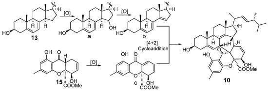
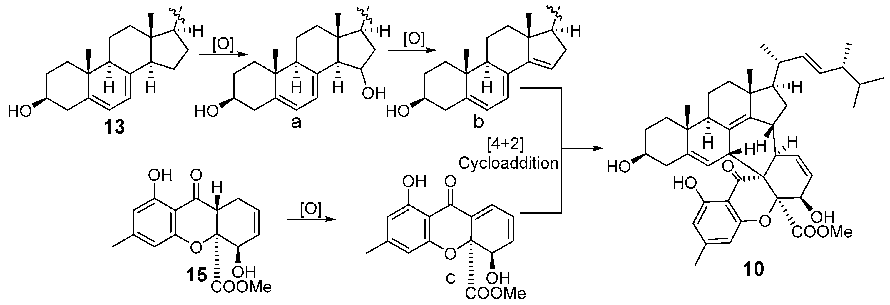
It should be noted that compound 10 may be an artifact formed during the extraction and isolation procedure. Its biogenesis is proposed to proceed via the convergent assembly of ergosterol (13) and the tetrahydroxanthone (4R,4aS,9aR)-1,9a-dihydronidulalin A (15) (Scheme 1), analogous to the proposed biosynthesis of the rare ergosterol derivative rubensteroid from Penicillium rubens AS-130 [10]. On this basis, compound 13 undergoes sequential enzymatic oxidations, including regioselective hydroxylation at C-15 followed by enzymatic desaturation, to furnish the highly reactive dienophile b bearing the double bond Δ14. Concurrently, 15 is oxidatively dehydrogenated to generate the conjugated diene c. These highly activated intermediates undergo a stereospecific intermolecular [4 + 2] cycloaddition, constructing the bridging six-membered ring and ultimately affording the octacyclic hybrid 10.
Scheme 1.
Putative pathway for the biogenesis of 10.
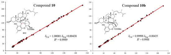
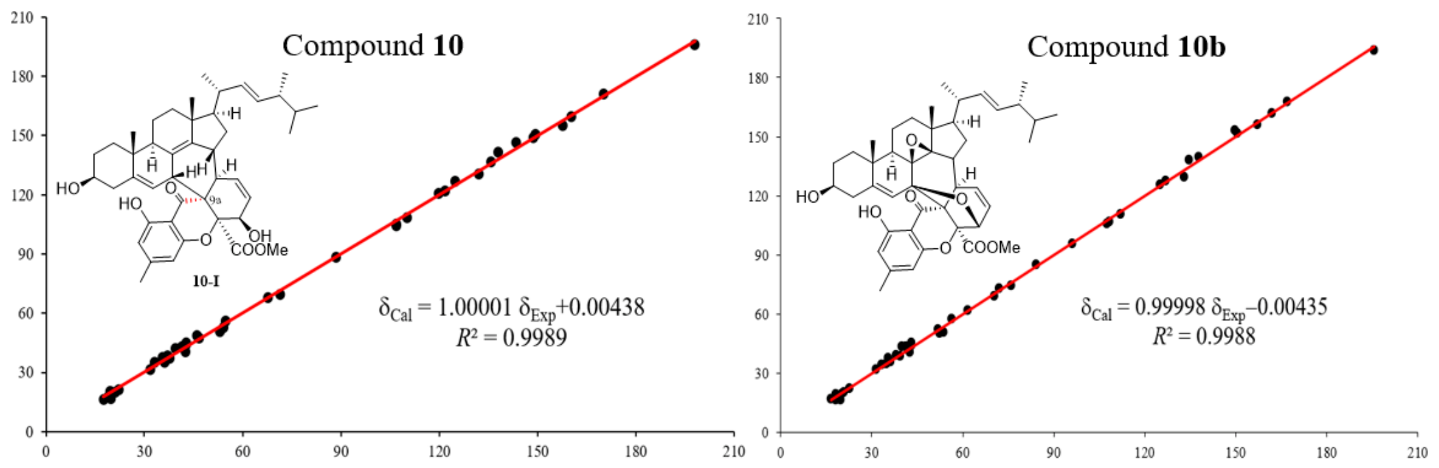
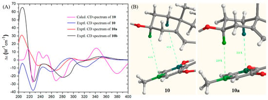
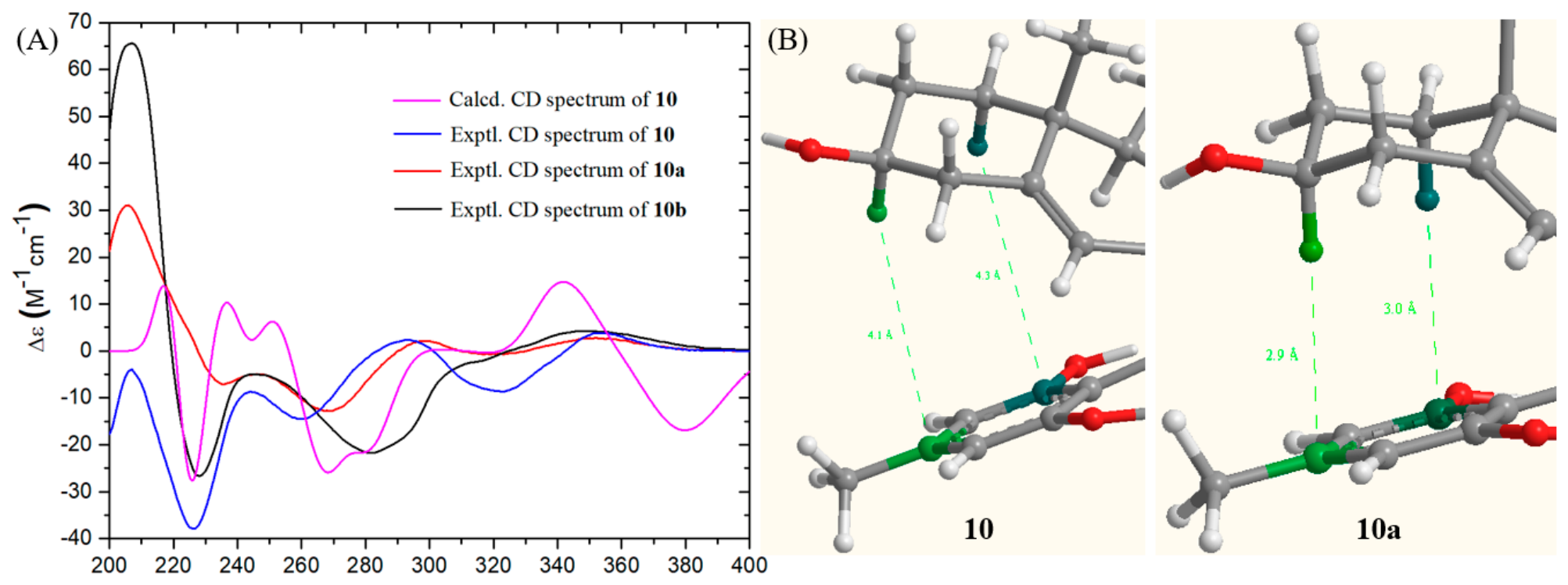
The relative configuration of the octacyclic core in 10, except for that at C-9′a, was established by analysis of its NOESY data (Figure 7 and Figure S48). The absolute configurations at C-20 and C-24 in the sterol side chain were proposed to be identical to those of 13 on the basis of biogenetic considerations. Particularly, the NOESY cross-peaks from H-15 (δH 2.10) to H3-18 (δH 0.92) and H-7 (δH 3.34) indicated that these protons are β-oriented, whereas the correlation between H-15 and H-2′ (δH 5.95) suggested that H-1′ is α-oriented. However, the configuration at C-9′a could not be determined by the NOESY data. Consequently, 10 may be either of the two C-9′a epimers (9′aS or 9′aR). To differentiate between these two structural candidates, we performed quantum chemical 13C NMR chemical shift calculations to assign the correct configuration at C-9′a. Comparison of the experimental and calculated 13C NMR data revealed that 9′aS-epimer (Figure 8, R2 = 0.9989, Table S3) exhibited markedly better agreement with the experimental values than 9′aR-epimer. Notably, for the 9′aR-epimer (Table S4), the calculated chemical shifts for C-3′ (131.4) and C-4′ (75.0) displayed significant deviations from the experimental data (119.8 and 67.7, respectively), with discrepancies of approximately 10 ppm. In addition, the significant upfield-shift in H-3, compared with those of 11, 13 and 14 (δH 2.82 vs. 3.75, 3.64 and 3.97), is likely attributed to the shielding effect exerted by the aromatic ring of the xanthone moiety. This spatial proximity, which can only be achieved in the 9′aS-epimer, provided further indirect evidence supporting the assignment of 9′aS-configuration. Thus, the structure of 10 was established as depicted. The absolute configuration of 10 was confirmed by ECD calculation (Figure 9A).
Figure 8.
Regression analysis of experimental and calculated 13C NMR chemical shifts (ppm) of 10 and 10b.
Figure 9.
(A) Experimental and calculated ECD spectra of 10 and experimental ECD spectra of 10a and 10b. (B)The distances between H-1α (cyan sphere) and H-3 (green sphere) and the benzene ring of the xanthone moiety are indicated by dashed green lines. In 10, the distances from H-3 and H-1α to the xanthone benzene ring are 4.1 Å and 4.3 Å, respectively. (B) In 10a, the distances are shortened to 2.9 Å and 3.0 Å, respectively.
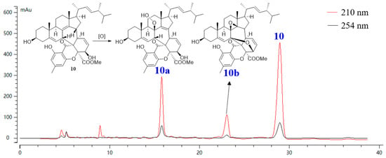
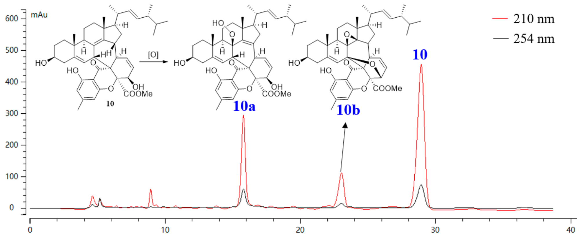
In addition, two minor related analogues were isolated, but their high-quality NMR data were unavailable owing to the limited sample amounts. It is worth noting that TLC and HPLC analyses of compound 10, which was kept at 4 °C over a period of four years (Figure 10), showed the presence of two significant impurities. These two impurities were further isolated, and their 1H NMR spectra proved to be identical to those of the two minor analogues mentioned above.
Figure 10.
HPLC profiles of compound 10 and its two oxidative storage artifacts (10a and 10b) detected after 48 months of preservation as a neat solid at 4 °C.
Compound 10a was assigned the molecular formula C44H56O9. Comparison of its NMR data with those of 10 indicated that 10a contains one additional oxygenated quaternary carbon and one fewer methine carbon. HMBC correlations from H-6, H-7, H-9, and H2-11 located this oxygenated carbon at C-8. The double bond originally between C-8 and C-14 was found to have migrated to C-14 and C-15, as established by the HMBC correlations from H-9, H2-16, H-17, and H3-18 to C-14, and from H2-16, H-1′, and H-2′ to C-15. Consequently, the remaining HO2 unit required by the molecular formula was assigned to a hydroperoxy group at C-8, which was further corroborated by the characteristic hydroperoxy proton signal at δH 7.19 (br s) and the carbon resonance at δC 84.4. The β-orientation of the hydroperoxy group at C-8 was assigned based on a combination of NMR spectroscopic evidence and molecular modeling. Compared with compound 10, the upfield shifts in H-1α (0.50 vs. 0.72) and H-3 (2.41 vs. 2.82 ppm) in 10a suggested that the benzene ring of the xanthone moiety is in closer proximity to the leftmost ring of the steroid moiety. This spatial compression effect is fully consistent with the β-configuration of the hydroperoxy group, as revealed by Chem3D energy-minimized models, which showed a significantly shorter interring distance (cal. 3.0 in 10a vs. 4.2 Å in 10) when the hydroperoxy group adopted the β-orientation (Figure 9B).
Compound 10b was assigned the molecular formula C44H54O8, indicating that 10b possessed one more degree of unsaturation than 10. The NMR data revealed that 10b lacked the Δ8(14) present in 10, and instead contained three additional oxygenated non-protonated carbons (δC 84.2, 61.7, and 72.0), which were assigned to C-7, C-8, and C-14, respectively, based on HMBC correlations (Figure 7). The remaining two degrees of unsaturation were accounted for by two additional rings. The C-4′-O-C-7 ether bridge was established by the strong HMBC correlation from H-4′ to C-7 and the downfield shift in C-4′ (δC 67.7 vs. 76.0). In addition, the chemical shifts in C-8 (δC 61.7) and C-14 (δC 72.0), together with the remaining degree of unsaturation, supported the presence of an epoxy ring between C-8 and C-14. In precursor 10, the 4′-hydroxy group was spatially close to C-7, which was activated by the adjacent two double bonds. This favorable arrangement facilitated an intramolecular nucleophilic attack of the 4′-hydroxy oxygen on C-7 upon oxidative activation, leading to the formation of the β-oriented ether bridge in 10b. The β-orientation of the epoxy moiety was proposed to be governed by the steric hindrance on the α-face of the C-8 and C-14, which was provided by H-9, H-12α, H-17, H-1′, and the carbonyl group of the xanthone moiety. The relative configuration of 10b was confirmed by the good correlation between the experimental and calculated 13C NMR chemical shifts (Figure 8).
2.2. Biological Activity Assessment
The isolated compounds were evaluated for their anti-ferroptotic and cytotoxic activities.
2.2.1. Compound 15 as a Ferroptosis Inhibitor
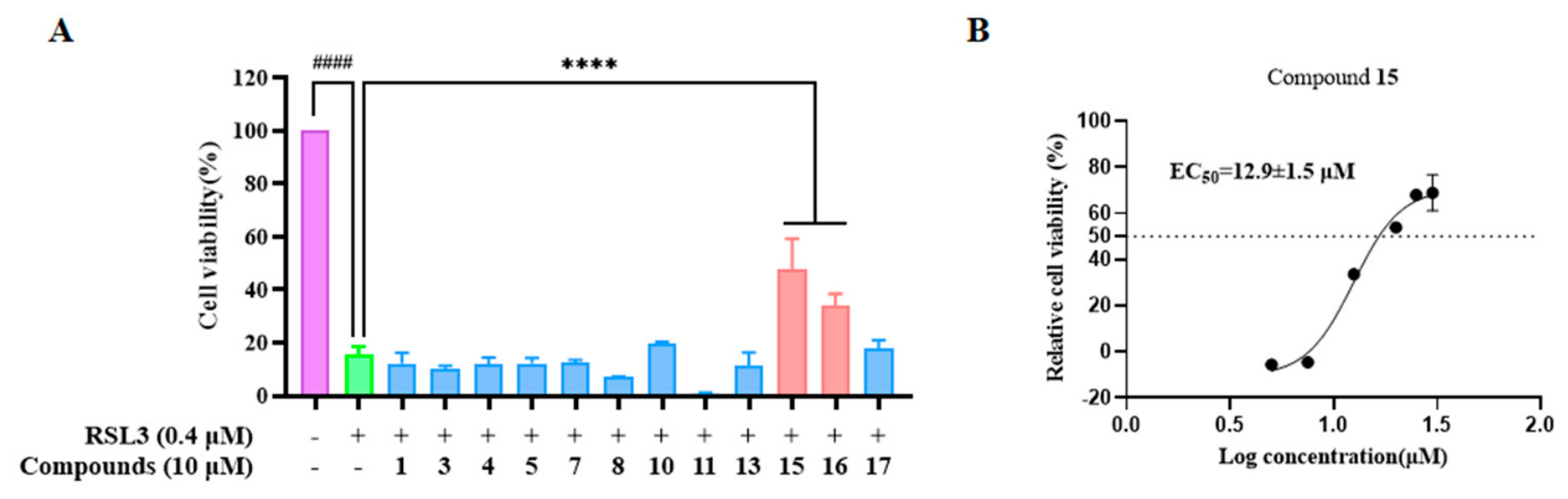
The anti-ferroptotic activity of these compounds was evaluated in RSL3-induced HK-2 cells, with the exception of compounds 2, 6, 9, 12, and 14 due to insufficient amounts. As shown in Figure 11A, treatment with 0.4 μM RSL3 markedly reduced cell viability to 15% of untreated controls. Notably, at 10 μM, compounds 15 and 16 exhibited cytoprotective effects, restoring viability to 47.8% and 34%, whereas other compounds showed negligible protective effects (<30% viability). To further characterize the potency of 15, concentration-response experiments (5–30 μM) were conducted. Ferrostatin-1 (Fer-1), a canonical ferroptosis inhibitor, was used as the reference. As depicted in Figure 11B, 15 exerted a dose-dependent protective effect, with cell viability increasing from 24% (5 μM) to 78% (30 μM) (EC50 = 12.8 ± 1.6 μM). Notably, its maximal efficacy at 30 μM restored viability to 78%, positioning 15 as a promising ferroptosis inhibitor. Unfortunately, the limited quantity of purified 16 prevented us from conducting further biological testing.
Figure 11.
Compound 15 inhibited ferroptosis in HK-2 cells. (A) Viability of HK-2 cells treated with tested compounds (10 μM) and RSL3 (0.4 μM). (B) Dose-dependent protective effect of 15 (5-30 μM) in RSL3-induced HK-2 cells. Data are presented as mean ± SD (n = 3). #### p < 0.0001 vs. Control; **** p < 0.0001 vs. RSL3-treated groups. DMSO: vehicle control; Fer-1 (1 μM): positive control.
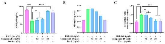
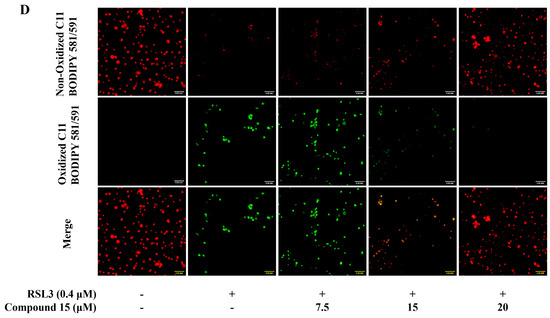
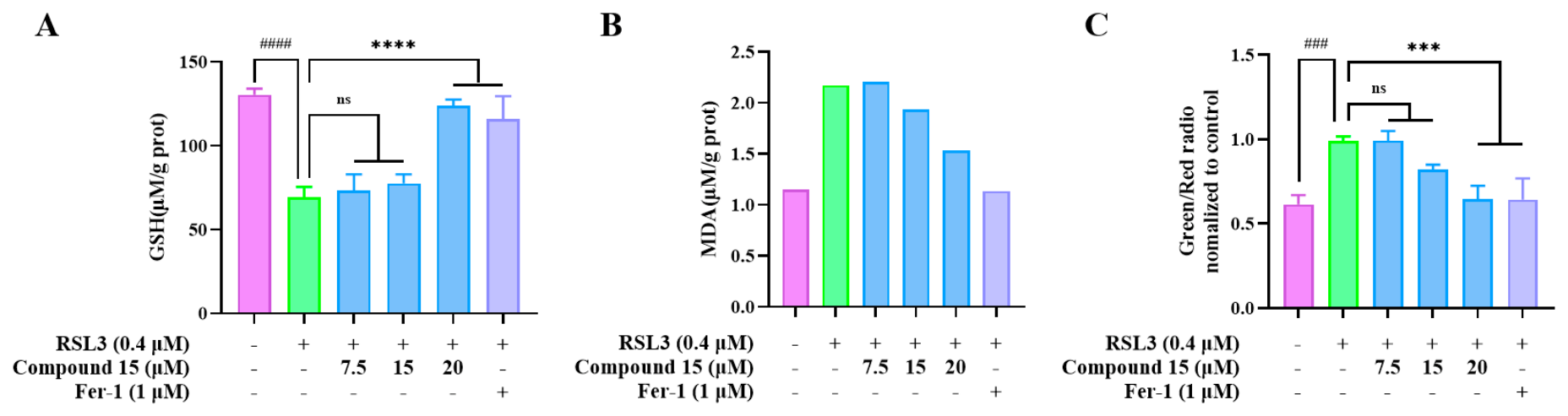
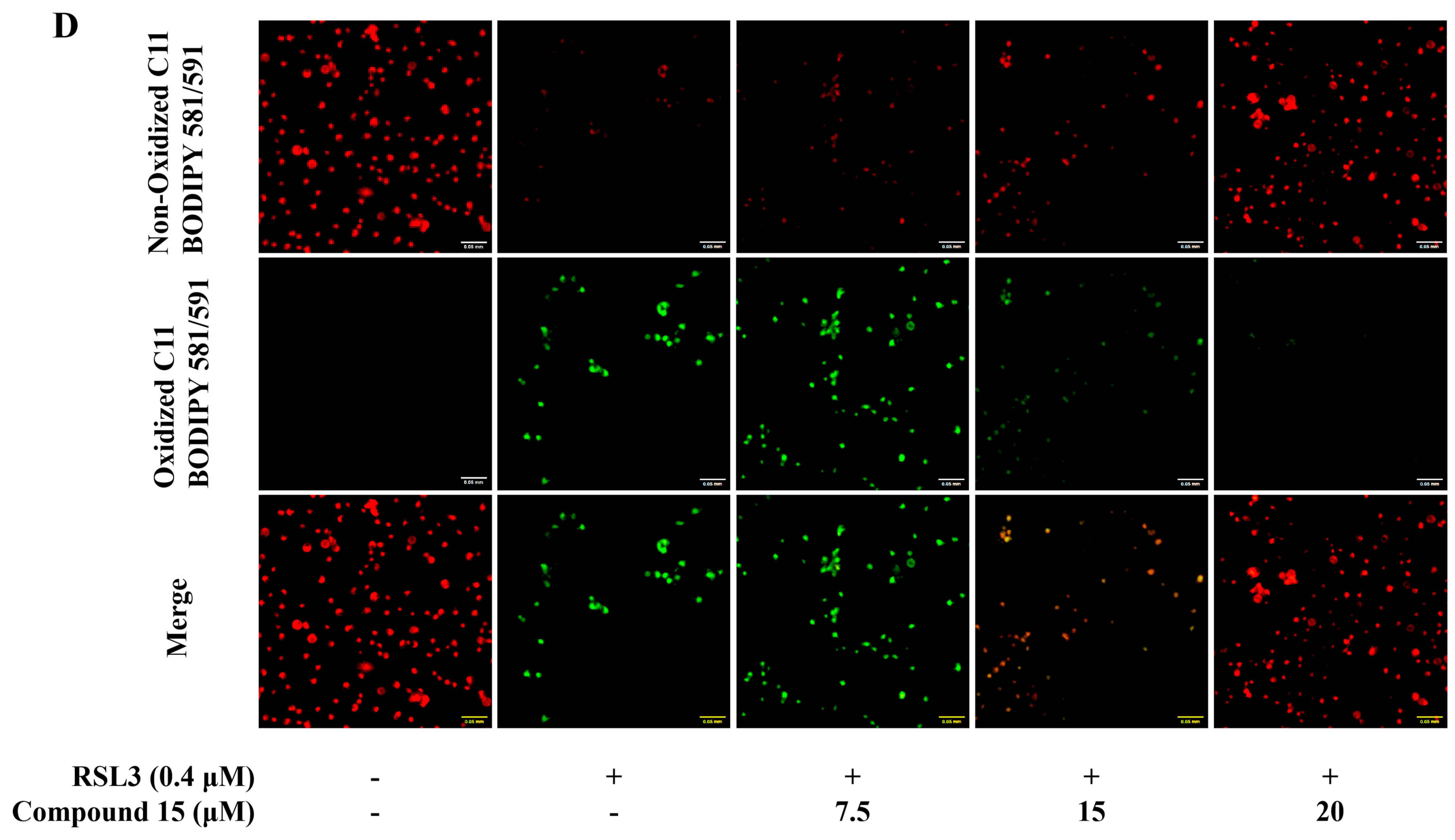
Given the ferroptosis-inhibitory activity of compound 15 (Figure 11), we further explored the underlying mechanism through comprehensive analysis of oxidative stress markers and lipid peroxidation dynamics. As shown in Figure 12A,B, treatment with 0.4 μM RSL3 for 24 h induced pronounced lipid peroxidation in HK-2 cells, and intracellular glutathione (GSH) levels decreased to 53.1% of control levels, concurrently with a 1.9-fold increase in malondialdehyde (MDA) levels compared to untreated controls. Notably, cotreatment with 20 μM 15 significantly reversed these effects, normalizing MDA levels to 1.33-fold that of the control and restoring GSH to 94.8% of control levels. These data indicate that 15 attenuates RSL3-induced oxidative stress. Further analysis using fluorescence imaging with the oxidation-sensitive fluorescent probe C11-BODIPY581/591 revealed that RSL3 markedly intensified lipid peroxidation, increasing the green/red fluorescence ratio to 1.61-fold of the control (Figure 12C,D). However, cotreatment with compound 15 dose-dependently attenuated this ratio to 1.3-fold at 15 μM and to the control level (1.0-fold) at 20 μM, clearly indicating suppression of lipid peroxidation.
Figure 12.
Compound 15 attenuated ferroptosis by inhibiting lipid peroxidation. (A) GSH content; (B) MDA levels; (C,D) C11 BODIPY fluorescence ratio (green/red) reflecting lipid peroxidation. Data are presented as mean ± SD (n = 3–6); ### p < 0.001, #### p < 0.0001 vs. control; *** p < 0.001, **** p < 0.0001 vs. RSL3; ns, not significant.
2.2.2. Compounds 11 and 16 Exhibited Cytotoxic Activity Against MCF-7 Cells
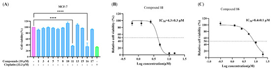
The same set of compounds was tested for cytotoxicity against MCF-7 cells (Figure 13), with compounds 2, 6, 9, 12, and 14 excluded for the same reason. Single-concentration cytotoxic screening revealed that compounds 11 and 16 exhibited antiproliferative effects against MCF-7 cells at 10 μM. After treatment with compound 11, the cell viability decreased to approximately 36%, while compound 16 reduced cell viability to around 34%. Their inhibitory effects were comparable to that of cisplatin (33.3 μM), the positive control. Subsequent dose–response assays further confirmed the cytotoxic activity, and the IC50 values of compounds 11 and 16 were calculated to be 4.3 ± 0.3 μM and 8.4 ± 0.1 μM, respectively. Compound 11 displayed stronger cytotoxic activity than compound 16 against MCF-7 human breast cancer cells. Cytotoxicity against non-cancerous cell lines was not assessed in the current study.
Figure 13.
Cytotoxic evaluation of selected compounds in MCF-7 human breast cancer cells. (A) Cell viability of MCF-7 cells treated with the tested compounds at 10 μM. Cisplatin (33.3 μM) served as positive control. (B,C) IC50 curves of compound 11 and 16. Data are presented as mean ± SD (n = 3). **** p < 0.0001 vs. vehicle control.
3. Materials and Methods
3.1. General Experimental Procedure
Specific rotations were determined on a Rudolph Autopol VI polarimeter (Rudolph Research Analytical, Hackettstown, NJ, USA) at the sodium D-line (589 nm) and 25 °C. UV spectra were recorded on a UV-2600 spectrometer (Shimadzu Co., Kyoto, Japan) at room temperature over 190–400 nm. ECD spectra were recorded on a Chirascan V100 spectrometer (Applied Photophysics, Surrey, UK) at room temperature over 200–400 nm with a bandwidth of 1.0 nm, a scan rate of 100 nm/min, and a response time of 1 s. The NMR spectra were acquired on Bruker AVANCE III HD 400 MHz and 600 MHz spectrometers (Bruker, Fällanden, Switzerland) using solvent signals (Methanol-d4: δH 3.31/δC 49.0; CDCl3: δH 7.26/δC 77.0) as references. All spectra were recorded at 298 K. The relaxation delay (d1) was 1.5 s for 1H and 2.0 s for 13C. The number of scans (NS) was 16 for 1H, 1024 for 13C, and 8–64 for 2D experiments. HRESIMS spectra were acquired on an Agilent 6546A Q-TOF mass spectrometer (Agilent Technologies, Singapore) and a Bruker FTMS spectrometer (Bruker Daltonics, Bremen, Germany), both equipped with ESI sources and operated in both positive and negative ion modes over m/z 100–1000. Mass resolution was 30,000 (FWHM at m/z 118) with mass accuracy < 0.8 ppm. Samples were separated on a C18 column (2.1 × 50 mm, 1.8 μm) eluted with 80% aq. acetonitrile isocratically at 0.3 mL/min over 5 min, with 5 μL injection. Semi-preparative high-performance liquid chromatography (HPLC) was undertaken on a Shimadzu LC-6AD pump (Shimadzu Co., Kyoto, Japan) equipped with a UV detector, employing a YMC-Pack ODS-A HPLC (YMC Co., Ltd., Kyoto, Japan) column (250 mm × 10 mm, S-5 μm, 12 nm). Single-crystal X-ray diffraction data were collected on two diffractometers: Cu Kα data were acquired on a Rigaku XtaLAB Synergy-R diffractometer (Rigaku Corporation, Tokyo, Japan; Cu Kα, λ = 1.54184 Å, 1.2 kW) and reduced using CrysAlisPro (version 1.171.44.91a); Mo Kα data were collected on a Bruker D8 VENTURE diffractometer (Bruker AXS Inc., Madison, WI, USA; Mo Kα, λ = 0.71073 Å) and reduced using SAINT (version 8.40B). All structures were solved using SHELXT (version 2014/5) and refined using SHELXL (version 2019/3) within the Olex2 (version 1.5) interface.
3.2. Fungal Strain and Identification
The strain MABC05-5-M4 was isolated from the deep-sea water sample (−5644 m) collected from the Western Pacific Ocean (DY27I-MABC05). The strain was identified as Penicillium rubens by Changxu Huang (from Prof. Zhuhua Luo’s group) based on microscopic examination and internal transcribed spacer (ITS) sequencing. Its ITS sequence was deposited in GenBank under accession number KP200040, and the strain was preserved at the Key Laboratory of Marine Biogenetic Resources, Third Institute of Oceanography, Ministry of Natural Resources (MCCC 3A00591).
3.3. Fermentation, Extraction, and Isolation
The fermentation was carried out in 50 Fernbach flasks (500 mL), each containing 80 g of rice. Artificial seawater (100 mL) was added to each flask, and the contents were soaked for three hours before autoclaving at 15 psi for 30 min. After cooling to room temperature (r.t.), each flask was inoculated with 3.0 mL of the spore inoculum and incubated at r.t. for 30 days. The fermented material from all flasks was combined, soaked in EtOAc (5 L), and ultrasonically extracted for 1 h. The EtOAc solvent was concentrated to give 7.21 g of crude extract.
The extract was fractionated by MCI gel column chromatography (CC) with MeOH/H2O (20:80 → 100:0) as eluent to yield 9 fractions (Fr.1–Fr.9). Fraction 5 (2.7 g) was separated by an ODS silica gel CC (55 g) using a gradient elution of MeOH/H2O (10:90 → 60:40, 500 mL per gradient) to yield 6 subfractions (Fr.5.1–Fr.5.6). Fr.5.3 (0.15 g) was purified by HPLC (YMC-Pack ODS-A column, 250 × 10 mm, S-5 μm, 12 nm) using MeCN/H2O (52:48, 2 mL/min) as the mobile phase to afford compound 1 (20.0 mg, tR 29.0 min, yield 0.28%). Compounds 2 (3.1 mg, tR 31.6 min, yield 0.04%) and 6 (1.3 mg, tR 39.7 min, yield 0.02%) were obtained from fractions Fr.5.4 (0.38 g) and Fr.5.5 (1.37 g) by HPLC using MeCN/H2O (65:35, 2 mL/min). The fraction Fr.5.6 (0.97 g) was further separated on an ODS silica gel CC using MeOH/H2O (40:60 → 90:10) as eluent to yield 5 subfractions (Fr.5.6.1–Fr.5.6.5). Fr.5.6.2 (30.0 mg) was purified by HPLC using MeCN/H2O (56:44, 2 mL/min) as the mobile phase to afford compound 8 (3.7 mg, tR 37.9 min, yield 0.05%). Fr.5.6.4 (78.2 mg) was purified by HPLC using MeCN/H2O (47:53, 3 mL/min) as the mobile phase to afford compound 5 (3.5 mg, tR 32.8 min, yield 0.05%) and compound 11 (6.2 mg, tR 47.2 min, yield 0.09%). Fr.5.6.5 (87.6 mg) was purified by HPLC using MeCN/H2O (42:58, 2.5 mL/min) as the mobile phase to afford compound 13 (5.5 mg, tR 20.3 min, yield 0.08%) and compound 12 (2.6 mg, tR 36.5 min, yield 0.04%). Fr.6 (2.3 g) was separated on an ODS silica gel CC using a gradient elution of MeOH/H2O (40:60 → 90:10) to yield 12 subfractions (Fr.6.1–Fr.6.12). Fr.6.3 (91.8 mg) was purified by HPLC using MeOH/H2O (57:43, 3 mL/min) to afford compounds 3 (4.3 mg, tR 23.1 min, yield 0.06%) and 7 (3.6 mg, tR 34.1 min, yield 0.05%). Fr.6.4 (81 mg) was purified by HPLC using MeOH/H2O (60:40, 2 mL/min) to afford compounds 4 (5.3 mg, tR 23.6 min, yield 0.07%) and 9 (1.2 mg, tR 40.9 min, yield 0.02%). Fr.7 (1.1 g) was purified by ODS silica gel CC using a gradient elution of MeOH/H2O to yield 7 subfractions (Fr.7.1–Fr.7.7). Fr.7.4 (0.61 g) was purified by HPLC using MeOH/H2O (47:53, 3 mL/min) to afford compound 16 (15.6 mg, tR 16.9 min, yield 0.22%) and compound 17 (6.3 mg, tR 25.1 min, yield 0.09%). Fraction F.8 (1.7 g) was purified on an ODS silica gel CC eluting with MeOH/H2O (30:70 → 100:0) to yield 9 subfractions (Fr.8.1–Fr.8.9). Fr.8.7 (109.3 mg) was purified by HPLC using MeCN/H2O (47:53, 3 mL/min) to afford compounds 14 (1.3 mg, tR 23.1 min, yield 0.02%) and 15 (3.6 mg, tR 34.1 min, yield 0.05%). Fr.8.8 (62.6 mg) was purified by HPLC using 100% MeCN (3 mL/min) as the mobile phase to afford compound 10 (13.8 mg, tR 29 min, yield 0.19%). After storage for 48 months, compound 10 was repurified by HPLC using 100% MeCN (3 mL/min) to afford compounds 10a (1.6 mg, tR 14.6 min), 10b (0.9 mg, tR 22.3 min), and 10 (4.1 mg, tR 28.4 min). All yields were calculated based on the initial crude extract (7.21 g).
Penirubenoid A (1): Colorless crystals (MeOH); [α]25D +476.5 (c 0.1, MeOH); UV (MeOH) λmax 219, 281 nm; ECD (c 2.9 × 10⁻⁴ M, MeOH) λmax (Δε) 290 (+10.20), 266 (−0.43), 233 (+4.36) nm; 1H and 13C NMR data, see Table 1; ESIMS m/z 345.2054 [M + H]+ (calcd for C21H29O4+, 345.2066).
Penirubenoid B (2): Colorless oil; [α]25D +45 (c 0.05, MeOH); ECD (c 5.3 × 10⁻⁴ M, MeOH) λmax (Δε) 333 (−0.93), 267 (+2.71), 234 (+2.28) nm; 1H and 13C NMR data, see Table 1; ESIMS m/z 361.2126 [M + H]+ (calcd for C21H29O5+, 361.2015).
Penirubenoid C (3): Colorless crystals (MeOH); [α]25D +240.0 (c 0.1, MeOH); UV (MeOH) λmax 258 nm; ECD (c 2.8 × 10⁻⁴ M, MeOH) λmax (Δε) 325 (−1.91), 235 (+14.52) nm; 1H and 13C NMR data, see Table 1; ESIMS m/z 359.1845 [M + H]+ (calcd for C21H27O5+, 359.1853).
Penirubenoid D (4): Colorless oil; [α]25D +193.2 (c 0.1, MeOH); UV (MeOH) λmax 260 nm; ECD (c 2.9 × 10⁻⁴ M, MeOH) λmax (Δε) 240 (+4.04) nm; 1H and 13C NMR data, see Table 1; ESIMS m/z 363.2158 [M + H]+ (calcd for C21H31O5+, 363.2166).
Penirubenoid E (5): Colorless oil; [α]25D +206.5 (c 0.1, MeOH); UV (MeOH) λmax 271 nm; ECD (c 2.8 × 10⁻⁴ M, MeOH) λmax (Δε) 290 (+11.93), 266 (−0.98), 240 (+4.69) nm; 1H and 13C NMR data, see Table 1; ESIMS m/z 421.2218 [M + H]+ (calcd for C23H33O7+, 421.2221).
Penirubenoid F (6): Colorless oil; [α]25D +42 (c 0.1, MeOH); ECD (c 5.6 × 10⁻⁴ M, MeOH) λmax (Δε) 335 (−0.55), 240 (+2.41) nm; 1H and 13C NMR data, see Table 1; ESIMS m/z 361.2013 [M + H]+ (calcd for C21H29O5+, 361.2010).
Peniruxanthosterol A (10): Colorless oil; [α]25D +206.5 (c 0.1, MeOH); UV (MeOH) λmax 338, 282 nm; ECD (c 2.9 × 10⁻⁴ M, MeOH) λmax (Δε) 354 (+3.81), 323 (−8.70), 259 (−14.54), 226 (−37.84) nm; 1H and 13C NMR data, see Table 2; ESIMS m/z 695.3953 [M − H]− (calcd for C44H55O7−, 695.3953).
Peniruxanthosterol A1 (10a): Colorless oil; [α]25D −93.0 (c 0.1, MeOH); ECD (c 2.8 × 10⁻⁴ M, MeOH) λ max (Δε) 352 (+2.78), 299 (+2.13), 269 (−12.81), 236 (−7.08), 206 (+31.08) nm; 1H and 13C NMR data, see Table 2; ESIMS m/z 727.3850 [M − H]− (calcd for C44H55O9−, 727.3852).
Peniruxanthosterol A2 (10b): Colorless oil; [α]25D −154.0 (c 0.05, MeOH); ECD (c 2.8 × 10⁻⁴ M, MeOH) λmax (Δε) 347 (+4.27), 282 (−21.73), 228 (−26.68), 207 (+65.61) nm; 1H and 13C NMR data, see Table 2; ESIMS m/z 709.3739 [M − H]− (calcd for C44H53O8−, 709.3747).
The known compounds were identified as talarolutin D (7) [1], talarolutin B (8) [1], talarolutin A (9) [1], 3β,15β-dihydroxyl-(22E,24R)-ergosta-5,8(14),22-trien-7-one (11) [16], 15β-hydroxyl-(22E,24R)-ergosta-3,5,8,22-tetraen-one (12) [17] ergosterol (13) [18], ergosterol peroxide (14) [18], (4R,4aS,9aR)-1,9a-dihydronidulalin A (15) [19], 1-hydroxy-3-hydroxymethyl-9H-xanthen-9-one (16) [20], nidulalin B (17) [21].
3.4. X-Ray Crystallographic Analysis
Colorless crystals of penirubenoid A (1) and C (3) were obtained by slow evaporation from MeOH/H2O (8:1 and 7:1, v/v), respectively, while colorless crystals of penirubenoid F(6) were obtained by slow evaporation from methanol. Suitable crystals were selected and mounted on loops for diffraction experiments. Data were collected at 150.15 K for 1 and 3, and at 100.00(10) K for 6. All structures were solved using intrinsic phasing with the SHELXT structure solution program and refined with the SHELXL refinement package (least-squares minimization) within the Olex2 interface.
Crystal Data for penirubenoid A (1). C21H28O4, Mr = 344.454, orthorhombic, space group P212121 (no. 19), a = 6.0607(3) Å, b = 14.2909(9) Å, c = 21.6010(12) Å, α = 90°, β = 90°, γ = 90°, V = 1870.92(18) Å3, Z = 4, T = 150.15 K, μ(Mo Kα) = 0.083 mm−1, Dcalc = 1.223 g/cm3, 17,570 reflections measured (5.7° ≤ 2θ ≤ 56.7°), 4626 unique (Rint = 0.0932, Rsigma = 0.0752), which were used in all calculations. The final R1 was 0.0699 (I > 2σ(I)) and wR2 was 0.1741 (all data). Crystallographic data have been deposited at the Cambridge Crystallographic Data Centre (CCDC 2581952).
Crystal Data for penirubenoid C (3). C21H26O5, Mr = 358.437, orthorhombic, space group P212121 (no. 19), a = 8.8159(6) Å, b = 11.6877(7) Å, c = 17.6814(14) Å, α = 90°, β = 90°, γ = 90°, V = 1821.8(2) Å3,Z = 4, T = 150.15 K, μ(Mo Kα) = 0.092 mm−1, Dcalc = 1.307 g/cm3, 21,798 reflections measured (4.18° ≤ 2θ ≤ 56.6°), 4531 unique (Rint = 0.0826, Rsigma = 0.0673), which were used in all calculations. The final R1 was 0.0639 (I > 2σ(I)) and wR2 was 0.1412 (all data). Crystallographic data have been deposited at the Cambridge Crystallographic Data Centre (CCDC 2581951).
Crystal Data for penirubenoid F (6). C21H28O5, Mr = 360.43, triclinic, space group P1 (no. 1), a = 5.9146(2) Å, b = 8.9798(4) Å, c = 9.1091(2) Å, α = 83.391(3)°, β = 72.379(3)°, γ = 78.477(3)°, V = 451.03(3) Å3, Z = 1, T = 100.00(10) K, μ(Cu Kα) = 0.760 mm−1, Dcalc = 1.327 g/cm3, 19,569 reflections measured (10.07° ≤ 2θ ≤ 148.884°), 3324 unique (Rint = 0.0470, Rsigma = 0.0273), which were used in all calculations. The final R1 was 0.0460 (I > 2σ(I)) and wR2 was 0.1190 (all data). Crystallographic data have been deposited at the Cambridge Crystallographic Data Centre (CCDC 2584211). Flack parameter = 0.1(3).
3.5. Biological Study
The ferroptosis-related assays (cell culture, cell viability, MDA and GSH levels, and C11-BODIPY581/591 staining) were performed as described previously [12]. Additionally, the cytotoxicity assay against MCF-7 cancer cell lines was evaluated following the reported method [22]. Detailed protocols are provided in the Supporting Information.
Supplementary Materials
The following supporting information can be downloaded at https://www.mdpi.com/article/10.3390/md24090326/s1: Figure S1: 1H NMR spectrum of the EtOAc extract of Penicillium rubens MABC05 in DMSO-d6 (400 MHz); Figures S2−S37, S43−S60, S74−S82: 1D and 2D NMR, and HRESIMS spectra of 1–6, 10, 10a and 10b; Figures S38−S42, S61−S73: 1H and 13CNMR spectra of 7–9 and 11–17; Tables S1–S5 Calculated NMR data of 2 and 10, Tables S6–S12: Optimized cartesian coordinates of 2–6 and 10, and details for biological study.
Author Contributions
Investigation, L.L., X.Y., C.H., P.W., Q.L. and Y.Z.; resources, Z.L. and Z.C.; writing—original draft preparation, L.L. and X.Y.; writing—review and editing, Z.C.; project administration Z.C.; funding acquisition, Z.C. All authors have read and agreed to the published version of the manuscript.
Funding
This research was funded by grants from the National Natural Science Foundation of China (82460683 to Z.C.) and the Fundamental Research Funds for Hainan University (KYQD(ZR)-23061 to Z.C.).
Institutional Review Board Statement
Not applicable.
Data Availability Statement
Data is contained within the article and Supplementary Materials.
Conflicts of Interest
The authors declare no conflicts of interest.
References
- Kaur, A.; Raja, H.A.; Swenson, D.C.; Agarwal, R.; Deep, G.; Falkinham, J.O.; Oberlies, N.H. Talarolutins A–D: Meroterpenoids from an endophytic fungal isolate of Talaromyces minioluteus. Phytochemistry 2016, 126, 4–10. [Google Scholar] [CrossRef] [Scilit]
- Liao, L.; Lee, J.H.; You, M.; Choi, T.J.; Park, W.; Lee, S.K.; Oh, D.C.; Oh, K.B.; Shin, J. Penicillipyrones A and B, meroterpenoids from a marine-derived Penicillium sp. fungus. J. Nat. Prod. 2014, 77, 406–410. [Google Scholar] [CrossRef] [Scilit]
- Sun, J.; Yi, C.; Yang, X.; Li, F.; Zheng, X.; Xue, Z.; Yang, Y.; Ding, Z.; Yang, X. Novel sesquiterpenes and polyketides from the fungi Corynespora sp. YG-11 and Penicillium rubens YG-2 promote tobacco growth and enhance its stress resistance. Ind. Crops Prod. 2026, 245, 123318. [Google Scholar] [CrossRef] [Scilit]
- Revathy, M.R.; Mohan, A.S.; Kesavan, D.; Dhaneesha, M.; Santhini, P.V.; Vanuopadath, M.; Nair, B.G.; Poulin, R.X.; Sajeevan, T.P.; Philip, R. Citrinin (CIT), a polyketide mycotoxin from a mangrove endophytic fungus Penicillium rubens EF 363 and its bioactive potential. Vegetos 2026, 39, 1077–1086. [Google Scholar] [CrossRef] [Scilit]
- Shyong, J.; Tran Huynh, Q.D.; Dziedzic, S.; Aguirre, E.; Rabot, C.; Yuan, B.; Herrero-MacKenzie, H.E.; Stajich, J.E.; Lee, C.K.; Kenkel, C.D.; et al. Activation of the trichodimerol pathway through deletion of mcrA in marine Penicillium rubens YAP001. ACS Chem. Biol. 2025, 20, 823–829. [Google Scholar] [CrossRef] [Scilit]
- Xu, X.; Dong, Y.; Yang, J.; Wang, L.; Ma, L.; Song, F.; Ma, X. Secondary metabolites from marine-derived fungus Penicillium rubens BTBU20213035. J. Fungi 2024, 10, 424. [Google Scholar] [CrossRef] [Scilit]
- Shi, W.; Chen, J.; Liao, F.; Li, L.; Yang, Y.; Yang, X.; Cai, L.; Ding, Z. The cryptic metabolites and anti-phytopathogenic activities from Nigrospora lacticolonia and Penicillium rubens uncovered by the synergism with host Paris polyphylla, monoculture, and co-culture. Bioorg. Chem. 2024, 148, 107438. [Google Scholar] [CrossRef] [Scilit]
- Li, W.X.; Zhou, X.Q.; Ji, S.D.; Wang, Y.N.; Sun, Z.F.; Huang, Z.Y.; Zhou, Z.M.; Hui, Y.; Chen, W.H. Two new lactam derivatives from a Sphagneticola trilobata derived fungus Penicillium rubens PQJ-2. Nat. Prod. Res. 2024, 38, 372–378. [Google Scholar] [CrossRef] [Scilit]
- Ying, Z.; Li, X.M.; Yang, S.Q.; Wang, B.G.; Li, H.L.; Meng, L.H. New polyketide and sesquiterpenoid derivatives from the magellan seamount-derived fungus Penicillium rubens AS-130. Chem. Biodivers. 2023, 20, e202300229. [Google Scholar] [CrossRef] [Scilit]
- Ying, Z.; Li, X.M.; Wang, B.G.; Li, H.L.; Meng, L.H. Rubensteroid A, a new steroid with antibacterial activity from Penicillium rubens AS-130. J. Antibiot. 2023, 76, 563–566. [Google Scholar] [CrossRef] [Scilit]
- Zhang, S.C.; Wang, H.N.; Sai, C.M.; Wang, Y.; Cheng, Z.B.; Zhang, Z. The cytotoxic activity of secondary metabolites from marine-derived Penicillium spp.: A Review (2018–2024). Mar. Drugs 2025, 23, 197. [Google Scholar] [CrossRef] [Scilit]
- Zhang, Y.; Wang, H.; Xiao, J.; Xu, W.; Liu, W.; Luo, Z.; Wu, P.; Yang, X.; Zhang, Z.; Cheng, Z.B. Agathic acid derivatives including a novel ferroptosis inhibitor from the fungus Penicillium thomii. Bioorg. Chem. 2025, 164, 108867. [Google Scholar] [CrossRef] [Scilit]
- Xiao, J.; Wu, P.; Zhang, Y.; Lv, Q.; Chi, Y.; Xu, W.; Lin, W.; Cheng, Z.B. New polyketides and a ferroptosis inhibitor from the marine-derived fungus Diaporthe searlei CS-HF-1. Mar. Drugs 2025, 23, 402. [Google Scholar] [CrossRef] [Scilit]
- Zhang, Z.; Li, Y.; Wang, H.; Xu, W.; Wang, C.; Ma, H.; Zhong, F.; Ou, J.; Luo, Z.; Luo, H.B.; et al. Ergone derivatives from the deep-sea-derived fungus Aspergillus terreus YPGA10 and 25,28-dihydroxyergone-induced apoptosis in human colon cancer SW620 cells. J. Nat. Prod. 2024, 87, 1563–1573. [Google Scholar] [CrossRef] [Scilit]
- Li, Y.; Shi, J.; Liu, R.; Liu, Y.; Liu, R.; Wu, Z.; Xu, W.; Ma, H.; Luo, H.B.; Cheng, Z.B. Structure revisions of phenolic bisabolane sesquiterpenes and a ferroptosis inhibitor from the marine-derived fungus Aspergillus versicolor YPH93. J. Nat. Prod. 2023, 86, 830–841. [Google Scholar] [CrossRef] [Scilit]
- Wang, F.; Fang, Y.; Zhang, M.; Lin, A.; Zhu, T.; Gu, Q.; Zhu, W. Six new ergosterols from the marine-derived fungus Rhizopus sp. Steroids 2008, 73, 19–26. [Google Scholar] [CrossRef] [Scilit]
- Zhang, C.Y.; Ji, X.; Gui, X.; Huang, B.K. Chemical constituents from an endophytic fungus Chaetomium globosum Z1. Nat. Prod. Commun. 2013, 8, 1217–1218. [Google Scholar] [CrossRef] [Scilit]
- Ondeyka, J.G.; Jayasuriya, H.; Herath, K.B.; Guan, Z.; Schulman, M.; Collado, J.; Dombrowski, A.W.; Kwon, S.S.; McCallum, C.; Sharma, N.; et al. Steroidal and triterpenoidal fungal metabolites as ligands of liver X receptors. J. Antibiot. 2005, 58, 559–565. [Google Scholar] [CrossRef] [Scilit]
- Fujimoto, H.; Asai, T.; Kim, Y.P.; Ishibashi, M. Nine constituents including six xanthone-related compounds isolated from two ascomycetes, Gelasinospora santi-florii and Emericella quadrilineata, found in a screening study focused on immunomodulatory activity. Chem. Pharm. Bull. 2006, 54, 550–553. [Google Scholar] [CrossRef] [Scilit]
- Zhou, Q.; Snider, B.B. Synthesis of hexacyclic parnafungin A and C models. J. Org. Chem. 2010, 75, 8224–8233. [Google Scholar] [CrossRef] [Scilit]
- Kawahara, N.; Sekita, S.; Satake, M.; Udagawa, S.I.; Kawai, K.I. Structures of a new dihydroxanthone derivative, nidulalin A, and a new benzophenone derivative, nidulalin B, from Emericella nidulans. Chem. Pharm. Bull. 1994, 42, 1720–1723. [Google Scholar] [CrossRef] [Scilit]
- Ding, W.; Li, Y.; Gao, Z.; Shi, S.; Tian, X.; Xiao, M.; Jiang, Y.; Zhang, S.; Yin, H. Strepactones A–C: Unprecedented 6/8/5-tricyclic polyketides and their derivative from a coral reef-derived Streptomyces sp. with antibacterial and antitumor activities. Mar. Drugs 2026, 24, 255. [Google Scholar] [CrossRef] [Scilit]

Disclaimer/Publisher’s Note: The statements, opinions and data contained in all publications are solely those of the individual author(s) and contributor(s) and not of MDPI and/or the editor(s). MDPI and/or the editor(s) disclaim responsibility for any injury to people or property resulting from any ideas, methods, instructions or products referred to in the content. |
© 2026 by the authors. Licensee MDPI, Basel, Switzerland. This article is an open access article distributed under the terms and conditions of the Creative Commons Attribution (CC BY) license.